The work being done on the prevention of serious fractures in the racehorse
Fractures: are they inevitable or preventable?

The incidence of serious fractures in horses racing on the flat is low (around one case every one to two thousand starters); but when fractures occur the consequences are often severe for the horse and, sometimes, the jockey. Both the severity and the dramatic nature of these injuries cause a significant negative impact to everyone connected with the horse, as well as spectators.
The International Federation of Horseracing Authorities (IFHA) convened the Global Summit on Equine Safety and Technology at Woodbine Racecourse, Toronto, in June 2024, to focus on how current research into causes of racehorse fatalities on the course, including fractures, can be advanced and translated into action, to better understand the factors leading to fatalities and potentially mitigate them. At the meeting there were two workshops where specialist veterinary clinicians, pathologists and researchers came together to focus on Exercise Associated Sudden Death incidents and fractures.
1: Why do fractures occur?
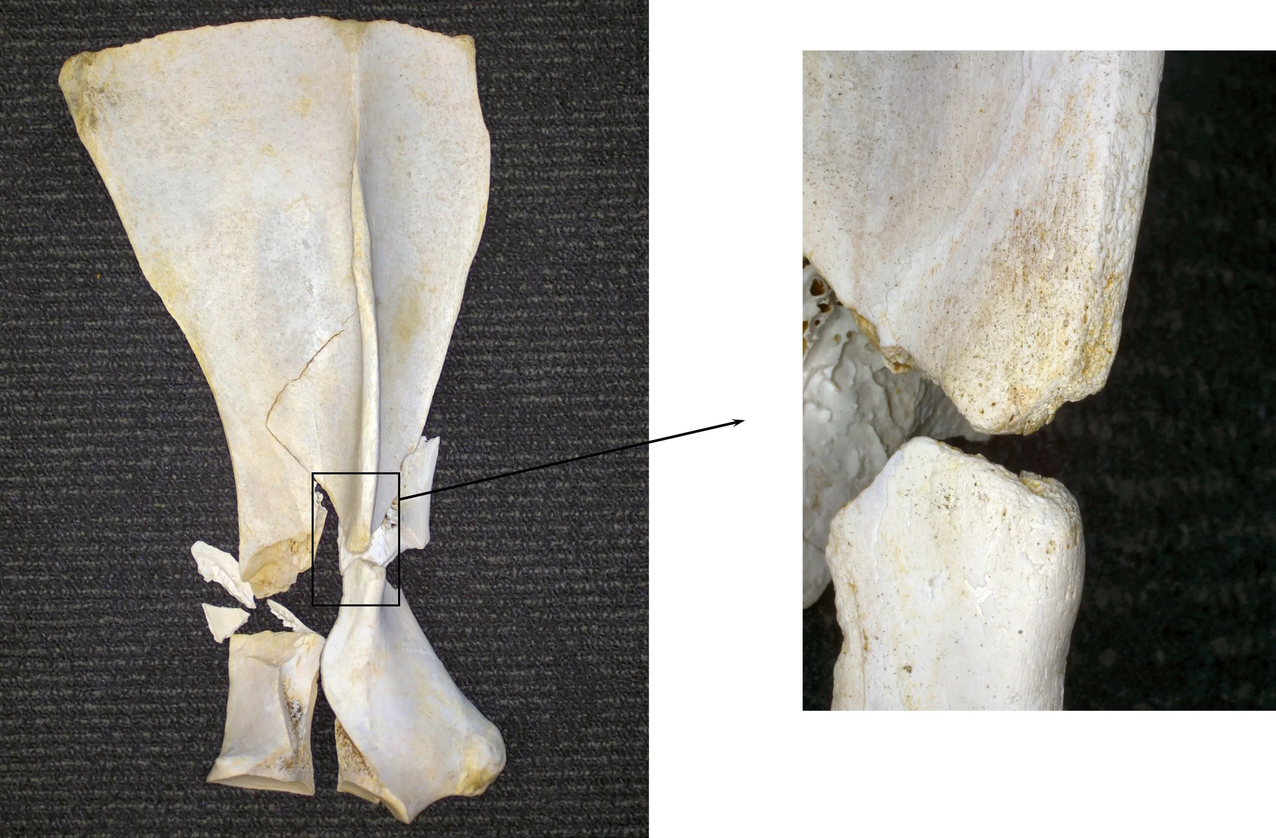
While most fractures that affect racehorses on the flat appear to arise spontaneously, while the horse is racing, and in the absence of any obvious trauma, we now know that the majority are the consequence of structural fatigue, involving a process that started weeks or even months earlier. Just as a paper clip will break if it is repeatedly bent once too often, a bone will fracture if it is loaded recurrently beyond certain limits. Microscopic damage accumulates in the bone tissue until a small fissure develops, which can extend to a severe fracture if it is not detected and the horse continues to race.
The fatigue life of bone decreases exponentially as the magnitude of load on the bone during each loading cycle increases. Loads on the skeleton are directly related to the speed of exercise, work at a fast gallop causes fatigue damage at a rate many orders of magnitude greater than when the horse is exercised at slower speeds. Unfortunately, fatigue damage, and even formation of small fissures in the bone, often proceed without the horse showing any outward signs and so, historically, affected horses have often gone undetected until it is too late.
2: Are there any natural biological mechanisms that protect against fracture?
An ability to run fast has been of evolutionary benefit to horses by enabling them to escape predators. Humans have refined this characteristic in specific breeds, notably the Thoroughbred, through targeted breeding and training practices. The need for a horse to undertake a fast gallop in the wild is likely to be relatively infrequent and well-within the bounds that are likely to cause fatigue damage. Conversely, society’s expectations of racehorses, and the training imposed on them, place loads on the skeleton that may be excessive.
Some readers may be surprised to learn that bone is a remarkably “smart” tissue. It is so much more than the equivalent of the “concrete structural beam” laid down to support man-made structures. While bone is composed partly of minerals, it is still very much a living tissue, packed with blood vessels, nerves and cells. Furthermore, the cells are all physically interlinked through microscopic projections, and can communicate with each other through their interconnections. This biological network allows bone tissue to detect the effects of loads applied to the bone as whole, and to initiate a cellular response to model the bone structure if those loads change.
For instance, increase in the magnitude of prevailing loads (e.g. through increase in an animal’s weight or the introduction of high-speed work) will cause greater deformation of the bone and this will stimulate a process that results in the formation of additional bone mass and, if beneficial, subtle reshaping of the bone’s geometry. The bone will be stronger as a result and deform less for the given load. Conversely, if the prevailing loads decrease (e.g. during a period of rest) bone deformation is reduced, and bone may be removed through a process of resorption. This overall mechanism of remodelling is called “adaptation”. Bone adaptation facilitates the formation and maintenance of a skeleton that is continually fit for purpose, even as that purpose changes.
Beyond this, there are biological mechanisms that detect when a bone is damaged (e.g. small cracks develop) and will initiate repair. The restoration process involves removal of microscopic packets of bone tissue, including the damaged portion, and its replacement with fresh, healthy material. This process means that fatigue-related damage of bone can be resolved and, theoretically, that a bone can tolerate infinite cycles of load. However, the process of repair has been shown to be inhibited if the bone is still regularly subjected to cycles of high load (i.e. the horse remains in training) and it can also be overwhelmed if the rate of damage accumulation is too high.
3: Are fractures something that racing just has to live with or can we do something about them?
Applying the knowledge we have gained from research over the past forty years has already had an impact on reducing the risk of fractures in some situations, and there are good reasons for optimism that we will be able to prevent a much greater proportion of racing fractures in the future. For example, epidemiological studies can help to identify certain management practices and design features of racecourses associated with an increased risk of fracture and these can be modified.
The introduction of strict rules governing the validity of sale of a horse in a claiming race in some jurisdictions in the USA, depending on the health status of the horse immediately after the race, has had a significant impact on reducing the incidence of fractures. Over 300 potential risk factors have been examined and those found to have a significant association with fracture, modelled in numerous studies.
Applying the findings to individual racecourses still requires much work and, ultimately, almost half of the variation in risk of fracture appears to be due to factors associated directly with the horse itself. Epidemiology has also been applied to identify characteristics of a horse’s history that may indicate that it is at higher risk of fracture. These studies depend on access to detailed and accurate data and recent work has highlighted the importance of including veterinary records. Access to these records, suitably anonymised to protect confidentiality, will have a profound impact in the development of more accurate models that can be used to predict the risk of fracture.
Studies into the genetics of Thoroughbreds have identified particular genes that are associated with a predisposition to fracture. This is a highly complex field and interaction of the environment and genetics ultimately determines the fracture status of an individual animal. However, genetic screening will help to identify horses that may benefit from closer monitoring. Clearly this is a sensitive topic, especially to breeders, although hopefully stakeholders will make choices that will be in the best interest of Thoroughbreds and racing in the long term.
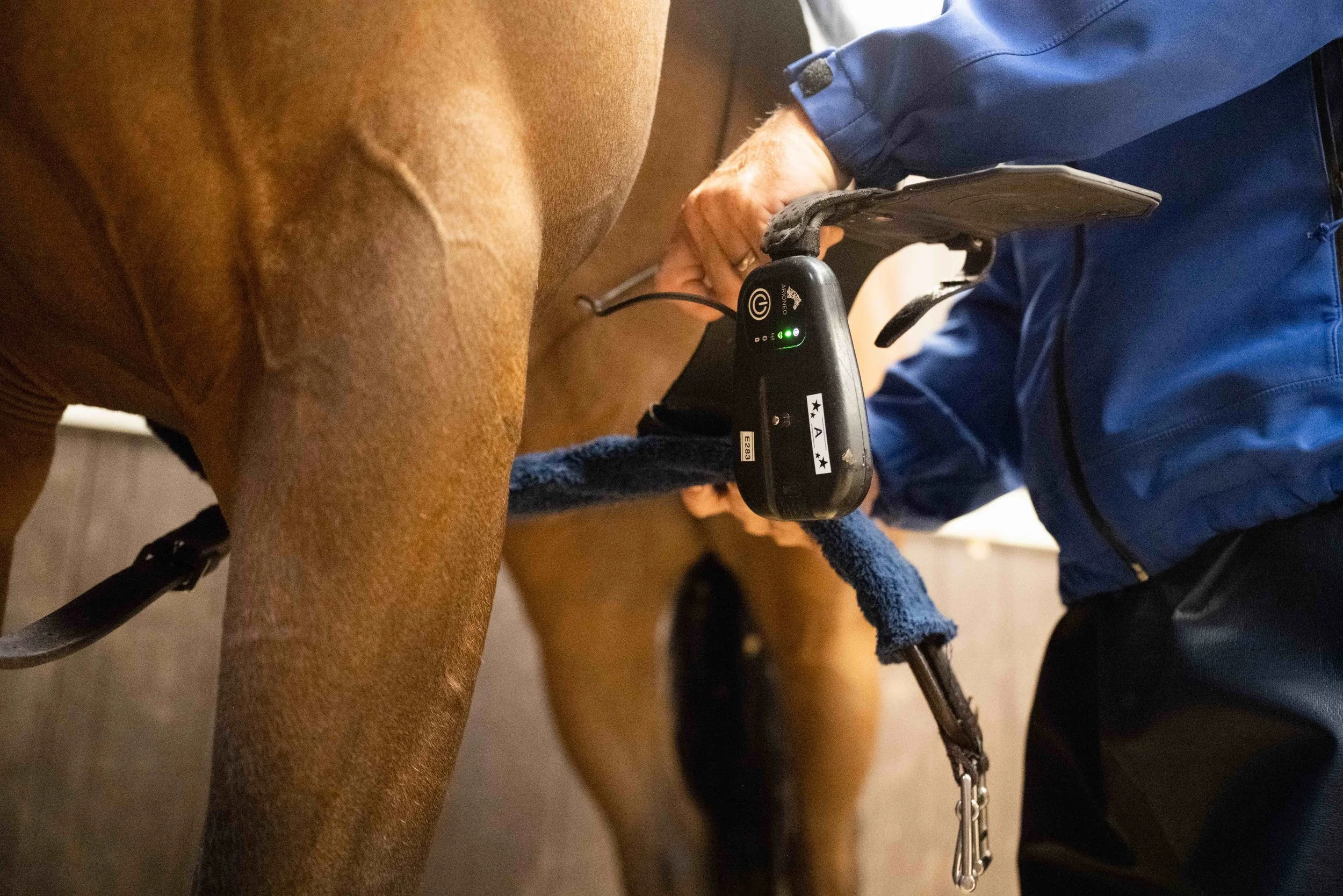
Wearable technology, carried on the horse to record cardiac and stride data shows promise in being able to identify horses at early stages of skeletal injury. Preliminary results suggest that this is possible by identifying changes in stride characteristics, such as stride length, in individual animals in races leading up to the horse sustaining an injury. There has been a lot of interest and publicity associated with wearable technology and its use to identify horses both at imminent risk of fracture as well as those with irregularities of cardiac rhythm which may lead to exercise associated sudden death, although work still needs to be undertaken to substantiate their use in both these contexts.
These devices are also proving increasingly valuable as tools to record actual workload undertaken by individual horses in training. This is important information that is needed for researchers to accurately model associations between workload and risk of fracture, and for trainers to use to apply such models in the future.

Modern clinical imaging technology, especially that which allows detailed scrutiny of areas of bones in the locations where fractures commonly originate, facilitates identification of bone damage due to fatigue much earlier than was previously possible. The increasing availability of computed tomography (CT) and positron emission tomography (PET) machines that can be used to image the lower limb of the standing horse has made their application more practical in horses that are in active training.
There is potential for this technology to be used to screen horses prior to racing, as is currently undertaken before some race meetings in the state of Victoria, Australia. The enhanced resolution of CT helps to identify subtle fissures in bone that may be overlooked by conventional x-ray procedures and in such cases its value as a screening tool is indisputable. However, work is still ongoing to decipher the importance of even more subtle changes in bone structure. Undertaking imaging studies carries expense, and is not without some risk, and the concept of regularly imaging the relevant anatomical regions of racehorses to screen for risk of fracture clearly has challenges.
A simple blood test for “biomarkers” that could identify animals at an early stage of pre-fracture pathology would be a helpful tool. Even if the accuracy of such a test was insufficient to make important decisions about whether or not to race a horse, it could at least be used to reduce the number of animals that would need to be subjected to imaging studies.
Research into changes in the patterns of arrays of many molecules in the blood (so called “omics” studies) have led to many breakthroughs in screening systems in humans and animals. Preliminary studies give hope that this technology can be used to assist in the detection of horses at increased risk of fracture, although a practical test is still likely to be several years off.
It is important to acknowledge that all screening tests carry some risk: they may falsely identify normal horses to be at increased risk (false positives) and fail to recognise horses that have a problem (false negatives). Careful development and thorough testing of screening systems is therefore essential, and this takes time and money. Furthermore, no test is ever 100% accurate, and the practical application of screening designed to prevent fractures will require racing to strike a balance between stopping some normal horses from running and failing to identify some of those likely to suffer a fracture. This will require informed and engaged discussion between researchers and all relevant industry stakeholders, and strong leadership and clear communication from racing’s regulators.
Perhaps an even greater question is whether we can recommend ways of exercising horses in training that minimise the risk of fatigue damage that leads to fracture in the first place. Research conducted in Melbourne, Australia demonstrated that trainers who worked their horses at high speed in training less frequently than others experienced a lower incidence of fracture.
A very important additional bit of detail was that, excepting the extremes, their racing results and performance outcomes were no different. Recognition that bone is like other tissues, in that it needs time to respond to changes in physical activity and requires a training programme that accounts for this, is important. We are all well aware of the need to develop muscle to improve power and endurance in training, but the concept of training bone is less familiar.
Fortunately, an adaptive response of bone has been shown to occur reasonably quickly to only a few cycles of altered load. Stimulating young, healthy racehorses to increase the strength of their bones requires only a short distance of gallop work. Therefore, introducing a very small amount of fast work at the early stages of a training programme can be sufficient to prepare the bones for more sustained work without the risk of causing substantial fatigue damage. However, if the horse is rested from high-speed work for more than a few days, the process reverses, and bone mass will start to be resorbed (in evolutionary terms, there is no point wasting energy carrying around bones that are bigger and heavier than you need them to be).
So, when a horse is brought back into work after a period of rest, the process of training the skeleton has to be considered all over again. Periods of rest in themselves are also important to allow the bone to repair because even a sensitive training programme will result in some fatigue damage, and the accumulation of this damage may eventually lead to fracture. The innate, biological mechanism of bone repair is switched off (inhibited) while horses remain in active training and a period of rest is required for this process to be reactivated and for repair of fatigue-damaged tissue to take place. Therefore, periodic periods of rest from training are required in order to give the skeleton the best opportunity to heal and “reset” itself.
In summary, a training programme that:
reduces the distance of work (especially high-speed work) that a horse undertakes in its racing career to optimal levels that promote bone health (while racing and training);
stimulates appropriate development of bones through short bursts of fast work early on in a training campaign;
builds periods of rest into a horse’s life span in training;
will all reduce the risk of accumulated fatigue damage that predisposes to fractures developing.
To move forward, the industry needs to invest in research so that metrics can be developed to help trainers to apply these concepts to their own training techniques in a practical way. There will be a range of durations and speeds of work that will reduce the risks of fracture across different populations of Thoroughbreds, and these will only be demonstrated by investing in studies measuring the effects of different training regimes in significant numbers of horses.
Scientists and Racing Working Together
Developing and applying measures that are designed to reduce the risk of fracture will require the commitment of all stakeholders. We also need to be realistic in acknowledging that, due to the athletic nature of racing, the risk of a racehorse sustaining a fracture will never be eliminated and, sadly, for some of these injuries euthanasia will be the only humane option.
When racing authorities communicate to the public that the safety of equine athletes in racing is a priority, their credibility is clearly demonstrated by investing in work to bring the risks of injury, including fractures, down to the lowest possible level.
There will inevitably be an element of “pain” associated with the work and changes required to reduce injury rates. Screening systems will disrupt normal cycles of work, there will be frustration as horses that appear fit are withdrawn from races on the basis of findings of “risks” that we might find difficult to conceptualise, there will be additional financial demands to fund studies, and a sense of exposure as records that are necessary for researchers to do their work are shared, even with promises of confidentiality and anonymity.
At the Toronto meeting, the IFHA has already committed to supporting global scientific research efforts to reduce the incidence of racehorse injuries, including fractures. I look forward to the benefits this co-ordinated approach to research can bring by both reducing injury rates in equine athletes, and at the same time, demonstrating to society the priority racing gives to equine welfare.
Assessing the approaches to diagnosing and treating proximal suspensory desmitis
Article by Connor Parsons DipWCF

Diagnosing proximal suspensory desmitis in the hind limb can be difficult. However, the modern diagnostic modalities available to the industry today makes it possible to isolate injuries, allowing both veterinarians and farriers to work together to achieve the best diagnosis and prognosis possible for the equine in question.
In this article, Connor Parsons reviews the anatomy and function of the suspensory ligament, causes and signs of proximal suspensory desmitis and whether there is an ideal procedure for diagnosing, treating and formulating a prognosis for the horse as part of his DipHE Farriery studies.
ANATOMY
The equine limb is complex yet effective. The suspensory ligament is made up of dense white fibrous connective tissue which suspends the fetlock and prevents hyperextension.

Originating at the proximal, plantar aspect of the third metatarsal/carpal attaching to two palmar depressions distal to the carpometacarpal and tarsometatarsal joints descending the channel formed by the 2nd, 3rd and 4th metatarsal/carpal, bifurcating two thirds of the way down the 3rd metatarsal/carpal, making a firm attachment to the palmar aspect of the proximal sesamoids, pulling the sesamoids proximally, then travelling dorsally and distally at an oblique angle to merge with the common digital extensor tendon. This forms a sling to support the fetlock joint. The ligament and its branches are strong but only slightly elastic (Devereux, 2006).
The suspensory ligament also forms a part of the hindlimb stay apparatus which is a system of ligaments, tendons and muscles that work together to allow the horse to stand and doze with minimal muscular effort. Also known as the fright and flight mechanism (Colles & Ware, 2020).
DAMAGE TO THE SUSPENSORY LIGAMENT
Suspensory ligament damage can affect horses of all breeds and ages. However, it is most common in competition horses. Proximal suspensory desmitis (PSD) is inflammation or damage of the main body at the origin of the ligament at the proximal end of the third metacarpal/metatarsal.
The suspensory ligament can be inflamed or there can be changes to the fibre pattern of the ligament. These cases will present with lack of performance, being worse on soft surfaces. In more severe cases a core lesion (hole) can be seen on an ultrasound scan, where a number of fibres have ruptured. This type of injury will have a more sudden onset of lameness (Dyson, 1994). Injury can be solely within the ligament, involve tearing of the fibres of the ligament or be connected to avulsion fractures at the origin, involving the proximal 3rd metacarpal/tarsal (Baxter, 2020). Complete rupture is possible, however, very rare. The prognosis for a complete rupture is not favourable (Dyson, 1994).
Although the suspensory ligament has a slight elasticity to its make-up, if it is stretched it tends to heal with a loss of elasticity making it susceptible to recurrent damage (Colles & Ware, 2020).
SIGNS OF PROXIMAL SUSPENSORY DESMITIS

Proximal suspensory desmitis is a difficult condition to diagnose as the hind limb is complex and many of the functioning structures work in unison. A horse suffering with inflammation or damage to the main body of its hind suspensory can present one of three ways. It may have a unilateral lameness, a bilateral lameness or just a general decrease in performance (Dyson,1994).
CAUSES OF PROXIMAL SUSPENSORY DESMITIS OF THE HINDLIMB
Although there has been extensive research into proximal suspensory desmitis, there is no primary cause in all cases.
Proximal suspensory desmitis is a common injury in both front and hind limbs of the equine athlete. Usually bilateral in the hind limb (Dyson, 2016). All types and breeds of horses are susceptible to this type of injury. Poor conformation is a contributing factor to proximal suspensory desmitis.
Conformational defects such as straight hocks, sloping pasterns and long-toe, low-heel conformations would be at higher risk to injury. These conformational defects will all apply unnecessary pressure to the suspensory ligament. Horses that have suffered with this condition will be predisposed to a repetitive strain injury of this ligament (Devereux, 2006). Overextension of the tarsus as a result of overextension of the fetlock has been linked to proximal lesions. The higher the severity of trauma, the higher the severity of ligamentous lesion. Working horses on deep, soft surfaces will increase the risk of this injury (Baxter, 2020).
The hindlimbs are more frequently affected with this condition than the forelimbs with a much lower success rate of the horse returning back to performance prior to rest (69% hind vs 80% forelimb) (Colles & Ware, 2020).
DISCUSSION

In a study of six horses, this is an extremely small cohort of horses to be able to state an average age a horse is likely to present with this condition. This study also shows that all of the horses studied were of varying fitness levels, therefore stating that this does not affect the likelihood of injuring the hind suspensory ligament. There was only one horse in this study that was unfit and overweight. The rest were all competition fit with good muscle mass, showing that fitness doesn’t necessarily decrease the risk of this injury happening. The case history of the six horses studied did not include which discipline or level the horse was working at. This would be an interesting factor to consider when looking at which horses would be more susceptible to proximal suspensory desmitis.
Each individual case was being looked after by different veterinarians, giving a clear picture of different approaches on how to diagnose and treat this condition. Although for the purpose of a study the varying opinions will make the comparison more difficult. All horses presented with a reduction in performance prior to veterinary contact. Only one horse was reported with a bilateral lameness behind. Flexion testing appeared to aggravate the lameness making it more prominent to see. Local analgesia has been shown to be effective in isolating the area to be investigated. Also, showing lameness on the other hind once the worse limb has been blocked out.
Using digital diagnostic modalities such as ultrasonography to diagnose this condition allows the veterinarian to study the changes in the fibre pattern of the suspensory ligament. This will allow the veterinarian to see the severity of damage caused and allow them to provide the best treatment plan possible. In this study only one horse had a lesion while the other five horses had thickening and slight changes to the fibre pattern. Horse 2 had lesions on both hind limbs however the veterinarian didn’t medicate, box rest was recommended. His prognosis was guarded.
Although radiographs of the feet don’t directly help with the diagnosis of proximal suspensory desmitis, they do allow the farrier to trim accordingly to restore the hoof back to correct hoof pastern axis and mediolateral foot balance. This will reduce lever arm forces thus reducing any unnecessary pressures on the plantar aspect of the limb.

Horses were radiographed for foot balance to aid with remedial trimming and shoeing. This will increase the equines prognosis allowing the farrier to have a clear picture of what is being dealt with. All of the horses that were radiographed presented with a negative sole plane and weak heels.
The question is whether this foot conformation is because the horses are wanting to apply more pressure to the caudal aspect of the hoof in the landing phase, reducing the movement of the metacarpophalangeal articulation. This is an attempt to reduce the loading forces applied to the suspensory ligament. However, it will also cause the heels to become weak. Or, if this conformational defect has caused the suspensory ligament to become inflamed or damaged, thus causing proximal suspensory desmitis.
Proximal suspensory desmitis can be secondary to other conditions such as hock conditions or sacroiliac problems which cause the horse to adopt a different gate. Therefore causing unnecessary loading on the suspensory ligament. It is important that the primary cause is diagnosed and treated when treating proximal suspensory desmitis. This is where scintigraphy can be a useful tool to get a clear picture of the cause involved in individual cases. Scintigraphy is an expensive diagnostic modality which carries significant health and safety risks, this must be taken into consideration when dealing with cases.
All horses studied were worse on a soft surface where it is harder for the horse to guard itself from soft tissue injuries. Horses that are worse on soft surfaces generally are suffering from soft tissue pain. However, nerve blocks will help the veterinarian pinpoint the structures involved when diagnosing lameness.

Although it is possible to have a unilateral lameness with proximal suspensory desmitis in the hind limb it is most common for the lameness to be bilateral. All of the horses in this study had a bilateral lameness, generally worse on one limb than the other. Although presenting prior to veterinary contact as lack of power or struggling to strike off on the correct canter lead.
When a veterinarian is deciding on a treatment plan, the horse is looked at carefully including its previous history as some treatments come with higher risks, although can be extremely effective for reducing inflammation. Shockwave treatment comes with minimal risks involved and is effective; however, many racing authorities require a mandatory 5 day Stand-Down period from racing following the administration of extra-corporeal shockwave therapy. Findings from this study show that the horses with the best prognosis of getting back to competitive work have undergone surgery. Understandably this is the last resort treatment as it is invasive and expensive for the client.
Only one horse from this study did not have any medical intervention and this horse had the least favourable prognosis. This would suggest that box rest alone is not generally enough if the horse is expected to get back to full athletic fitness. The most common veterinary treatment is steroidal injections into the area of interest and shockwave therapy with rest. However, the use of corticosteroids in horses in training often adopt a clear 14-day exclusion on the use of intra-articular (joint) injections before racing in line with different racing authority regulations.

Water based therapy can also be considered as part of the recovery process when bringing the horse back into work. It’s known to reduce limb oedema, stimulate nerves, and improve circulation, which speeds the healing process and provides pain relief. It also aids in joint stability, providing all-around support to the limbs.
Cold water therapy is typically prescribed when the goal is to reduce heat and inflammation. Applying cold water or ice reduces the amount of accumulating fluid to an injured area and can somewhat numb the area, causing a topical analgesic effect.
Underwater treadmills are often used for horses with tendon and ligament injuries to provide a gradual transition back into exercise and regain the range of motion. Swimming is also used to condition the horse without putting a load on the skeletal system. It is often used in the early stages of tendon and suspensory injuries due to no pressure being placed on the lower limb. Trainers who use swimming as part of their routine often find that, in addition to the cardiovascular workout, it also helps the horse relax and settle its mind.
This is not always successful and horses are then admitted for surgery. While the surgery for this condition is successful, there must be consideration taken into the fact that it is not legal to compete at certain levels once this surgery has taken place.
The study shows that the farriery treatment involved when dealing with this condition is varied, depending on which veterinarian the horse is being looked after by. However, the author has had positive results from many different shoeing styles. The main importance of trimming and shoeing for this condition has been shown to restore the best possible hoof pastern axis through trimming, supporting the entire limb and fitting a shoe with an early breakover. This will reduce the lever arm on the metacarpophalangeal articulation, thus minimising unnecessary pressure on the suspensory ligament.
CONCLUSION
Having such a small cohort of horses in a study makes it difficult to finish with a conclusive result. This small study however, has given a positive result in the diagnosis stages of dealing with this condition. At this stage nerve blocks are invaluable along with ultrasonography. In less obvious cases MRI is useful to gain a diagnosis and occasionally scintigraphy will be used to locate the problem. Radiography is a useful tool when dealing with PSD and checking the origin area for avulsion fractures.
This study has also shown that there is a link between a negative solar angle and proximal suspensory desmitis. However, this would need to be studied further and on a greater scale to determine why there is a link between this conformational defect and this condition.
It is paramount that correct foot balance is achieved by the farrier. To achieve this foot balance radiographs are required. This study has shown that there is no definitive way to shoe for this condition, however it has shown a positive result from an early breakover shoe, allowing the horse relieve pressures on the caudal aspect of its hoof. Horses that had the best prognosis underwent surgery, allowing them to get back to competitive fitness.
REFERENCES
Baxter, G. M., 2020. Adams and Stashak's Lameness in Horses. 7th Edition ed. Hoboken, NJ: John Wiley & Sons.
Colles, C. & Ware, R., 2020. The Principles of Farriery. 2nd edition ed. Marlborough: J.A.Allen.
Devereux, S., 2006. The Veterinary Care Of The Horse. 2nd Edition ed. London: J.A.Allen. Dyson, S., 1994. Proximal suspensory desmitis in the hindlimb: 42 cases. British Veterinary Journal, 150(3), pp. 279-291.
Dyson, S., 2016. American Association of Equine Practitioners. [Online] Available at: https://aaep.org/horsehealth/lowdown-high-suspensory-disease-proximal-Suspensory-desmitis [Accessed 19 11 2022].
Smith, M., 2022. Newmarket Equine Hospital. [Online] Available at: https://www.newmarketequinehospital.com/media/pm1beabc/hah349-Vet_susp_desmitis-final.pdf [Accessed 9 April 2023].
The X Factor - Growth spurts in young horses: What can we learn from 'human' research into growth and maturation in sport and exercise?
By Alysen Miller
Ask anyone to list five famous Belgians, and odds are that Kevin De Bruyne’s name will make an appearance. The Manchester City midfielder is widely regarded as one of the best footballers of his generation. Yet you might not have heard of him at all were it not for an innovative talent development scheme in his home country that could influence the way we select, train and manage racehorses.
Traditionally young footballers, like racehorses, are grouped age. By contrast, bio banding is the process of grouping athletes on the basis of attributes associated with growth and maturation, rather than chronological age. “Whether you mature earlier or later has quite a lot of bearing in sport, where greater speed, strength or power can be important,” explains Professor Sean Cumming, an affable Orkney Islander based at the University of Bath who studies growth and maturation. “When you look at children in sport, we group them by age for competition and for training. And while age groups are great in so far as it allows you to match kids of similar cognitive development, motor skills and experience, the challenge is that kids can vary hugely in terms of their biological maturity.” Although the effect of this ‘maturity bias’ doesn’t kick in until pubertal onset at around 11 or 12 years of age, the variance in biological maturity can already be anything up to five or six years by that point.
The concept that relative age can play a determinative role in future sporting success is not new. It explains why broodmares are covered in spring to produce foals in February and March. A winter-born colt running in the Derby in early June of its three-year-old year may be up to 10% of its life older than a spring-born animal—an unquestionable advantage. Or is it?
Indeed, it’s not only in horse racing where the orthodoxy around the so-called ‘relative age effect’ holds sway. In his book Outliers, Malcolm Gladwell notes that a disproportionate number of elite Canadian hockey players are born in the earlier months of the calendar year.
The reason, he posits, is that since youth hockey leagues determine eligibility by calendar year, children born in January are pitted against those born in December. Because the earlier-born children are likely to be larger than those born later (at least until somatic factors kick in), they are often identified as better athletes.
This, in turn, gives them more exposure to better coaching, and the gap between the two groups widens. Sociologist Robert K. Merton has dubbed this the ‘Matthew Effect’ after a verse in the Gospel of Matthew: "For unto everyone that hath shall be given, and he shall have abundance. But from him, that hath not shall be taken away even that which he hath.”
But, cautions Professor Cumming, this only tells part of the story: “What even a lot of the academics get wrong is that relative age and maturity are not one and the same. In fact, our data shows that only about 8% of the relative age effect in academy football can be explained by physical maturity. It’s quite possible to be the oldest kid in the age group but also the least mature, or the youngest kid in the age group but also the most mature.”
The focus on relative size and strength alone, in other words, can create a bandwagon effect. “If you’re looking to identify and develop the most talented young athletes, then it’s going to cloud your vision. It’s going to make some kids look fantastic and some kids look quite poor.” Perhaps tellingly, the last January-born Derby winner, Pour Moi, came in 2008. The youngest winner of the last 10 years, Anthony Van Dyck, was born in mid-May.

Machester City and Belgium superstar midfielder Kevin De Bruyne is the Royal Belgian Football Association’s Programme of the Futures’ most famous graduate
Enter De Bruyne. The Royal Belgian Football Association’s Programme of the Futures, as it is known, allows late-developing players to hone their skills by playing mostly friendly matches against teams of the same physical maturity level, irrespective of age. De Bruyne is the scheme’s most famous graduate. Other members of the late-developer club include Dries Mertens, Thomas Meunier and Yannick Carrasco. By deliberately creating a climate in which late-maturing players get a second bite at the cherry, a country with a population of just 11 million has become a global footballing superpower. Unsurprisingly, other nations are starting to catch on, and several similar programmes have sprung up across the UK and Europe.
Every professional football club has a story about the one who got away—the player that was cut from their programme for being too physically small, from Jamie Vardy (released by Sheffield Wednesday at 15) to Harry Kane (the now 6’2” striker was released by Arsenal at the age of nine). But the consequences are more far-reaching than just missing out on the next footballer superstar. There is compelling evidence to suggest that tailoring the training load to the stage of the athlete’s biological maturity can reduce injuries. The amount of time spent off through injury during an athlete’s formative years is thought to be one of the single biggest factors that determines future professional success.
Since overuse injuries and stress fractures all peak when the athlete is going through their pubertal growth spurt, it is important to identify when an athlete is entering this phase and adjust the load accordingly. As Professor Cumming explains, “Because we know the growth spurt typically takes off at around 85-86% [of the athlete’s predicted adult height] and peaks at around 90-91%, as soon as they move into that phase we can change the training prescription to more developmentally focused stuff—coordination, balance, core strength—all things that are going to help the child transition to a phase when their body is changing rapidly, when they’re more at risk of certain types of injuries.” Early evidence from clubs using the method has pointed to a 72% reduction in injuries.
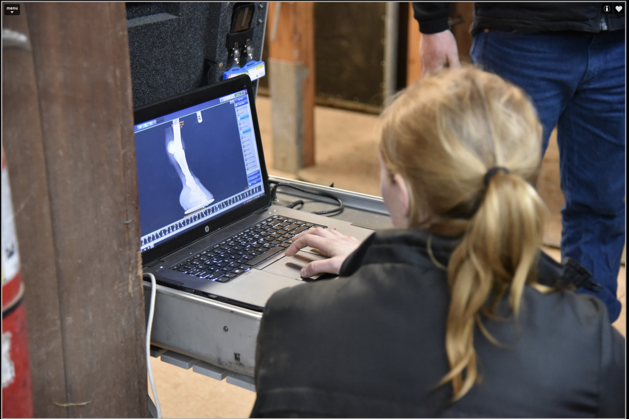
Daniel & Claire Kubler have been bio-banding their horses using knee x-rays, among other metrics, to determine when to increase a horse’s workload
And it’s not just football clubs that are starting to understand the benefits of bio-banding. Daniel and Claire Kübler have been bio-banding their horses using knee x-rays, among other metrics, to determine when to increase a horse’s workload. “We back most of our own horses and train them away to where they can canter relatively comfortably at a normal speed,” says Daniel. “Once a horse can canter away, that’s when we go in and do that first set of x-rays.” The horses are given a grade based on the degree of fusion in the growth plates in the knee, with A being an open growth plate, B being partially closed and C being a closed growth plate. “Those really open ‘A’ horses, you might say, ‘OK, there’s no point—give it a break,’” says Daniel. The C’s, likewise, tend to be easy cases. “It’s really the B horses that are the interesting ones, where you have to make a bit more of a decision,” says Daniel. “What we don’t want to be doing is increasing the workload on a horse that’s relatively immature.”
Although the growth rate in horses varies somewhat by breed, most horses do not reach full physical maturity until around six years of age, with larger breeds like draft horses still growing until eight years of age. A two-year-old horse is an adolescent; it has reached approximately 97% of its mature height by 22 months but critically, its bones will not fully fuse for another four years.

@Equine partnership - equinepartnership.ie
Like humans, horses grow distal to proximal—that is, from the feet up—with the pasterns developing first, fusing at around six months, followed by the cannons at around the one-year mark. The pelvis and spine fuse last. It is during the horse’s two-year-old year that the major leg bones—the radius, ulna and tibia—will fuse. It is therefore important to understand when a horse is entering its growth spurt and tailor its regime accordingly. “It’s about injury reduction,” argues Daniel. “Young athletes are highly susceptible to injury, and by recognising and identifying the growth spurt, you’re massively reducing the injury rate by adapting the training load.”
“The knees are the most delicate bit,” he goes on. “That’s where most of your injuries occur that can cause problems down the line. When you’ve got one with poor grading on its knees, it’s being pre-emptive in your training,” he continues. “You would train that horse a bit more conservatively and not push it quite as hard. You might spend more time on an incline gallop, or you might introduce swimming into the horse’s routine so that you’re putting a bit less concussion through those joints. And hopefully you’re getting the benefit down the line, because they haven’t been pushed too hard, too young.”
Joint licence-holders Daniel and Claire have long advocated for the role of science in training racehorses. “We’re not scared of it,” says Claire, who holds a degree in physiology from Cambridge University. “Having the additional awareness of it gives you a greater understanding,” she asserts. Coming from a non-racing background, meanwhile, has allowed Daniel to approach training with something of a fresh perspective: “It’s the critical questioning. A lot of things in racing are done because that’s the way they’ve always been done, and you can work backwards and find that the reason they work is because, scientifically, it stacks up. But there’s other things where you actually go and look at the science, and it doesn’t make any sense to do that.”
“I love reading about human sports science and listening to podcasts to get ideas,” he explains. “Essentially we’re all mammals, and although there are some differences, there are also a lot of similarities.”
Following the science has not only allowed the Küblers to produce happy, healthy horses—“I’d like to say our horses are very sound and durable,” notes Claire—it has helped them manage owners’ expectations. “Owners enjoy the insights and better understanding themselves as to how the horses progress and develop,” she says.

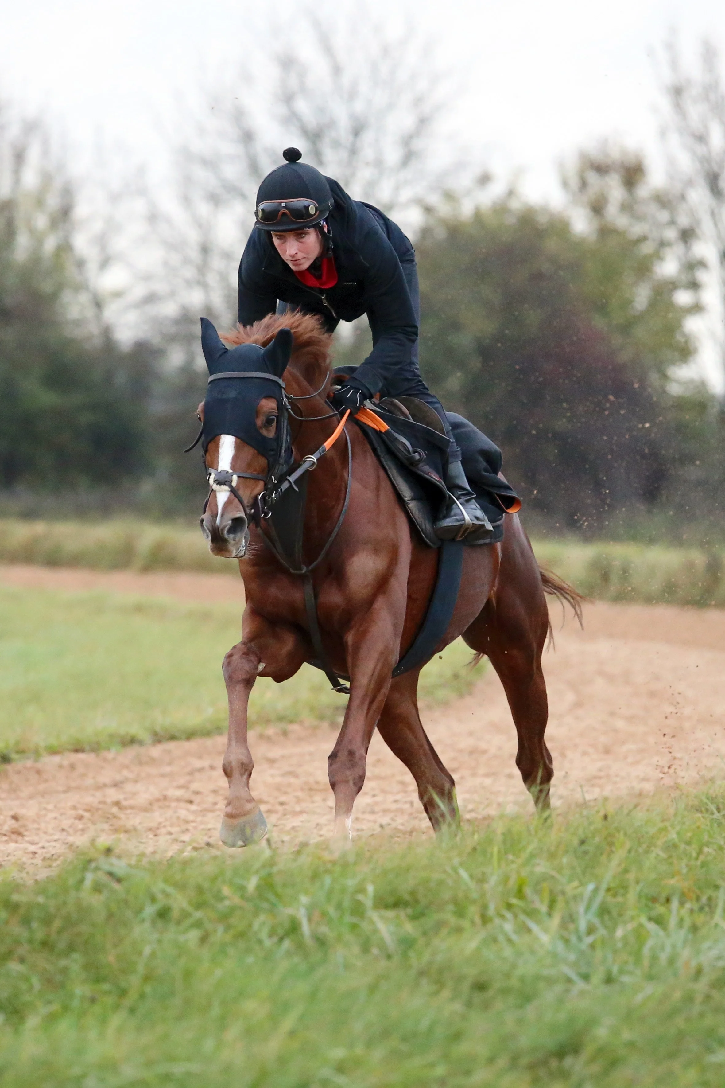
Feedback from work riders is just as important as the science and can provide and can provide as much insight into the horse’s state of growth as an x-ray
“As a trainer, sometimes you can look at a horse and you can see it’s backwards and it needs time,” says Daniel. “What’s helpful about having the knee x-rays is that it’s a very visible thing to show to someone who doesn’t necessarily understand horses particularly well or isn’t used to them. It’s a simple way to say, ‘Your horse is immature.’ That’s a helpful tool as a trainer in terms of being able to communicate very clearly with your owners.” Posting regularly on social media, meanwhile, has attracted interest from outside the sport—including from Professor Cumming himself, who reached out to Daniel through Twitter.
The science is certainly compelling. But, emphasises Daniel, you cannot rely on data alone. “You can’t solve the challenge of training racehorses purely with numbers in the same way that I don’t think you can solve it purely just by looking anymore, because you’re not looking at bits of information. It’s an example of using a scientific, data-driven, analytical approach to enhance the welfare and time the horse’s development in the right way for that individual,” he says.
“The numbers don’t lie, but still you need the horsemanship,” agrees Claire. Feedback from the work riders, she says, can provide as much insight into a horse’s state of growth as an x-ray. “They can pick up on the horse, whether it’s still maturing and doesn’t quite mentally understand what it’s doing. Then you can come up with ideas together as a team,” she says.
In a climate where racing, and equestrian sport in general, is the subject of increasing scrutiny—both from outside the sport and from within—t is submitted that any sports science techniques that can deliver tangible welfare benefits to the horse should be embraced.
“At the end of the day, they have to go out and race, and they all have to be sound enough to do that,” says Daniel.
“You’re always trying to find ways to help get an edge on the track—to get more winners,” agrees Claire. “But you also just want to do the best for the horse so you’re getting a sound horse to achieve its optimum best.”
SUBSCRIPTION OPTIONS
4 x print issue and online subscription to European Trainer & online North American Trainer. Access to all digital back issues of both editions.
Your subscription will start with the October - December 2025 issue - published at the end of September.
If you wish to receive a copy of the most recent issue, please select this as an additional order.