The On-going Effort to Minimise the Rate and Impact of Fractures
Published in European Trainer, January - March 2018, issue 60.
In thoroughbred racing, musculoskeletal injury is a major safety concern and is the leading reason for days lost to training. Musculoskeletal injury is the greatest reason for horse turnover in racing stables, with financial implications for the owner and the racing industry. Injuries, particularly on race day, have an impact on public perception of racing.
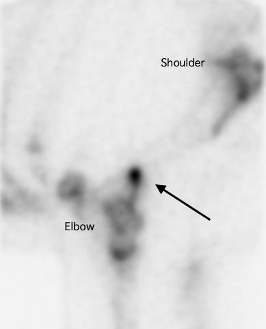
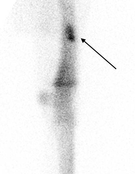
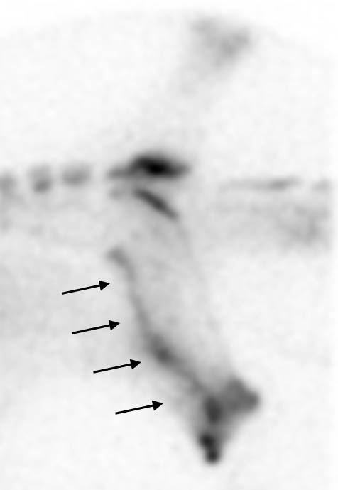
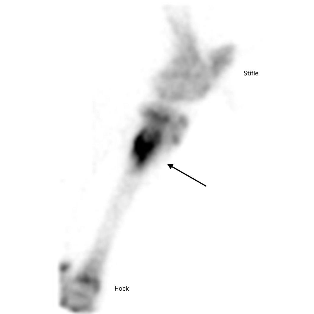
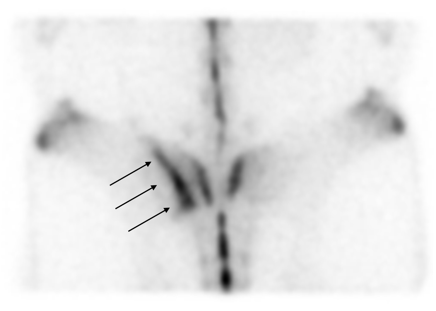
Upper limb and pelvis fractures are less common than lower limb fractures, but they can lead to fatalities. Reducing the overall prevalence of fractures is critical and, at the very least, improving the rate of detection of fractures in their early stages so the horse can be withdrawn from racing with a recoverable injury will be a big step forwards in racehorse welfare. Currently, we lack information on the outcomes following fracture, and an article recently published in the Equine Veterinary Journal (EVJ) from the veterinary team at the Hong Kong Jockey Club (HKJC) addressed this important knowledge gap.
Hong Kong Fracture Outcome Study
The HKJC veterinary team is in a unique position to carry out this work because their centralised and computerised database of clinical records, together with racing and retirement records, allows them to document follow-up, which is all but impossible elsewhere in the world. Dr Leah McGlinchey, working with vets in Hong Kong and researchers from the Royal Veterinary College, London, reviewed clinical records from 2003 to 2014 to identify racehorses that suffered a fracture or fractures to the bones of the upper limb or the pelvis during training or racing, confirmed by nuclear scintigraphy, radiography, ultrasonography, or autopsy....
To read more - subscribe now!
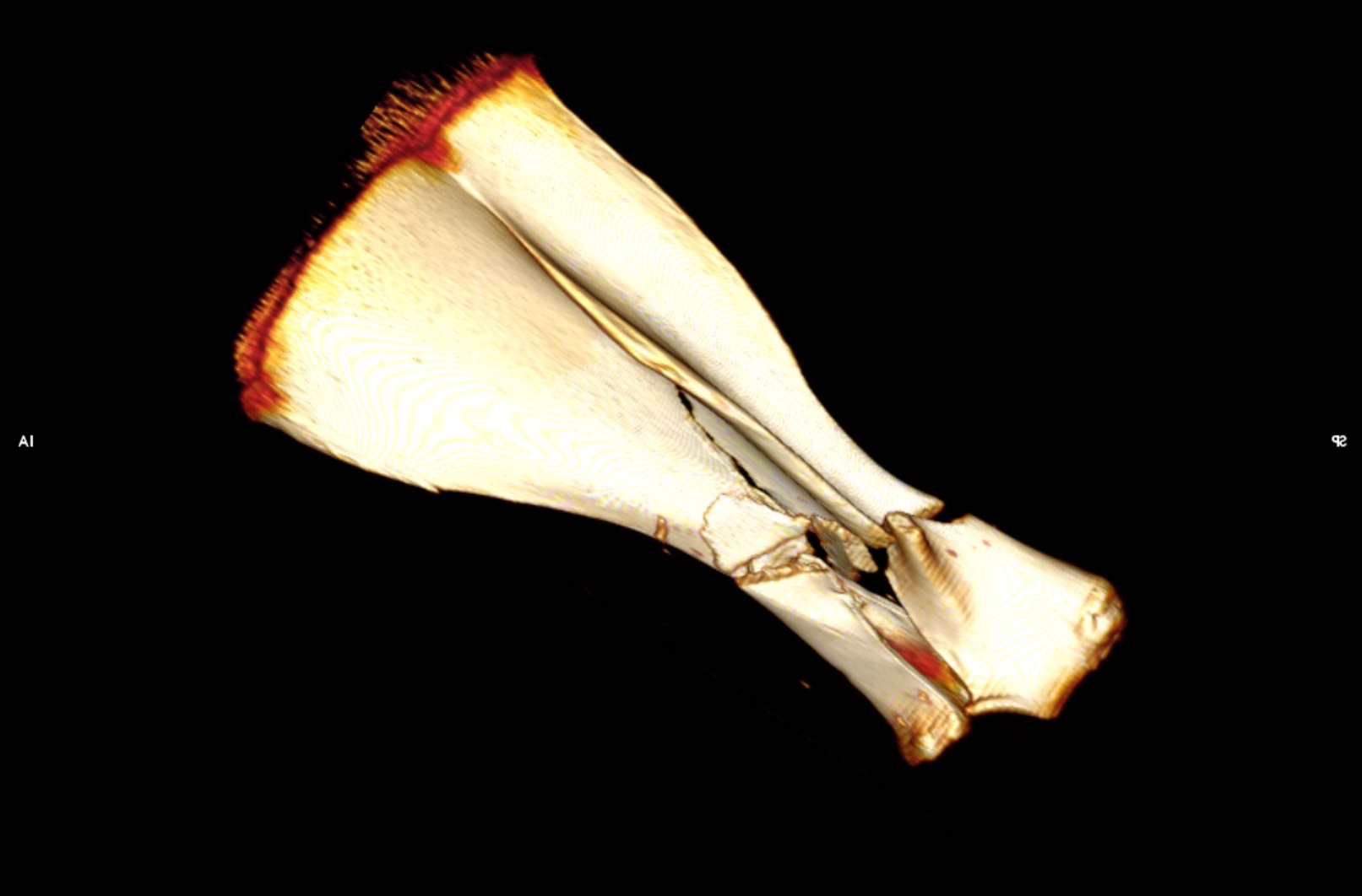
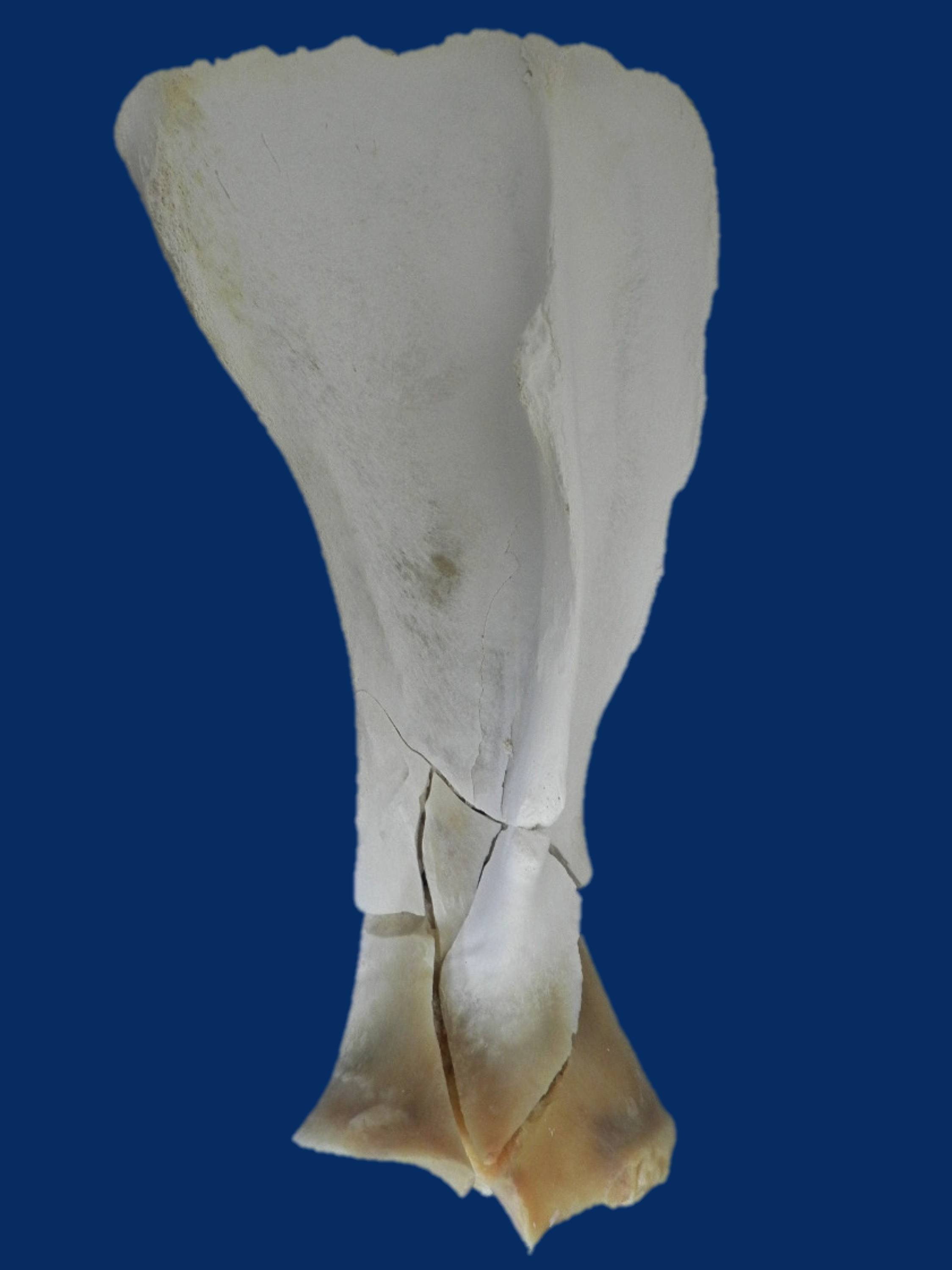
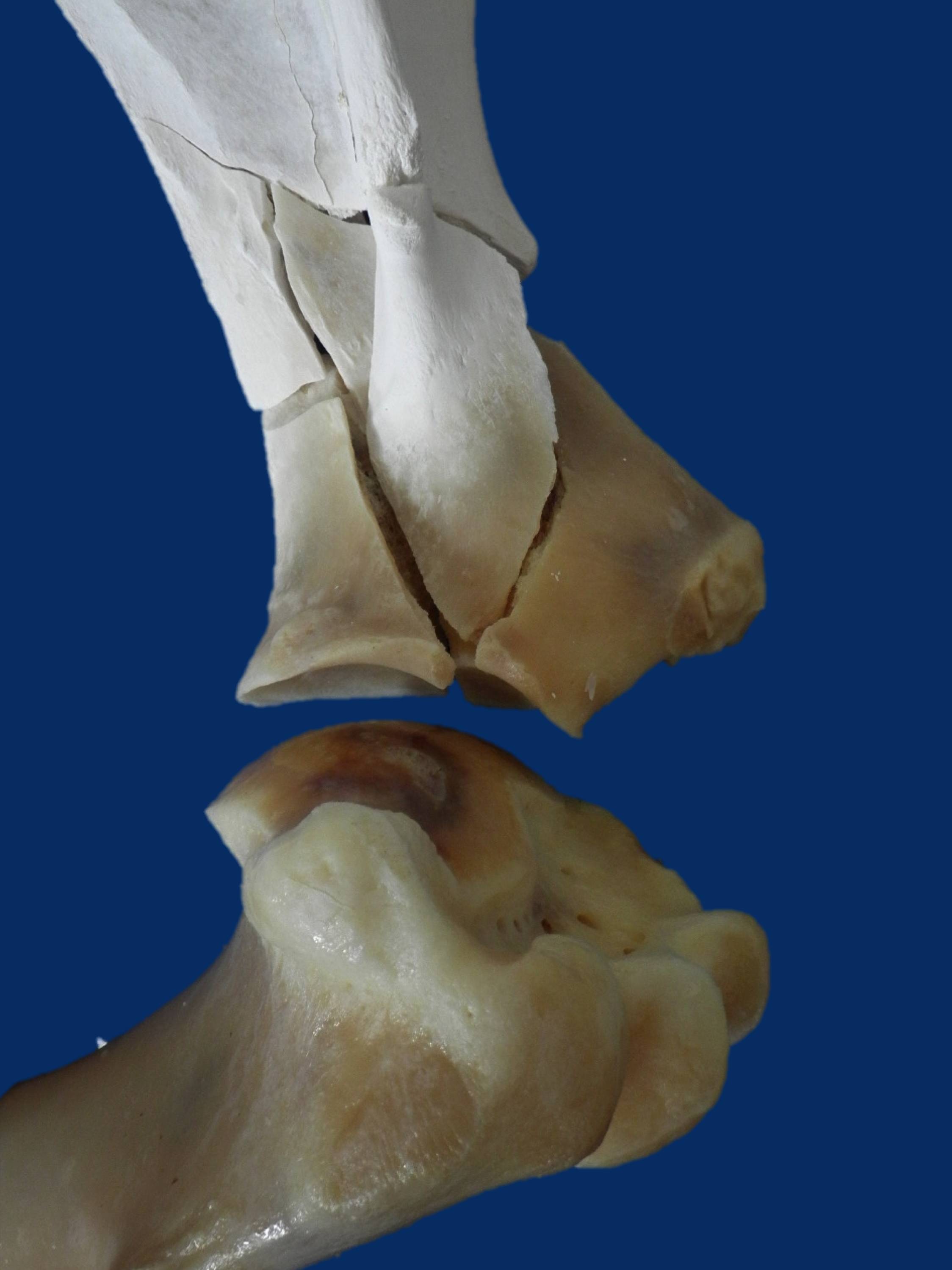
Gallery