A Look at Learning Theory for Reducing Stress and Developing Top Performers

Who doesn’t want to produce a performance athlete who is less stressed, experiences fewer setbacks and enjoys improved welfare? It has been shown that correct application of learning theory principles, starting from a young age, can clear the track.
Learning theory explains how each horse acquires, processes, and remembers the knowledge they need to perform as a racehorse. For handlers this means developing a deep understanding of how a horse learns. Naturally gifted horsepersons are already employing some of the principles, often without even knowing it, with their impeccable timing of cues.
In the past two decades, both social licence to operate and equine welfare have come to the forefront. Failing to grasp how the horse’s brain works (both their capabilities and limitations) can lead to confusion, unnecessary stress, and dangerous behaviors. Conversely, understanding equine learning theory can streamline training, lessen the chances of injury to both horse and handler and improve efficiency in training.
How Foal NZ is Using Learning Theory for the Win
Globally recognized for their success training thoroughbred foals, Foal NZ has been achieving remarkable results in New Zealand. Through utilizing learning theory they have completed over 35,000 training sessions without injury for the past two decades. Yearlings fetching million-dollar price tags and Group One race champions such as So You Think, Military Move and Jimmy Choux emerged from the program, earning acclaim in Thoroughbred racing circles.
Learning theory is a way of explaining the different types of training typically used with horses and contains four main quadrants that explain how consequence is used to shape behavior. Sally King of Foal NZ explains how they use learning theory to create confident, capable young athletes.
The Foal NZ team use primarily positive and negative reinforcement to encourage the foals to learn the desired behavior while becoming confident in both their ability to learn and their relationships with people. Using negative reinforcement (removing a cue the horse doesn’t enjoy as soon as the horse responds), the handler will ask the foal to move forward using pressure from a rope around the foal’s rump, releasing the pressure as the foal moves the first foot off the ground.
Once the foal is confident about being touched by people, then they will start to include positive reinforcement (doing something the horse likes) by offering the foal neck scratches once a desired behavior is performed. “This encourages the foal to try to find the solution to what we are asking as they feel relaxed and confident in their abilities – the perfect mental state for accelerated learning,” says King.
Positive punishment (adding something the horse does not like such as vocal or physical reprimand) is less effective than other methods. An example of positive punishment is when a horse rears and ‘shanking’ the horse’s face is employed to punish them. While this may work temporarily as the horse attempts to avoid pain, numerous studies in children have shown that using positive punishment creates anxiety and fear and reduces brain function. Likewise, if the horse is afraid, they are hindered from using their brain to find solutions. Handlers that can recognise stress from facial expressions, muscle tension, and behaviours can pre-empt the rear by changing the situation to reduce tension.
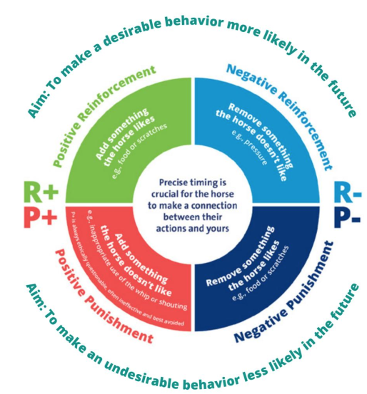
Negative punishment (taking something away that the horse enjoys or values) is generally not recommended as this may cause stress and increase anxiety.
Desensitizing and flooding are two learning concepts that have also been used in training. Desensitizing involves gradually getting the horse accustomed to something, while flooding entails exposing the horse to a frightening stimulus in an intense and unavoidable manner. For instance, a young horse may spook or hesitate at a particular part of the track during their morning workout due to something new or unusual appearing in that area.
If a colt spooks and stops, and the handler was to tie them or hold them so they were forced to stay near what was frightening them, this would be called flooding. Eventually the horse would stop showing the fear behaviours but only because they have learned that nothing they do will make the scary object go away. It is not because the horse now feels comfortable in that area. Flooding can have a compounding effect called ‘trigger stacking’. Initially, the horse may suppress signs of fear, but as stress accumulates and the threshold is crossed, it can lead to sudden, intense reactions or behavioral outbursts.
In contrast, desensitization could involve putting more distance between the horse and the ‘scary’ area at first. They may pass that area alongside another horse to gradually increase comfort with that part of the track, enabling pace to be maintained in future laps. A jockey that can sense his mount starting to hesitate or veer away can use their powers of prediction to desensitize and foster confidence rather than risk escalating stress.
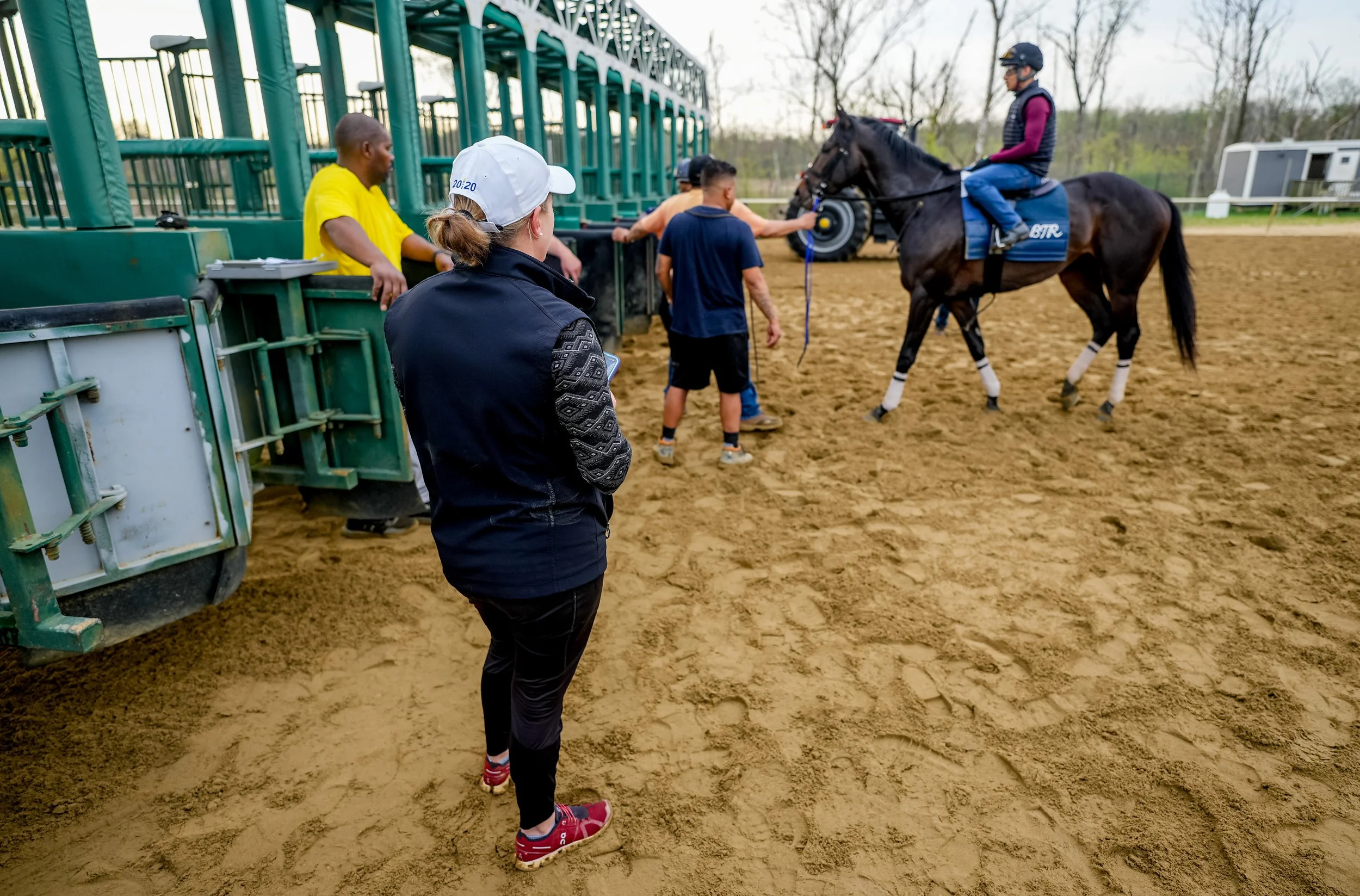
Another well-applied example of learning theory is how trainers typically ‘shape’ responses when introducing horses to the starting gates. Trainers typically break down the elements of being able to use starting gates successfully into multiple parts, gradually going from walking past the gates to walking through open gates following a lead horse, to being beside the lead horse, to stopping inside the open gates, to stopping with one gate closed, then two, then waiting inside, then breaking at a walk and subsequently faster gaits. This way of training called ‘shaping’ also considers the horses ethology in understanding that they are social, prey animals and can feel uncomfortable being restricted in small spaces.
Timing and consistency are arguably the most important tenets of effective training; if the horse can predict what the handler or rider wants and knows they will consistently ask for the behavior in the same way each time, they are much more likely to perform successfully and confidently.
Effective training relies on the simple relationship between the cue, the response and the reinforcement and being able to read stress levels. “In the bloodstock industry, young horses are more likely to be exposed to a wider range of handlers and environments than sport horses but will typically have to perform a smaller range of behaviors than a sport horse,” says King. Thorough training of cues and responses will set the horse up for future success when a wide range of handlers, with varying experience ask the horses to perform behaviours during varying states of arousal.
Using a training system that uses clear principles of learning and lessens the occurrence of conflict behavior, avoidance and escape behaviors has positive outcomes for both horse and human safety and welfare. A young horse that has been trained this way will be more compliant, better able to cope with environmental and social changes and consequently safer. Not only that, but they will feel like they can predict their world, succeed at their job and have some sense of control over what happens to them – all things that increase self-confidence and thus optimize performance.
Within a busy yard, there are time pressures often resulting in limited time to achieve results. Using clear and consistent approaches based on learning theory results in quicker and more robust training, more efficient use of staff time and therefore increased productivity; and for most organizations, improved commercial viability.
Study on Humans Reading Equine Behaviour
This brings us to the next question – How well can you tell? Can a wide range of horse handlers accurately gauge a horse’s behavior as positive, neutral or negative? Dr. Katrina Merkies, a professor in the Department of Animal Biosciences at the Ontario Agricultural College has collaborated on numerous horse behaviour studies and recently published a paper on this very topic.
As it turns out, most of us might not be as perceptive as we think. Merkies’ recent study explored how accurately people can interpret horse-human interactions by looking at photos and watching videos. On average, participants correctly identified whether a situation was positive, negative or neutral only about 52% of the time, which is barely better than chance.
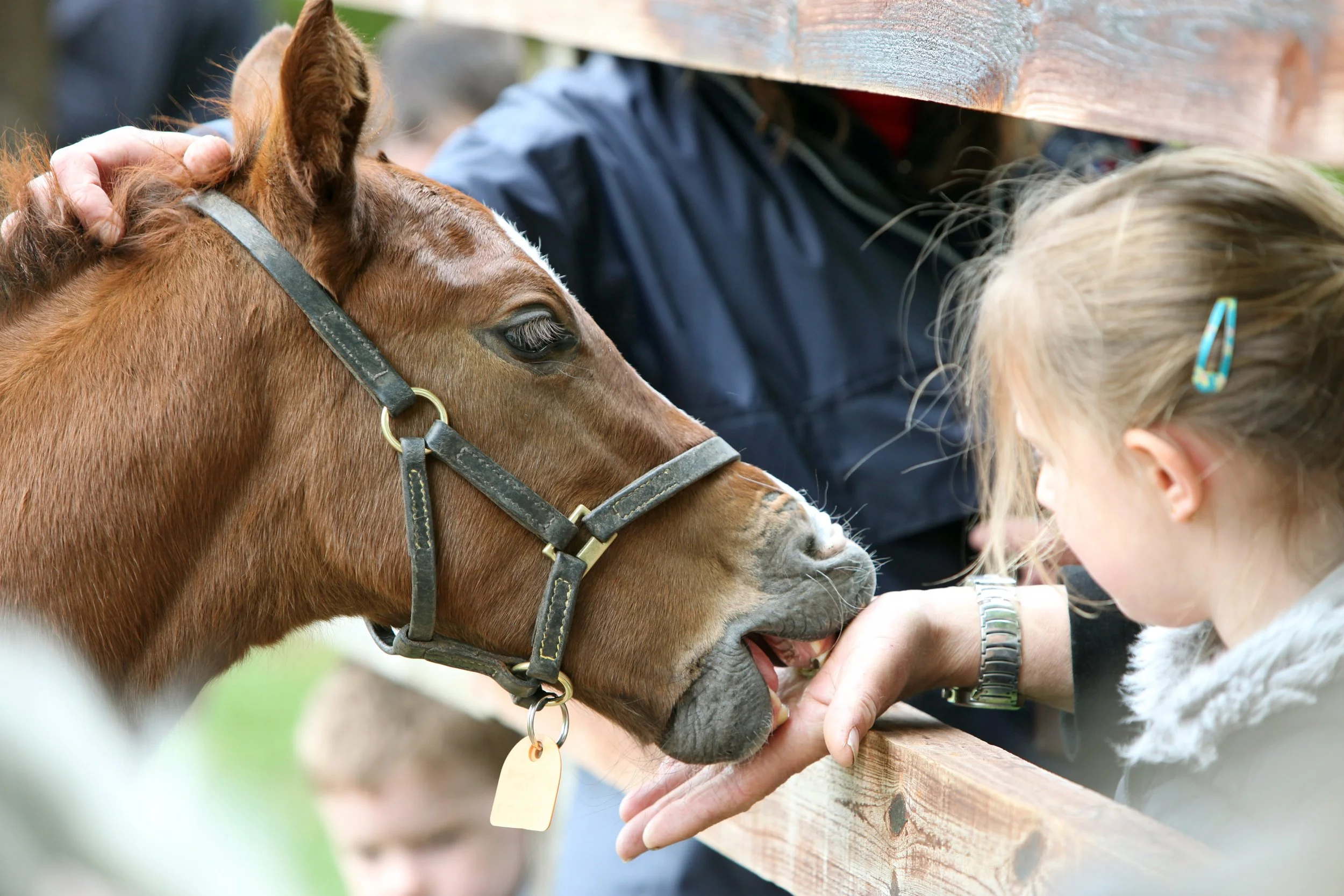
To establish a benchmark, the researchers first had equine behavior specialists evaluate the same media. Their assessments were treated as the gold standard. When the study participants viewed these clips, their interpretations often missed the mark—unless the emotional cues were especially obvious. For instance, people were more likely to recognize a negative scenario when a horse clearly refused to walk across a tarp, or a positive one when a foal willingly approached a person for attention.
These results raise important questions about how well we understand the emotional lives of animals, and how that understanding—or lack thereof—can impact their welfare and how we approach training.
Subtle Signs
While people were somewhat successful at identifying obvious emotional cues in horses, the study revealed a significant gap in recognizing more subtle indicators. According to Dr. Merkies, many of these nuanced signals are found in the horse’s facial expressions. Participants often reported focusing on the horse’s face to gauge their emotional state but frequently overlooked finer details.
Some of the key subtle cues included the direction of the horse’s ears, tension lines around the eyes, and the flaring of nostrils. These small but telling signs can reveal a lot about how a horse is feeling—whether they are anxious, curious, or relaxed. Unfortunately, these indicators are not always easy to spot, especially for those without specialized training in equine behavior.
Improving our ability to recognize these subtle cues could raise the bar for increasing positive human-animal interactions and improving the chances of early intervention at the first sign of physical issues.
Does Self-Awareness Help Us Understand Horses?
One of the central questions of the study was whether people who are more in tune with their own bodily sensations—such as heartbeat, breathing, or muscle tension—are also better at interpreting the emotional states of horses. This idea stems from human psychology research, which shows that individuals with greater internal awareness, or interoception, tend to be more empathetic toward others.
To explore this in the context of human-horse interactions, the researchers used a validated tool called the Multidimensional Assessment of Interoceptive Awareness (MAIA-2). This questionnaire measures how aware people are of their internal bodily states across eight dimensions, including emotional awareness, attention regulation, and body listening. Participants rated themselves on a scale from 0 (not at all) to 5 (very much) for each item.
Participants were asked to evaluate the various horse-human interaction clips before the MAIA-2 results were recorded to avoid skewing the results. Surprisingly, the results showed no significant correlation between a person’s interoceptive awareness and their ability to accurately assess the horse’s emotional state.
This unexpected outcome raises several possibilities. It could mean that interoceptive awareness simply doesn’t translate across species, or that the MAIA-2 isn’t the right tool for this kind of cross-species empathy. Another possibility is that the participants—many of whom were highly experienced with horses—relied more on their practical knowledge than on emotional intuition when evaluating the clips.
Learning to See What Horses Are Telling Us
The study highlights a clear need for improvement in how people perceive and interpret subtle equine behaviors. So how can we get better at this? According to Dr. Merkies, education is the obvious starting point—but it’s not the whole solution. “We can learn about these cues,” Merkies explains, “but being able to apply that knowledge in real-life situations is a different challenge.”
One promising approach is the use of tools like the Horse Grimace Scale which can help observers assess facial expressions and other subtle signs of discomfort. These tools are gaining traction in professional settings; for example, the Hamilton Mounted Police unit uses facial grimace scoring as part of their daily horse care routine. Incorporating such practices into everyday horse management can train people to notice and interpret the finer details of equine behavior.
Dr. Merkies emphasizes the importance of shifting our perspective: “We need to stop, listen, and pay attention—not from an anthropomorphic viewpoint, but by trying to understand how the horse is experiencing the situation.” This means resisting the urge to project human emotions onto horses and instead learning to see the world through their eyes.
Challenging Industry Norms
Another barrier to better understanding horses is the normalization of certain behaviors within the equine industry. “There are a lot of myths that get passed down and accepted as just the way things are,” says Dr. Merkies. Take, for example, a horse pinning their ears when the girth is tightened. This is often dismissed as the horse being a grouch or even 'normal' behaviour for that horse, but this mindset can prevent us from asking deeper questions: Why is the horse reacting this way? What are they trying to communicate? Is there a physical reason for this reaction.
Another great example of negative feedback, often ignored or normalized, is a horse that displays discomfort while being groomed by constant fidgeting, head tossing or grimacing. Again, the discomfort should be acknowledged and addressed, perhaps with softer brushes or counter-condition using positive reinforcement. Is there a physical issue that requires veterinary intervention?
By challenging long-held assumptions and encouraging critical thinking, the equine community can move toward more benevolent and informed interactions with horses.
Positive Reinforcement - Building Better Bonds
One of the most promising ways to improve horse training and welfare is through the use of positive reinforcement—a method that rewards desired behaviors to encourage their repetition. Dr. Merkies emphasizes that this approach not only works but often leads to better outcomes than traditional methods that rely on punishment or pressure.

Despite its effectiveness, positive reinforcement is sometimes misunderstood. Common myths suggest it might make horses ‘mouthy’ or lead to weight gain from treat overuse. Others argue it’s unnecessary because a horse should obey out of affection or loyalty. These misconceptions can discourage people from adopting more compassionate and effective training techniques.
Positive reinforcement doesn’t have to be complicated—or even food-based. While treats are a common and convenient reward, other reinforcers can include scratches, companionship, or access to a favorite location. The key is understanding what motivates your individual horse.
Dr. Merkies offers a simple but powerful example: “When you go to halter your horse, do they come to you, ignore you, or turn away?” These responses are forms of feedback. Even subtle behaviors—like a horse turning its head slightly away when approached—can signal discomfort or reluctance.
Recognizing and responding to these cues can transform training into a more cooperative and enjoyable experience for both horse and handler.
Ultimately, positive reinforcement fosters a relationship built on trust and mutual respect. “It’s super satisfying,” says Dr. Merkies, “when they come running up to the gate or whinny from the field. Then I know they’re looking forward to the training session.”
The benefits of a horse experiencing more positive interactions with humans than negative ones are obvious from a welfare and safety standpoint. When a horse is repeatedly exposed to negative interactions with humans, they may develop fear or resistance, which can make handling more challenging and increase the risk of injury for both the horse and the handler. If you are looking for ways to use more carrot and less stick to reduce stress and setbacks, consider applying the principles of learning theory to your horse training program.

2025 Gerald Leigh Memorial lectures where we learn about the latest research in laryngeal surgeries and tendon rehabilitation
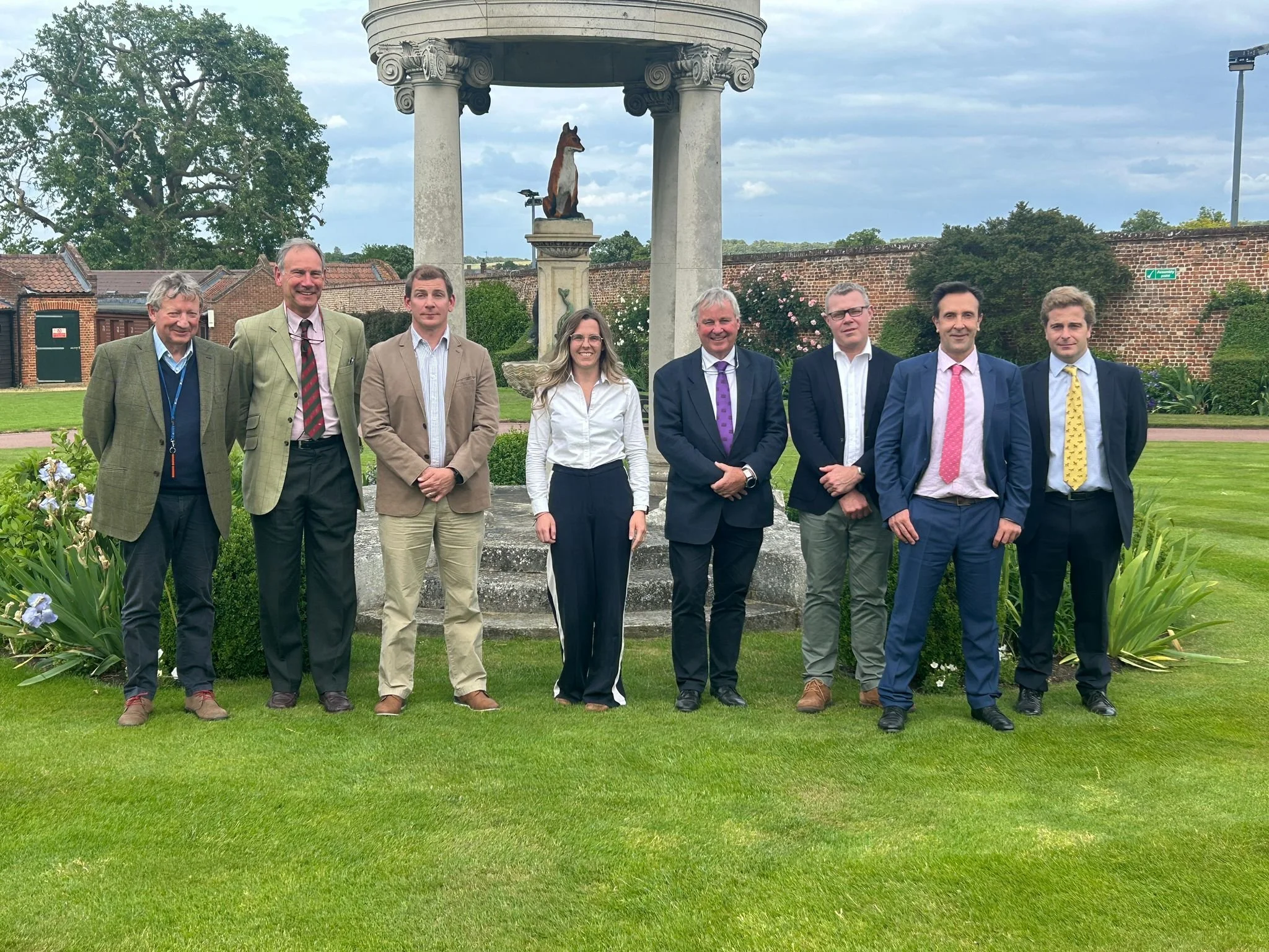
The seventh renewal of the Gerald Leigh Memorial Lectures, in association with Beaufort Cottage Educational Trust took place at Tattersalls in Newmarket, England on June 4th.
The Gerald Leigh Charitable Trust was established in 1974, set up in memory of Gerald Leigh, a prominent owner breeder and best known for breeding the highly successful Barathea and Markofdistinction.
His legacy lives on through the trust, which not only reflects his remarkable achievements and lasting influence in the world of Thoroughbred breeding and racing, but also continues his deep passion for scientific advancement and the welfare of horses—both within the racing industry and the wider equine community. The trust stands as a testament to Gerald Leigh’s enduring commitment to excellence, care, and innovation in all aspects of equine life.
Wind ops- the decision making and diagnostics
Tim Barnett MRCVS of Rossdales Veterinary Surgeons, delivered two informative and interesting lectures on wind ops and the decision making and diagnostics relating to them. As we all know, wind surgery addresses upper airway conditions in horses that impair breathing and performance. Key anatomical structures involved include the arytenoid cartilage, vocal folds, epiglottis, and soft palate. Common issues include vocal fold collapse, often causing a whistling noise and linked to progressive recurrent laryngeal neuropathy “roaring”, which severely obstructs airflow. Another frequent problem is dorsal displacement of the soft palate (DDSP), where the soft palate flips over the epiglottis, blocking airflow and causing sudden loss of performance.
Palatal instability often precedes DDSP. Other conditions include medial deviation of the aryepiglottic folds, nasopharyngeal collapse, epiglottic entrapment, and ventral luxation of the arytenoid apex (VLAC). These disorders vary in severity and may be progressive or multifactorial. Barnett clarifies that although surgical interventions target these conditions, outcomes depend on the specific disorder and severity.
Upper airway conditions remain a major cause of poor performance in racehorses, with many requiring multiple surgical interventions. Accurate diagnosis, particularly via exercising endoscopy, is key, as many disorders only become apparent during physical exercise.
Tieback (prosthetic laryngoplasty) is the most common wind surgery but carries risks such as aspiration, pneumonia and swallowing dysfunction, despite efforts to improve surgical techniques. Newer techniques, like standing tiebacks and improved implants (e.g., titanium buttons, reinforced screws), aim to reduce complications and enhance results.
Other surgeries like Hobday (vocal fold removal via laser) were also discussed, emphasizing the delicate nature of airway surgeries and the ongoing challenge to balance treatment effectiveness against risks and complications in our racehorses.
For DDSP, tie-forward surgery, which mimics natural muscle action to restore laryngeal position, has shown positive results, while thermocautery remains controversial. Epiglottic entrapment can now be safely corrected in standing horses using lasers or scissors. Emerging therapies include laryngeal reinnervation and dynamic neuroprosthesis to restore muscle function, as well as vocal fold filling to reduce aspiration pressure.
Collagen cross-linking is also under investigation as a less invasive method for soft palate stiffening. Barnett concludes, precise diagnosis and tailored interventions are crucial for optimal results in treating upper airway disorders in racehorses. These advances reflect a growing push for safer, more effective airway interventions in the racehorse.
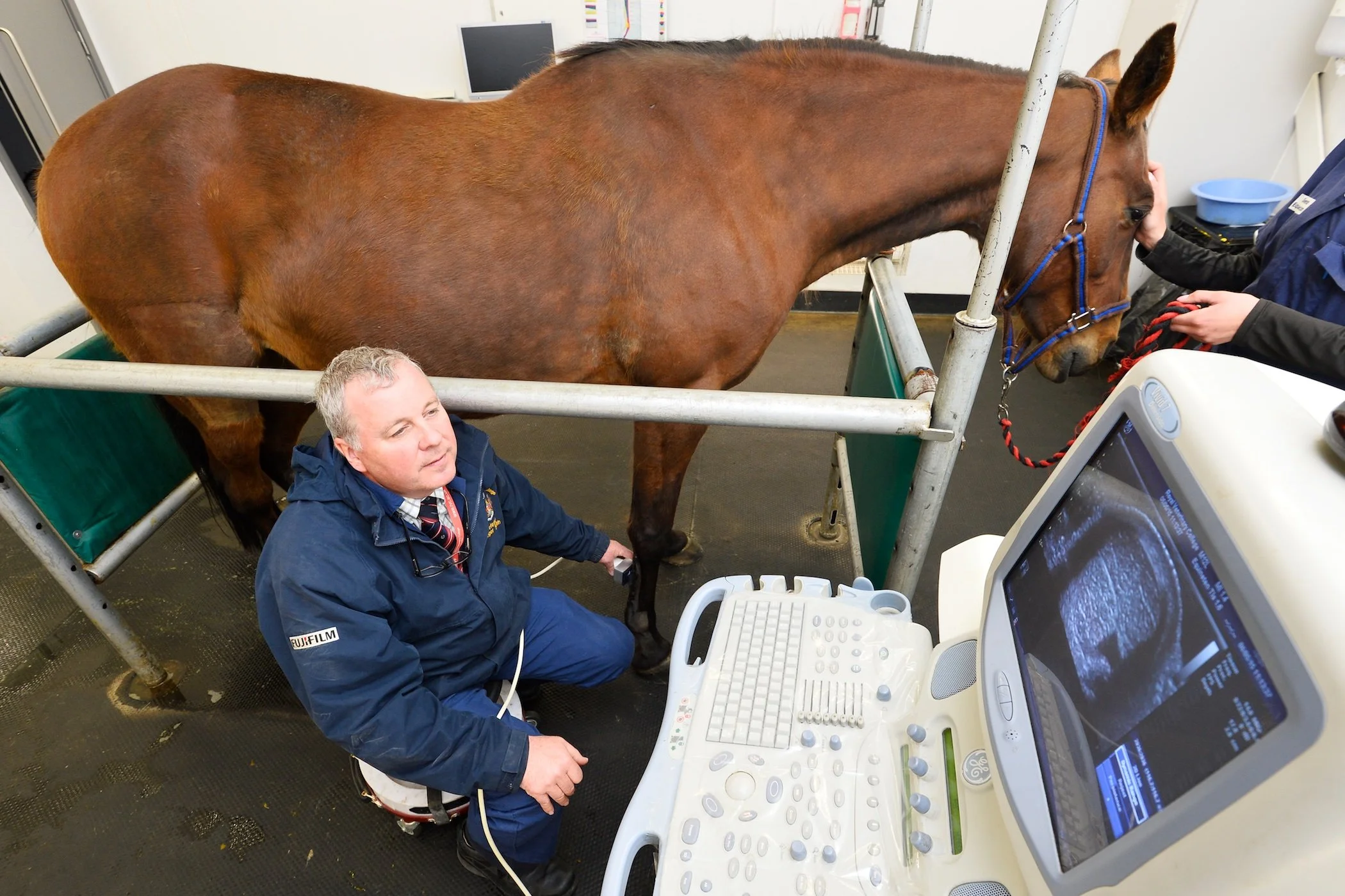
Barnett then moved onto discussing the critical role of exercise endoscopy in diagnosing upper airway dysfunction in Thoroughbreds, highlighting the limitations of resting endoscopy. While useful for detecting conditions like total RLN, epiglottic entrapment, or arytenoid chondritis, resting scopes often miss dynamic issues such as soft palate disorders and vocal fold collapse.
Recent developments in overground endoscopy which are battery-powered and rider-compatible, allow evaluation during real time exercise, providing accurate and practical diagnosis. This method has become the preferred standard, especially for assessing palatal instability and early RLN.
Clinical signs such as respiratory noise, poor performance, or sudden stops may indicate airway dysfunction, but accurate diagnosis requires proper exercise testing with horses cantering or galloping while synchronizing breaths per stride. Additional tools like laryngeal ultrasonography aid diagnosis and planning of treatment.
Barnett cautioned against performing airway surgery without thorough diagnostics, as multiple simultaneous conditions can exist, and treatments must be carefully targeted to improve outcomes. Around 25% of Thoroughbreds show clinical RLN, reinforcing the need for tailored, evidence-based treatment plans to support both welfare and performance.
Laryngeal surgeries - the evolution of research and engineering of the tie back and nerve graft
Dr Fabrice Rossignol, of Grosbois/Chantilly Equine Clinic discussed laryngeal surgeries, focussing on the evolution of research and engineering of the tie back and nerve graft. Rossignol’s specialist clinic is at the forefront of treating recurrent laryngeal neuropathy (RLN).
The condition is often linked to degeneration of the recurrent laryngeal nerve, affecting the cricoarytenoideus dorsalis (CAD) muscle, which is critical for opening the airway during exercise. Rossignol explains that this muscle contains few fatigue-resistant fibers, making it vulnerable to atrophy. Even minor narrowing of the airway significantly increases resistance, due to the exponential pressure effects described by Poiseuille’s law.
Diagnosis involves treadmill endoscopy and ultrasonography (caudal view of swallowing can be particularly useful) to assess dynamic airway collapse and muscle atrophy. Treatment is tailored to severity; advanced cases may require a tieback (laryngoplasty) using synthetic prostheses to partially open the arytenoid cartilage, though this risks complications like coughing. Newer techniques aim to restore function rather than replace it. One such innovation combines traditional tieback surgery with nerve grafting from the spinal accessory nerve, which activates during inspiration and contains fatigue-resistant fibers.

This hybrid approach improves airway opening and reduces side effects. Standing surgery under sedation allows more precise suture placement, minimizing anesthetic risk. Emerging technologies like 3D-printed implants and titanium screw anchors further enhance outcomes. Rossignol echoes Barnett’s earlier advice, that early intervention and careful case selection remain key to success.
Dr Rossignol continued on to discuss what and how we, as a racing industry, can learn from other disciplines.
Recent research in trotters and sport horses highlights how neck flexion contributes to dorsal and lateral pharyngeal collapse, likely due to nerve inflammation affecting muscles such as the stylopharyngeus. Nasal obstruction, including alar fold collapse and nasal muscle paralysis, also play a role in compromised airflow. Treatment options now include alar fold resection, nasal fenestration (widening), and innovative approaches like titanium mesh implants to replace lost muscular function.
Dr Rossignol explains that high-speed treadmill testing has proven critical in diagnosing dynamic airway conditions, while a multidisciplinary approach involving vets, trainers and farriers enhances management strategies. Use of nasal dilation devices, such as nasal strips, remains restricted under many jurisdictions' rules of racing.
It is clear that Rossignol champions cross-disciplinary learning, working with trotter trainers over decades has yielded practical insights, such as shoe removal to enhance performance. The methodical, detail-driven tack and equipment adjustments made in trotting disciplines provide valuable lessons in optimizing performance.
Dr Rossignol also shares advances in surgical techniques, including refined approaches to epiglottic entrapment, emphasizing the importance of collaborative care. Cross-disciplinary exchange continues to inform diagnosis, treatment and rehabilitation, enriching equine sports medicine and improving outcomes across disciplines.
An update on wind surgeries: what's new?
To conclude the lectures on wind ops, Mark Johnston, Dr Rossignol and Tim Barnett took to the floor to field audience questions. The discussion focused on recurrent laryngeal hemiplegia (RLN) in horses, highlighting its probable hereditary component but unclear linking between particular genes. Experts note the complexity of breeding influences and caution against oversimplifying genetic causes, as RLN will most likely be linked with other traits.
Surgery helps individual horses but may skew breeding populations, as generally only the more expensive stallions receive treatment. Disclosure of surgeries before breeding is debated but difficult to enforce. Non-surgical solutions like resistance masks are emerging but their impact on reducing surgery isn’t yet clear. Overall, understanding and managing RLN’s genetics and treatment remain challenging and unresolved.
Early diagnosis of recurrent RLN relies on ultrasonography to detect early muscle atrophy; surgery is recommended promptly to prevent irreversible damage. In contrast, dorsal displacement of the soft palate (DDSP) often stems from muscle fatigue, immaturity, or inflammation and is best treated medically with training and reinforcement until at least three years old. Surgery is a last resort if medical management fails.
Multiple surgeries can be ethical if done safely and explained clearly. Yearling wind testing is variable and challenging to interpret, complicating sales disclosures. The increase in buyers scoping foals’ pre-sale is seen as an invaluable and unpleasant practice due to solid evidence that a foal’s laryngeal physiology will and can change tremendously as they mature. Ongoing research explores novel therapies such as pacemakers and magnetic stimulation.
The practical management of tendon rehabilitation
There is no introduction needed for Mark Johnston, who kindly provided us with his insight on the practical management of tendon rehabilitation. A renowned trainer with decades of experience offered a pragmatic view on tendon injury rehabilitation in racehorses, challenging long-held optimism around recovery. Despite advancements in ultrasound imaging and a range of therapies, from anti-inflammatories to experimental interventions like carbon fiber implants, he is yet to witness a truly successful long-term return to peak performance in top-level racing following a diagnosed tendon injury.
While ultrasound provides valuable detail, he still relies most on visual and tactile assessment, particularly tendon profile and signs of ‘bowing,’ which he considers a critical turning point. In his experience, few flat horses make a full comeback; many may race again, but recurrent issues and shortened careers are the norm. Mark’s approach is rooted in realism: throw everything anti-inflammatory at the injury early, manage workload carefully, and temper expectations.
Long rest alone is rarely effective and controlled rehab and early, aggressive treatment are key. He notes that previous use of prophylactic anti-inflammatories post-race helped reduce injuries, and questions whether restrictions on racecourse treatments may hinder progress. Prevention, early detection, and practical management remain the trainer’s most reliable tools.
Tendon injuries in racehorses
Professor Roger K.W. Smith FRCVS presented a detailed lecture on tendon injuries in racehorses, focusing on the superficial digital flexor tendon (SDFT) and the role of science in improving prevention and rehabilitation strategies. As a key structure for locomotion, the SDFT functions as an energy-storing spring but operates near its mechanical limits, especially in Thoroughbreds, making it prone to injury from accumulated loading rather than acute trauma.
Research shows that degeneration often precedes clinical injury, particularly within the interfascicular matrix (IFM), which loses elasticity with age and training. Tendon cells also become less responsive with age, impairing repair. Matrix metalloproteinases (MMPs) are implicated in post-exercise matrix degradation, further weakening the tendon.
Professor Smith emphasized prevention through training adjustments: avoiding hard ground, spacing out intense work, and ensuring sufficient recovery of, ideally 72 hours. Early detection is critical. Diagnostic tools such as ultrasound, Doppler, and Ultrasound Tissue Characterization (UTC) can identify structural changes before injury becomes apparent.
When injury occurs, a prolonged, structured rehabilitation program guided by regular imaging is essential. Biologic therapies like mesenchymal stem cells (MSCs) and platelet-rich plasma (PRP) are showing encouraging results, improving tendon structure and reducing re-injury rates. A personalized, biologically informed approach remains key to safeguarding tendon health in racehorses.
Tendinopathy - its causes, treatments and parallels between equine and human medicine
Lt. Col. Dr Tom Clack delivered a comprehensive lecture on tendinopathy, highlighting its causes, treatments, and parallels between equine and human medicine. Tendinopathy, a chronic overuse injury, follows a three-stage progression: reactive tendinopathy (early inflammation), tendon disrepair (structural change and neovascularization), and degenerative tendinopathy (reduced symptoms but increased rupture risk).
Historically, eccentric loading exercises, which came about via human Achilles research, became the core treatment. Today, management is more tailored, focusing on biomechanics, load control, and personalized rehabilitation.
Diagnosis includes clinical evaluation and ultrasound, with advanced modalities like Shearwave elastography and UTC offering deeper insights into tendon integrity and healing.
Dr Clack advocated a multimodal treatment strategy: progressive loading, extracorporeal shockwave therapy (ESWT), and injectables such as corticosteroids (for short-term relief) and PRP, which supports healing through growth factors.
Crucially, he emphasised the value of Thoroughbred racehorses as models for human tendon injury. Their tendons endure similar high loads, and developments in imaging, PRP, and regenerative therapies in equine medicine are increasingly influencing human sports injury treatment.
Dr Clack echoed the importance of early detection, strategic recovery protocols, and ongoing collaboration between human and veterinary medicine to improve long-term outcomes in equine athletes.
Rehabilitating the equine athlete
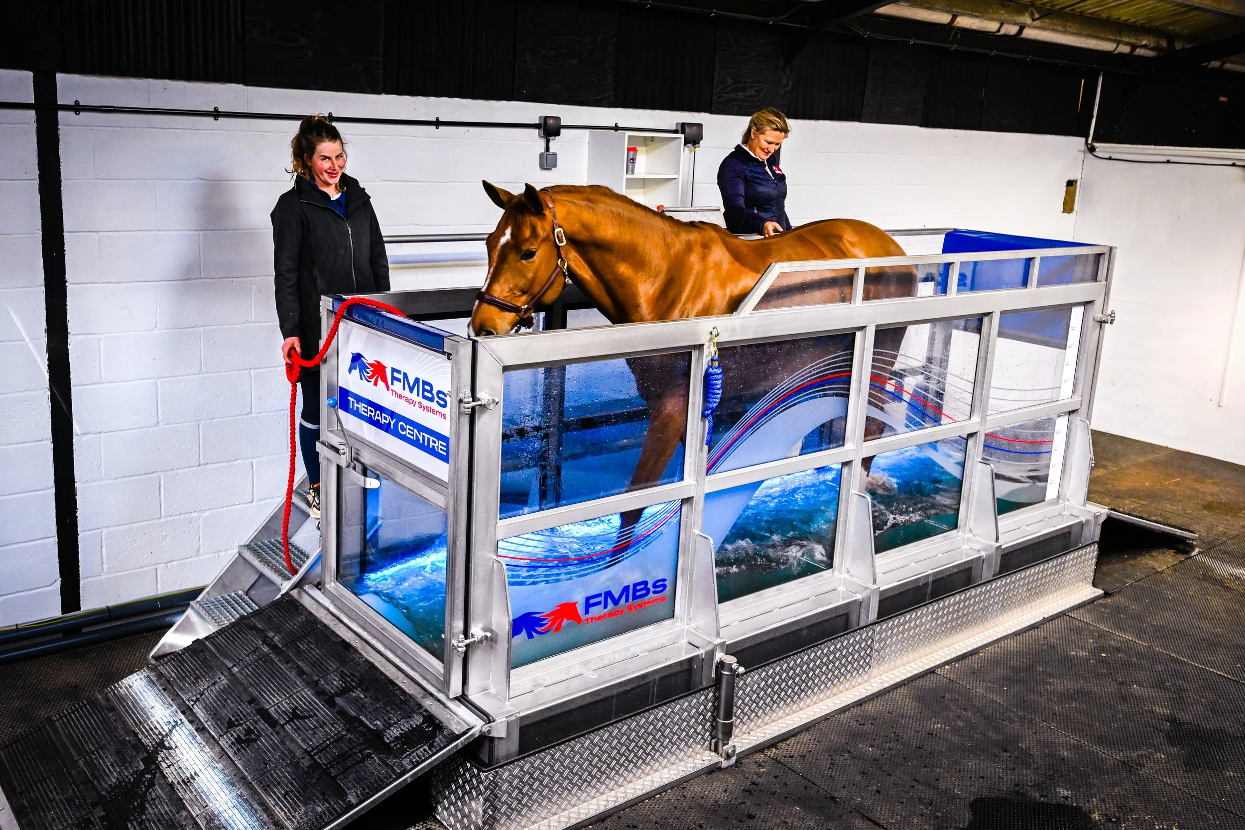
We were then treated to a lecture by Veterinary surgeon Amelia MacArthur, who provided a grounded and insightful view on equine rehabilitation, shaped by her hands-on experience running a specialist rehabilitation yard in North Yorkshire, England. Based at the former training yard of the Cheltenham Gold Cup winning trainer, Peter Beaumont, her facility includes a water treadmill, deep sand gallop, extensive hacking, and a quiet stable environment, all tailored to support recovery and performance conditioning.
It is clear that MacArthur advocates for a genuinely holistic approach, not rooted in fads, but in understanding the whole horse: injury history, temperament, conformation, previous management, and future athletic goals. Rehabilitation begins with controlled exercise, which is often hand-walking, though she acknowledges the safety challenges of managing fresh horses, advising use of protective gear and sedation when necessary. In-stable physiotherapy, such as weight-shifts and limb lifts, can supplement or replace walking early on.
She stresses that rehabilitation literature often lacks clarity, so individualized programs with regular reassessment, particularly ultrasound checks, are essential. Progressive loading, surface variation, and adapting treadmill use depending on injury type all help prevent reinjury. For tendon cases, treadmill work is delayed to avoid strain from reduced slip.
Crucially, MacArthur highlighted the impact of rider weight and balance, particularly for ex-racehorses, and the importance of body condition in supporting soundness. A striking case study showed how fat loss transformed a Highland pony’s tendon recovery and competitive ability.
Tendon injury; therapy and management
The final open floor discussion of the day took place between Mark Johnston, Professor Roger Smith, Dr Tom Clack and Amelia MacArthur. The topic of military-style training programs running parallel with equine management were discussed, particularly in managing overuse injuries like stress fractures. Key strategies include load management and gradual conditioning over 4–8 week cycles. It was noted that today’s horses, like modern human recruits, can often lack natural conditioning, especially in the feet, increasing injury risk.
Prevention is focused on structured training that supports both tendon and bone development, particularly in young horses (yearlings), where tendons must adapt before bones are heavily loaded. Ground conditions and surface variation also play a complex role in musculoskeletal health.
Rehabilitation and pre-training approaches remain debated, but there's agreement that progressive, controlled exercise is essential. Tendon injuries, especially in flat racehorses, are notoriously hard to overcome. Advances in ultrasound and imaging, such as UTC and shockwave elastography offer new promise, though they come with high costs and technical demands.
Steeplechase horses often return to competition successfully after injury, offering hope, but managing owner expectations remains key. Medication use, such as dexamethasone, is tightly regulated on racecourses to uphold integrity. Like elite human athletes, horses need carefully balanced workloads and rest to prevent chronic damage. While rehabilitation methods are improving, prevention remains the best strategy.
This year’s renewal of the Gerald Leigh Memorial Lectures was once again full to the brim with exciting new research and innovative thoughts from world leading experts. Attendees, all involved within various areas of the horseracing industry made for diverse and thought-provoking discussions.
The commonality amongst the lecturers and attendees alike was the undeniable commitment to ensuring the betterment of equine welfare in all avenues of bloodstock, racing and life after. Safe to say, all who attended are already looking forward to the 2026 lectures.
Tangible improvements to equine safety and welfare to reduce the prevalence of both EASD incidents and severe musculoskeletal conditions
In racehorses, exercise-associated sudden death – or EASD – is a very rare event but it can happen and this article is written to highlight a need for better understanding of why it happens as well as motivating vets, researchers and horsemen to do more to prevent it.
In June 2024, Woodbine Racecourse, Toronto hosted the International Horseracing Federation’s (IFHA) Global Summit on Equine Safety and Technology where EASD was one of two major workshop topics. This international event was sponsored by Cornell University’s Harry M. Zweig Memorial Fund for Equine Research, The Hong Kong Jockey Club Equine Welfare Research Foundation, and Woodbine Entertainment Group and specialist veterinary clinicians, pathologists and researchers spent two days sharing knowledge and ideas and debating how tangible improvements to equine safety and welfare in racing could be made towards reducing the prevalence of both EASD incidents and severe musculoskeletal conditions.
What is EASD?
The term EASD is used to describe a fatal collapse in a previously healthy horse, either during or shortly after exercise. Currently, across the world, different time-windows are used by regulators which makes quantification of the problem challenging. A benchmark definition is needed so that the occurrence rates can be audited and the EASD workshop team advised that an international definition be adopted to define EASD as within approximately one hour after exercise. Figures from the British Horseracing Authority (BHA) show that in Great Britain , the 2024 EASD incident rate was 0.04% or 4 horses per 10,000 starts. The British rate is comparable with other nations such as Australia and a little lower than the USA although the different definitions used in different racing jurisdictions make direct comparisons challenging.
Four broad EASD categories
The most authoritative international study looking at causes of EASD was performed with the British Horserace Betting Levy Board supported by a group in the University of Edinburgh’s Royal Dick School of Veterinary Studies. This report showed that determination of cause of death is significantly impacted by individual pathologist’s interpretation of findings, however, in broad terms about a quarter of cases EASD have a clear and definitive diagnosis of cardiopulmonary failure and a further 10-15% have necropsy findings which are strongly suspicious of cardiac or pulmonary failure; around 10% of EASD cases are due hemorrhagic shock brought on by rupture of a major blood vessel which is most commonly within the abdomen, while unfortunately around 20% of cases are unexplained despite detailed examination. A range of other rare conditions including brain and spinal problems, often relating to trauma, account for the remainder.
Within the cardiopulmonary failure category, it is generally accepted that the majority relate to cardiac arrest. This means that the cardiac rhythm is disrupted but, in fact it is actually very difficult to prove that a cardiac rhythm disturbance has been the trigger mechanism of death during a post-mortem examination. In the June 2024 IFHA summit, a significant amount of the workshop was dedicated to discussing current knowledge of cardiac rhythm disturbances, why they occur and how they might be detected in the future.
Cardiac arrest: a “perfect storm”
Cardiac arrest can be likened to a perfect storm where multiple adverse factors combine with devastating impact. Unlike catastrophic bone fractures or tendon injuries, cardiac arrest does not necessarily relate to an accumulating pathway of built-up microdamage and because of this, it is very difficult to predict cardiac arrest might occur. For a cardiac rhythm disturbance (aka an arrhythmia) to develop three elements are required: a substrate, triggers and, in some cases, one or more modulators. A substrate refers to the structure of the heart, this can be an area of scar tissue but the heart structure does not necessarily need to be pathological and the changes in muscle content which arise as a result of athletic training may also be a substrate.
A trigger reflects a change in the cellular and tissue environment such as alteration in concentrations of different electrolytes or development of low oxygen concentrations in the tissues yet changes in electrolytes and lowering oxygen concentrations occur every time a horse gallops. Modulators are an electrophysiological characteristic of the heart which might be a permanent feature of an individual’s cell make-up or more often might be a transient state such as a variation in the nervous system brought on by excitement, stress or perhaps pain.
The key point is all these independent factors have to combine to precipitate a cardiac arrest – indeed a horse might go through its life uneventfully despite the presence of a particular substrate or it may experience these triggers on a daily basis and come to no harm. It is the coalescing of multiple factors at a given moment that precipitates the rhythm disturbance that leads to cardiac arrest.
EASD at the molecular level
Arguably the biggest challenge we currently face in this arena is lack of knowledge of what is normal in the exercising horse. There is very little understanding of structural and electrical remodelling of the equine heart in response to exercise. We do know that the heart, just like any other muscle, will increase in size in response to training and we also know that in horses competing over longer distances such as steeplechasers, a big heart confers an athletic advantage. Exercise training can also lead to scar-tissue formation but in both human and equine athletes the importance of this pathology is uncertain. There is some evidence that fit horses also have altered cardiac electrical characteristics but again, knowledge in this field is very sparse.
Electrical activity in the heart muscle cells is controlled by ion channels – these are proteins that are sited within the cell membranes which effectively act as gates opening and closing to allow electrolytes such as sodium, potassium and calcium to move in and out of the cell and in doing so the electrolytes carry the electrical current.
Channelopathies – or abnormalities in these ion channels - have an important role in the development of rhythm disturbances but right now, research on equine ion channels has been limited…but that is changing rapidly. Researchers in Great Britain, Copenhagen and various US universities are working to understand equine channels and the genetic and acquired factors that determine how they function. As knowledge accumulates it may be possible to include tests for the molecular make -up of an affected individual in post-mortem exams – the so-called “molecular autopsy” which is improving diagnosis rates in human cardiac arrest suffers.
So far equine studies have not found conclusive evidence of genetic mutations associated with EASD. But there is evidence for heritability in the Thoroughbred: observations from Australia which have shown some stallions’ and at least one mare’s progeny have higher rates of EASD associations suggesting that it is likely that there are genetic elements at play in EASD. One of the key recommendations of the IFHA’s EASD workshop was that tissues from both horses impacted by EASD and those dying of other causes should be banked and shared amongst researchers to underpin and promote research studies in this area.
ECG is the cornerstone of arrhythmia diagnosis
Currently vets rely on resting and exercising electrocardiograms (ECG’s) to identify horses with arrhythmias. However, there are a number of limitations to using ECG as a screening and diagnostic tool:
ECGs can be technically difficult to perform during exercise as they are affected by motion artefact; leading to reduced quality of the trace.
ECGs currently must be manually interpreted, which is time consuming and leads to significant intra- and inter-observer variability.
There are no universal guidelines on how to perform the ECG; i.e. exactly where to place the electrodes, which affects the trace produced.
There is no consensus on interpretation of the results of an ECG examination in terms of the clinical significance of any abnormalities detected and whether the clinical presentation impacts criteria for interpretation. Indeed, we need to understand more about what is ‘normal’, before we can identify horses with an ‘abnormal’ trace.
Will wearables change the diagnostic landscape?
Over recent years, increasingly racehorse trainers have been using wearable devices during routine training. Generally, the trainer’s motivation is to collect data on speed and fitness variables in their horses to refine their training programs but several of these devices also have the capacity to include an ECG trace. The ECG can then be accessed if the horse has a problem during a training session and, usefully, the horse’s past record can also often be interrogated. The large numbers of recordings that are currently being made represents an untapped resource for collecting ECG information from large numbers of horses to better understand cardiac responses during exercise in both healthy and unhealthy individuals.
It has been known for some time that healthy horses frequently have mild rhythm irregularities – generally described as premature complexes or premature depolarizations – these minor fluctuations in rhythm occur at all phases of exercise and particularly as their heart rate is slowing rapidly at the end of a gallop. But the dividing line between what is normal variation and what is clinically concerning is not clear-cut. We do not know exactly how much beat-to-beat variation can be classed as normal versus a sign of significant arrhythmia and we have little understanding of the relationship between premature depolarizations and other factors such as stress, exercise intensity, medical interventions and adverse clinical events.
As a result, veterinary clinicians are looking forward to the ongoing expansion of wearables as an exciting new window into equine cardiac function. Yet, the scale of the unexplored data collection currently going on in training brings with it a challenge – with so many ECG traces being rapidly collected, how can we address the mammoth task of actually looking at them? Artificial intelligence (AI) is revolutionizing many aspects of modern life, including medical diagnosis. There is an urgent need to develop AI systems which can screen training ECGs to identify those that warrant further attention. And, although a large number of wearable devices are available on the commercial market, these products often lack validation which is needed before we can use the data they collect to make clinical decisions on individual animals and use the data as a research resource.
Could we deal with EASD cases better when they do occur?
Racetrack arrhythmia/collapse are, in reality, low probability but high impact events which can be difficult to manage due to their traumatic nature and the fact that they are often played out in the public eye. This is compounded by the availability of medical equipment and limited treatment options that may be futile.
However, when these events do unfortunately occur, they represent a golden opportunity to collect diagnostic information and biological samples which could be used to prevent future EASD events in other horses in the future. The combination of an ECG history, a video of the horse as it suffers the event, information from necropsy if the horse dies, and tissue banking offers valuable research insights.
The nearest parallel event from human sport is the cardiac arrests which are occasionally seen in footballers. Through the effort of football’s regulators, today pitch-side emergency medical facilities are excellent and large numbers of trained staff are in attendance, all leading to the best possible outcomes for sportsmen when medical problems arise. When looking to perform cardiopulmonary resuscitation and treatment attempts in the collapsed horse, the animals’ size is a major challenge; human defibrillators simply do not work in large animals.
We need more information on emergency medications that can be used in the presence of arrhythmias of unknown origin. These drugs need to be quick to administer, available and suitable to be carried by a racecourse vet, safe, effective and affordable. The IFHA’s EASD group identified that in pressurized situations, pre-determined protocol approaches to both emergency treatment and necropsy procedures are invaluable and the group is working to develop these protocols for dissemination across racing jurisdictions.
Will EASD risk always be present?
As EASD is such a rare event, it is impossible to believe that the risk of EASD can ever be removed entirely, but given the recent technological development in both veterinary science and wearables for training, there is reason to be optimistic that in the coming years, we will at last be able to improve diagnosis rates, identify some of the contributing risk factors and even potentially provide more effective emergency treatment options for these unusual but tragic episodes in our horses.
Be proactive rather than reactive with equine biosecurity

“In the equine industry, true biosecurity is hard to achieve because horses move around a lot, and many diseases are always present,” says Ontario Veterinary College infectious disease specialist Dr. Scott Weese. “However, it's still important to try to prevent diseases from entering and to have plans in place to manage any outbreaks.” With frequent horse movements, endemic pathogens and emerging diseases, there is a need for improved understanding and motivation to adopt better infection control practices.
Infection control begins in the barn and works best when the focus is pro-active rather than reactive. This includes having an access management plan, proper quarantine protocols for new and returning horses, and training EVERYONE who comes on to the property or handles the horses.
Access Management
Controlling how horses, humans, equipment and vehicles can move into and around your farm are all aspects of access management aimed to reduce the transmission of pathogens.
Access management begins at the entrance, where a training facility may use fencing and gated entries to restrict access to the stables and training areas, ensuring only authorized personnel can enter. Procedures at controlled access points such as hand sanitizing and boot cleaning help prevent the spread of infections. Both staff and service providers need to be made aware of any infection control measures in place. Clean outerwear that has not been worn to another barn are also recommended to prevent potential spread of disease.

A sign in procedure can be made mandatory for visitors. A log can be helpful to help trace the problem in the event of a disease outbreak. Providing guided tours can ensure they do not enter restricted areas. Additional signage can let visitors know where they can and cannot go.
Controlled access zones can designate specific areas for different activities, such as quarantine zones for new arrivals and separate zones for resident horses, with controlled access points to manage movement.
Isolation/Quarantine
When horses return home or new horses arrive, such as from a sale, it is a good idea to implement quarantine and/or isolation protocols. Ideally this involves housing in a separate building away from your resident horses, but it may be the end of an aisle with several empty stalls in between.
New and returning horses are kept separate and monitored for at least 14 days. This involves twice daily temperature checks and health checks including watching water consumption, appetite, urination, manure and any signs of illness.
Turn out paddocks should also be away from other resident equines, especially if that includes higher risk horses like broodmares and foals.
Effective quarantine includes using separate equipment for isolated or quarantined horses to avoid cross-contamination. This includes water buckets, feed tubs, grooming equipment as well as wheelbarrows, brooms, pitchforks and other cleaning tools.
Ideally, new and returning horses are handled by separate staff. Otherwise, quarantined horses are worked with last & hands are washed before & after each interaction. Strategically placed alcohol-based sanitizers can also be used. If wash stations are limited, this makes it easier for staff and visitors to follow infection control protocols. Disposable gloves, disposable shoe covers & protective clothing are also best practices. Barn cats and other pets should not be allowed to enter the quarantine area.
If you have a number new or returning horses in quarantine and one shows signs of illness, it should be further separated into isolation and seen by a veterinarian ASAP. Horses should remain in isolation until cleared by the vet, as the horse may have recovered from clinical signs but still be infectious. Signage once again should alert unauthorized persons at the entrance of any areas used for isolation or quarantine.
Not Sharing is Caring and Hygiene Practices

Of course, those new or returning horses should be housed in a stall that has been both cleaned and disinfected prior to their arrival.
Cleaning involves removing all visible manure, bedding and soil before washing the area with soap and water and then allowing it to dry. Then apply a disinfectant such as Virkon or other disinfectant recommended by your veterinarian. All disinfectants have strengths and weaknesses and are best used for specific purposes. Bleach has drawbacks as hard water can affect its effectiveness, it can be inactivated by organic material, and it can be irritating to the horse. Steer clear of pressure washers as they can aerosolize certain viruses.
An often-misused step, if you will pardon the pun, is the foot bath. One cannot just walk through without first going through the same routine as mentioned above, both cleaning and disinfecting. First remove debris from the footwear, including the soles using a brush or hose to get all the dirt out of the treads. Immerse the entire bottom of footwear in the disinfectant and scrub. Following the contact time on the product label is important and a dirty footbath does little in the way of boosting biosecurity. Then wash your hands. Other options include dedicated footwear and disposable shoe covers.
Hand hygiene cannot be overstated as one of the most important infection control measures. Best practices on application time for the soap or alcohol-based sanitizer is 20 – 30 seconds.

Everyone knows not to share communal water, but it is also important not to become blasé about biosecurity when it comes to filling or refilling water buckets. Submersing a hose from one bucket to the next or letting it touch the buckets can be a free ride for a pathogen looking for its next host. So instead of multi-tasking while filling buckets, one could be enjoying a beverage with their free hand.
Not sharing should extend beyond grooming equipment to tack, pads, blankets, and of course medical supplies like syringes, needles and dewormers.
More disease prevention measures include minimizing the presence of rodents and insects by keeping feed secure, eliminating standing water and regular removal of manure from stalls and paddocks and as well as management of manure storage areas.
Vaccination
Vaccination is a crucial aspect of equine healthcare, but vaccines do not provide immediate protection; it can take days or weeks for a horse to develop optimal immunity after vaccination, so timing is very important. Planning ahead will allow vaccines to be given well in advance of the next stressor such as travelling or competition.

While no vaccine boasts 100% immunity, horse owners can rest assured that they are taking proactive steps to maintain their horse's health, minimizing the risk of unexpected veterinary expenses. Vaccines significantly reduce the risk of disease which means if a vaccinated horses does get sick, they will generally experience milder symptoms and recover more quickly.
Working closely with a veterinarian to develop and maintain a vaccination program is an important step for optimal equine health. In addition to core vaccinations, your vet will know what diseases are endemic and emerging in your region or regions you will be travelling to. The frequency of your vaccinations or boosters will depend on a number of factors including special circumstances, such as an extended vector season or even a significant wound if it is incurred over 6 months after a Tetanus shot. The length of your competition season may also necessitate a booster of certain shots to maintain optimal immunity.
Emerging Diseases

Infection control specialist Dr. Weese says, “Understanding potential mechanisms of transmission is the basis of any infection control or biosecurity program.”
Most diseases in horses are caused by pathogens that mainly infect horses. They can spread continuously without needing long-term hosts (like the equine flu virus). They can remain in the horse without causing symptoms for a long time (like Strangles). Some cause infections that can come back at any time (like equine herpesvirus). Others may be part of the normal bacteria in horses but can cause disease if given the chance (like staphylococci and Enterobacteriaceae).
Horses can spread these germs even if they seem healthy, before showing symptoms, after recovering, or as part of their normal bacteria. This makes it hard to identify which horses are infectious. Some symptoms, like fever and diarrhea, strongly suggest an infection, but any horse can potentially spread germs. Therefore, it's important to have strong infection control practices to manage the risk.
In 2024, the Equine Disease Communication Center (EDCC) reported 577 Alerts for 813 confirmed cases of disease in North America. The most frequently reported disease was Strangles with 186 cases. Because Strangles is not reportable in all states or provinces the disease is likely much more prevalent than reported to the EDCC. Other frequently reported illnesses include: 153 West Nile Virus (WNV), 125 Eastern Equine Encephalitis (EEE), 109 Equine Infectious Anemia, 73 Equine Herpesvirus- Neurologic, 8 Equine Herpesvirus- Respiratory, 34 Equine Influenza.
Strangles: A bacterial infection caused by Streptococcus equi, leading to swollen lymph nodes and respiratory issues. It is highly contagious and spread through contact. This could be nose-to-nose between horses or via contaminated surfaces or equipment such as: shared halters, lead shanks, cross ties, feed tubs, stall walls, fencing, clothing, hands, the hair coat from other barn pets, grooming tools, water buckets, communal troughs.
After an outbreak, cleaning should involve removal of all organic material from surfaces and subsequent disinfection of water containers, feeders, fences, stalls, tack and trailers.
West Nile Virus (WNV): a mosquito-borne virus leading to neurological issues such as inflammation of the brain and spinal cord. WNV can be fatal and survivors can have residual neurological deficits for a period of months to permanent disability.
Eastern Equine Encephalitis (EEE): another virus transmitted by mosquitos Eighty to ninety percent of infected horses develop acute and fatal neurologic disease.
Equine Infectious Anemia (EIA): is a blood-bourne virus which can be transmitted by insects, medical equipment or passed from mare to foal in utero. With no treatment or cure, horses confirmed positive by a Coggins test can be quarantined for the rest of their life but are usually euthanized.
Equine Herpesvirus (EHV): This virus had multiple strains and can cause both abortion and neurologic symptoms. Spread via aerosol particles from nasal discharge or from contaminated surfaces. There are vaccines for respiratory and abortive strains but not the neurologic form of EHV-1 (EHM).

These diseases highlight the importance of biosecurity and vaccination in managing equine health. West Nile Virus and Eastern Equine Encephalitis are among the core vaccinations recommended by veterinarians.
In February 2025, Equine Guelph partnered with the Equine Disease Communication Center (EDCC), to help horse owners assess and manage infectious disease risks with the relaunch of Equine Guelph’s Biosecurity Risk Calculator (TheHorsePortal.com/BiosecurityTool). The interactive free tool is full of useful information from quarantine protocols, best practices for cleaning, and easy to understand practical access management tips. In just 10 minutes, you can assess and minimize biosecurity threats for your barn.
"Applying routine and basic biosecurity is the best way to prevent infectious diseases," says Dr. Nathaniel White the Director of the EDCC. "This includes isolation of new horses introduced to facilities, monitoring horses' temperature and preventing horse to horse contact while traveling and keeping vaccinations up to date. Being aware of disease prevalence using information from the EDCC and the updated "Biosecurity Risk Calculator" can help owners use management practices to decrease disease risk."

Equine Infection Control Measures During Transport
Pre-transport preparations entail more than just having your paperwork in order.
Taking the time to clean and disinfect the trailer or make sure the trailer you have hired always cleans between loads is of paramount importance. If the trailer smells like horses, it was not adequately cleaned. Perform a horse health check before you leave the property. It is not worth the gamble to stress a horse with travel when it is ‘not-quite right’.

Being particular about your horses traveling companions is just as important as the cleanliness of the trailer. Avoid travelling with horses from other locations as being in close quarters increases the risk of picking up an infectious disease.
Tie the horse loosely if possible. Horses tied short are less ability to lower their head to clear mucus. Allowing freedom of head movement can reduce stress and the bacterial load in the airways. Similarly, hay nets that are hung high, encouraging a high head position, and introducing dust and debris, can challenge mucous clearance.
Ventilation is another important consideration as improving air exchange can reduce the dust and mold spores hanging in the air. Drafts on the other hand can blow particles around in the trailer.

Many prefer shipping in leather halters because they will break in an emergency but there is a biosecurity benefit too as they are easier to clean. Bacteria can linger in the webbing of polyester halters.
Biosecurity is just as important on the road and when visiting other venues. Disease is easily spread through equipment sharing. While visiting venues off the farm be sure to bring your own broom and shovel for cleaning your trailer. Be sure to pack a thermometer along with your tack and other equipment. Clean & disinfect your equipment when you get ready to leave your off-site location.
Upon returning to the home farm, the cycle begins again, monitoring horses for possible delayed onset of symptoms.
To ensure effective infection control, it is crucial to maintain a proactive approach starting right in the barn with a plan. Implementing access management, enforcing proper quarantine protocols for new and returning horses, and thoroughly training everyone who enters the property or handles the horses are essential steps. By taking these practical steps, we can significantly reduce the risk of infections and promote a healthier environment for all.
Suppressing unwanted hormonal behaviors in breeding stock
Article by Kate Dugher

The desire to suppress unwanted behavior in the horse can present for many different reasons. The behaviors that we are talking about can be anything from poor performance to hyper-excitability, distraction, discomfort on girthing up, not responding to the jockey, bucking, rearing, squealing, kicking or aggression.
Is it hormonal?
Often it is assumed that overt behaviors are hormonally driven; however, it can be easy to discount many other possible causes of these behaviors, especially those that are related to pain. A full clinical examination by a veterinarian is always warranted when considering unwanted behavior in the horse in order to appropriately identify the cause and consider the most appropriate treatment options.
Common causes of abnormal/unwanted behavior can include:

Musculoskeletal pain (lameness)
Gastric ulceration
Dental disease
Poorly fitting tack
Stress
Hormonal influence
Learned behavior
There are also many reasons for normal and abnormal behaviors that can be associated with the reproductive system. Some of these could be identified as undesirable behaviors when associated with performance.
The equine reproductive cycle
Horses are seasonal long day breeders and are influenced by daylight length. This means that the majority of mares have inactive ovaries in the winter and do not exhibit estrus behavior during this time. In comparison, in the summer months, they exhibit a reproductive cycle that lasts an average of 21 days. They spend, on average, 5-7 days in estrus, ‘in season’, and 14 days in diestrus, ‘not in season’.

In the spring and autumn months the mare undergoes a transitional period. During this time, estrogen concentrations are variable, and estrus behavior can be seen irregularly. While stallions are also affected by seasonality, they still exhibit reproductive behavior all year round. The mare’s reproductive cycle can also be influenced by artificial light and therefore, it is worth considering that performance horses who are exposed to stable lights beyond the normal daylight hours in spring, autumn or winter may cycle for a longer period of the year or even throughout winter.
Puberty
Timing of puberty in the horse is varied and affected by both genetic and environmental factors. Not only by age but also by time of year in which they were born, body condition and social cues. Puberty in fillies is usually at around 12-19 months compared to colts at around 10-24 months, however, there are wide variations from these reference ranges.
Normal reproductive behavior in the mare
Normal estrus behavior occurs under high estrogen and low progesterone influence. Commonly associated behaviors include receptivity to stallions/geldings, vocalization, increased frequency of urination and presentation of hindquarters in a wide based stance.
Normal diestrus behavior under a dominant progesterone state includes repulsion to the stallion and can occasionally be associated with aggressive behavior to other horses. During pregnancy, the mare will also be under a dominant progesterone influence and is unlikely to exhibit estrus behavior particularly in the first trimester. Later in gestation a peak in testosterone and estrogen levels may be associated with changes in behavior.
Abnormal reproductive behavior in the mare
Ovarian pain
Many mares will show an obvious reaction upon rectal palpation of the ovary when close to ovulation, suggesting that the dominant follicle/ovary can sometimes be tender at this time. Comparatively, humans often describe some ovarian pain around the time of ovulation. Therefore, it can be assumed that some mares could also experience discomfort around the time of ovulation.
Other possible causes of ovarian pain that can occasionally occur in normal cyclicity include ovarian hematomas and haemorrhagic anovulatory follicles. It is also a consideration that external pressure placed onto the lumbar region close to the ovary around the time of ovulation could rarely elicit a painful response in some individuals.
Vaginal pain
Vaginal pain has occasionally been associated with conditions such as vaginitis and pneumovagina. These conditions describe inflammation and/or air in the vagina. These are most commonly associated with poor perineal conformation and can be evident in some performance mares.
If vaginal pain is suspected due to poor perineal conformation, then placement of a caslicks vulvoplasty may prove to be beneficial. If concurrent infection or urine pooling is suspected, then further intervention may be required.
Reproductive tumors

Reproductive cancer affecting the ovaries is one of the most common causes of cancer in the mare, the most common being the granulosa theca cell tumor (GTCT). These are generally locally invasive and are unlikely to cause any further health problems if the affected ovary is removed. They are often identified with a change in behavior. On rectal examination a common finding would be to identify one enlarged and one small ovary.
Depending on which reproductive hormones the tumor secretes is likely to influence the associated behavior. This can include stallion-like behavior, aggression, persistent estrus behavior or complete absence of reproductive behavior. The severity of this often depends on the stage at which this condition is identified. Other types of ovarian tumors are less common but depending on if/which hormones are secreted will dictate which hormonal behaviors are associated. It is suspected that occasionally there could be ovarian pain associated with some of these cases particularly when the ovary is very large in size.
Reproduction related treatment options
Mares
To have the most successful outcome in controlling reproductive hormonal behavior in the mare, it is important to understand whether the unwanted behavior is being exhibited all year round or just in the summer months and whether it is related to a particular stage of the estrus cycle.
Whilst it is commonly assumed that most behavior problems are associated with the mare being in season, occasionally some mares can show unwanted aggressive behavior under the influence of progesterone – when they are not in season.
Furthermore, it can be tricky to interpret this when trying to link hormonal behaviors to performance based unwanted behaviors and these signs can often be very individual. Keeping records of behavior versus stage of the reproductive cycle can help to try and decipher whether reproductive hormones are likely to be playing a part in the unwanted behavior. However, this does require careful monitoring and, most likely, multiple reproductive ultrasound examinations.
The other consideration is that unwanted behaviors are related to reproductive pain or abnormal hormone production due to pathological conditions of the reproductive tract as previously described.
Ways to mimic the diestrus state and suppress estrogen related behavior
Progesterone/Progestins
Progesterone is the dominant hormone produced by mares in diestrus. There are a multitude of systemic progestin (progesterone-like medications) available for use in horses in injectable and oral formulations.
Altrenogest is a synthetic progestin commonly used to suppress estrus behavior by acting as a progesterone agonist. This means that the horse is likely to exhibit normal diestrus behavior for that individual whilst it is being administered. Altrenogest is molecularly very similar to the anabolic steroids trendione and trenbolone. Occasionally the product may contain trace levels of these anabolic steroids. Therefore, its use for horses in training is to be taken with extreme caution and withdrawal times adhered to. It is banned for use in racing thoroughbreds in some countries.
There is also evidence to show that altrenogest can exhibit a reduced stress response and sedative-like effects in some horses, particularly mares. This effect may be beneficial in anxious individuals in training circumstances. However, arguably, dependent on the individual, a reduced stress response could have either a positive or negative effect on performance.
Injectable progesterone applications have been used in racing thoroughbreds with appropriate clearance times before racing. These are often available in oil-based preparations which are commonly associated with injection site reactions and therefore, many trainers would avoid administering these within 3 days of racing.
Upon cessation of progesterone supplementation, many mares will present with estrus signs 2-7 days after treatment, as this mimics normal luteolysis at the end of the diestrus phase. Therefore, the timing of administration and cessation of progesterone/progestin treatments is a crucial consideration when being used for the prevention of estrus behavior.
Intra-uterine devices (IUDs)
IUDs have been historically utilized to mimic early pregnancy in the mare with varying success. These require an ovulation to act upon to extend the life of the corpus luteum by blocking the hormonal release that normally brings them back into season. Therefore, they are only useful once the mare is already cycling.

Glass marbles have been the most used IUD historically; however, these are no longer recommended due to multiple evidenced side effects including risk of glass fragmentation in the uterus. The use of PMMA spheres or magnetic devices such as the iUPOD would be a preferable and safer alternative if an IUD was going to be used.
Interestingly, in the author’s experience speaking with clinicians who have administered these devices, there is surprisingly positive client satisfaction despite the inconsistent and variable evidence of the success of these devices in the literature.
Oxytocin
Administration of the hormone, oxytocin, at specific time points when the mare is in diestrus can extend diestrus by up to 60-90 days. This technique is evidenced by multiple studies. For optimal success, reproductive ultrasound would be used to identify ovulation and carefully plan the timing of injectable administration.
However, some studies have also evidenced successful extension of the diestrus phase without known timing of ovulation. The major downside of this technique is the need for administration of multiple injections/multiple reproductive examinations to time ovulation.
Immunological approach
Gonadotrophin releasing hormone (GnRH) is a hormone produced by the brain that is responsible for stimulating follicle growth in the ovaries and activation of a hormonal cascade to bring the mare into estrus.
GnRH vaccinations generate an immune response against GnRH, suppressing the hormonal cascade and ovarian activity and therefore, estrus behavior. An equine licensed product has previously been available in Australia. However, this is no longer in production. We have the option of a swine formulation, Improvac®, which has commonly been used in equids off license.

Major drawbacks for the use of this are common adverse injection site reactions, risk of anaphylaxis and concern over extended length of ovarian suppression. Therefore, this option would not be recommended in mares with a future breeding potential.
Surgical approach
Ovariectomy is a treatment option for hormonal behavior in mares. The ovary is the only supply of progesterone in the horse but is not the only supply of estrogen.
Ovariectomy has been associated with good client satisfaction in many cases to resolve unwanted hormonal behavior. However, in some mares, whilst removal of the ovaries would prevent cyclicity, it can occasionally result in persistent estrus behavior in the absence of progesterone produced by the ovaries. This is also a permanent option that will remove breeding potential.
The techniques discussed so far are not exhaustive and there are many other methods that have been used to affect cyclicity or hormonal behavior including pregnancy, induction of diestrus ovulation, GnRH analogue medication and infusion of intrauterine medical grade plant oils.
Colts/stallions
There are a few medicated options for hormonal manipulation in males. Progestagen administration e.g. oral altrenogest administration can quieten stallion like behavior in males but is banned for use in racing and training.
Immunization with off-license GnRH vaccines such as Improvac®, suppresses pituitary-gonadal hormone production aiming to cause a ‘chemical castration.’ However, results can be variable, particularly in mature stallions. As mentioned previously with mares, the downside of these vaccines are injection site reactions, risk of anaphylaxis and risk of prolonged sterility in future breeding animals.
Occasionally nutritional supplements have been used with effect in stallions such as L-tryptophan, a precursor of the neurotransmitter serotonin. This has induced calm and fatigue-like behavior in a number of species.
Synthetic preparations of calming pheromones based on an equine appeasing pheromone produced in perimammary gland secretions of lactating females have also been used with such success. Of course, the use of these to calm behavior vs the desire to generate an athletic performance animal is a consideration and results are likely to have wide individual variation.

Can Spirulina help horses recover faster from intense exercise?
Article by Jackie Bellamy-Zions interviewing Wendy Pearson and Dr. Nadia Golestani

Elevating performance and seeking the competitive edge is what makes equine supplements a billion-dollar industry, but what makes the difference between a supplement that simply creates ‘expensive urine’ and a nutritional supplement that could actually have an impact?
Associate professor at the University of Guelph, Wendy Pearson and Ph.D. candidate Dr. Nadia Golestani, answer this question and more in their quest to develop quality nutraceuticals with positive equine health benefits. Their latest study on Spirulina reveals potential for expediting recovery after intense exercise. It also holds promise supporting joint health and optimizing performance through enhancing oxygen delivery in the bloodstream.
Why Spirulina?
After becoming a DVM, Dr. Nadia Golestani began to pursue her goal of becoming an animal nutritionist, enrolling in the University of Guelph’s Master of Animal Biosciences program under the supervision of Dr. Wendy Pearson.
After attending Pearson’s lectures on exercise physiology, Golestani developed a good understanding of the controversy surrounding antioxidants and the lack of research as to whether they were good for exercise performance or not. For her Master’s, Golestani examined inflammatory response of cartilage during exposure to nutraceuticals that could potentially have a role in equine joint care.
Golestani had her eyes opened to the potential of Spirulina after reading a book named ‘Spirulina World Food.’ It was a gift from accomplished medicinal chemistry consultant, Ralph Robinson, which accompanied an award for Golestani’s research in equine nutrition and physiology.
Golestani wanted to explore ways Spirulina could be used in exercise physiology. Her Ph.D research, under Pearson, set out to study the effects of Spirulina as an antioxidant and how it could potentially modulate inflammation after high-intensity exercise in horses. It was made possible thanks to the support of Robinson, owner of Selected Bioproducts (Herbs for Horses) Inc., and funding from Equine Guelph.
What is Spirulina?

The blue-green algae is gaining popularity not just in human athletes but in equine ones as well. The nutritional profile contains C-phycocyanin and Beta carotene and 60 – 70% amino acids. It has vitamin B, iron, vitamin E and essential fatty acids, particularly gamma-linolenic acid (GLA) plus many more vitamins, proteins and minerals.
Golestani’s study focused on C-phycocyanin and Beta carotene in Spirulina with their potential antioxidant effects. New data shows antioxidants can be a double-edged sword with the capability of reducing inflammation as this may actually interfere with natural tissue adaptation after the rigors of exercise. Golestani’s study is looking for the best applications for Spirulina to optimize equine performance without interfering in the natural tissue adaptation process.
Enhancing recovery without interfering with transient inflammation
Some inflammation is normal after exercise and protects structures as they recover from the workout. Only when inflammation becomes excessive, does it become a concern in disrupting recovery.
“Transient inflammation is good and needed for recovery. Inflammation challenges the tissue, and the tissue responds by becoming stronger,” says Pearson. “What isn't good is chronic, sustained inflammation. We want to see if we can do something about the way tissue responds to an exercise bout, without interfering with transient inflammation.”
Golestani explains, when a horse undergoes exercise, their ATP (adenosine triphosphate) producing mitochondria are working hard. One of the natural byproducts is Reactive Oxygen Species (ROS are highly reactive molecules containing oxygen), which is good unless they are produced in excess leading to oxidative stress. When there is an imbalance between the production of ROS and the body's ability to detoxify them with antioxidants, this is when chronic oxidative stress can trigger vicious cycles of inflammation. Oxidative stress can also lead to cell death and therefore dysfunction and disease. Maintaining balance between ROS production and antioxidant defenses is essential for cellular health.
Golestani sums up, “Strenuous exercise, especially when it is high impact, is going to stress the horse’s joints and increase oxidative stress. Imbalance can damage the cells, proteins, lipids in the joint tissue and may lead to early onset of arthritis.”
Pearson adds, “Moderate intensity exercise is very good for protecting cartilage structure, but when you have repeated bouts of strenuous or very high intensity exercise it can tip the scales to more breakdown of cartilage than you have time to resynthesize. Tissue breakdown occurs when synthesis can't keep up, that's when you start to see declining structural integrity of the tissue.”
‘What is adequate recovery time’ becomes the million-dollar question with no definitive answer given a multitude of variables including the starting fitness level, type of activity, intensity of work, and other factors specific to each horse as an individual. This is where talented horse trainers excel. They can pick up on a change of behaviour in the horse in a workout even before physical signs of stress and adjust the training program accordingly.

You bet your biomarkers
Golestani researched the antioxidant effects of Spirulina, by looking at biomarkers associated with inflammation. In her study, biomarkers were measured before and after exercising horses that were given a Spirulina supplement against those who were not.
Results showed that exercise caused a temporary increase in nitric oxide (NO), a marker of oxidative stress, shortly after activity. This rise was discovered in both blood plasma and the synovial fluid. Horses given Spirulina had lower NO levels during recovery, indicating better management of oxidative stress. In joint fluid, NO levels increased 24 hours after exercise but were better controlled in the Spirulina group, with lower levels observed later in recovery. This signifies not only the potential for quick recovery from exercise but also properties that could promote joint health.
Another inflammation marker, prostaglandin E2 (PGE2), was also measured. PGE2 levels in the blood peaked eight hours after exercise and were higher in horses that received Spirulina, suggesting a stronger initial response to exercise. In joint fluid, Spirulina-supplemented horses showed lower PGE2 levels early in recovery, which may help reduce inflammation in joints over time and lower chances of early onset of arthritis.
A key finding was that Spirulina boosted levels of Resolvin D1 (RvD1). “RvD1 is so important in resolving the inflammation and promoting the clearance of inflammatory cells and for tissue repair,” said Goestani. RvD1 is a bio active lipid mediator derived from omega-3 fatty acid. Horses receiving Spirulina had consistently higher RvD1 levels in their blood and joint fluid during after exercise.

The rise in the RvD1 biomarker highlights how Spirulina has the potential to enhance this natural resolution pathway and its potential to protect against inflammation, speed up recovery and promote cartilage protection.
Pearson echoed the dramatic increase in Resolvin D1 in the horse’s receiving Spirulina to be pretty strong evidence that it could protect horses from bouts of transient inflammation from becoming chronic and contribute to faster recovery after exercise.
Horses fed Spirulina in the study also had higher hematocrit levels, which means their blood could carry more oxygen, translating into potentially enhanced performance. They also maintained higher glucose levels during recovery, providing more energy. Eight hours after exercise the control group had a drop in glucose, but the group fed Spirulina did not. Retaining glucose stores post-exercise is especially helpful for performance horses that need sustained energy and endurance during training or competition.
Importantly, there were no negative effects on cartilage biomarkers, further suggesting Spirulina may also promote joint health during recovery.
Look at the label and beyond the label
“Buying a quality product requires looking beyond the label,” says Pearson. “There are so many products on the market today that it is virtually impossible for even somebody like me, who spends my life looking at nutraceuticals, to look at a label of one product and tell the difference between that and the product on the shelf right next to it.”
Looking for third party quality assurance can be one indicator on the label that the product has some validity. Examples include:
ISO 22000 The International Organization for Standardization (ISO) provides standards to ensure the quality, safety, and efficiency of products.
HACCP Hazard Analysis and Critical Control Points principles identify, evaluate, and control hazards that are significant for food safety.
CCP Critical Control point is a step in the manufacturing process where control can be applied to prevent, eliminate, or reduce a food safety hazard to acceptable levels including: properly mixing ingredients to ensure uniform distribution and prevent contamination, applying heat or other sterilization methods to eliminate microbial hazards and ensuring the final product is packaged in a way that prevents contamination and preserves quality.
NASC The National Animal Supplement Council (NASC) Quality Seal is a mark of quality assurance for animal health supplements in the USA.
GMP (Good Manufacturing Practices) Certification: This certification ensures that the products are consistently produced and controlled according to quality standards. It covers all aspects of production, from the raw materials to the hygiene of staff.
UFAS (Universal Feed Assurance Scheme): This scheme is specific to the UK and ensures that animal feed and supplements are produced to high standards of safety and quality.
BETA NOPS (Naturally Occurring Prohibited Substances): This certification is particularly important for competitive horses. It ensures that the supplements are free from substances that are banned in equine sports.

Most importantly, Pearson implores horse owners to find out if the manufacturer has invested in research on the particular product they are marketing. “Lots of companies will talk about the fact that they're science based, but if you peel off a layer or two, you find that in fact the science they're talking about is science other researchers have done on ingredients that show up on the label on their product.”
Pearson emphasizes the importance of manufacturers conducting research of their products in the targeted species that supplement is created for. She takes a moment to lament the vast number of supplements on the market with no significant research behind them. It is much cheaper for companies to use anecdotal reports or sponsor a top rider to promote their products than conduct double blind studies with valid evidence-based results.
“The research is expensive,” says Pearson. “We are lucky to have funding from Equine Guelph for our latest study on Spirulina. If consumers prioritized purchasing products with research behind them, manufacturers that are not yet doing research on their products would have an economic reason to do so.”
So, horse owners have a bit of homework to do if they want a quality product and not just ‘expensive urine’. Asking to see the research on the product you intend to buy is the best bet for purchasing a product that is likely to deliver on its claims.
Nutraceuticals Increasing Popularity Raises Precautions
Pearson recalls when she started researching nutraceuticals for horses in 1997, “The word was not even well-known back then. It simply wasn’t a ‘thing’ at the time.” We have gone from whisperings of feed additives and ‘novel’ ingredients being the work of ‘witch doctors’ to the common place practice of adding supplements to feed.
“There is a night-and-day difference with upwards of 80% of horse owners adding something to their horses’ diet; whether that something is electrolytes or nutraceuticals or herbal supplements,” says Pearson. “These products can be really helpful in improving health, but they are not intended to be an opportunity for horse people to start to self-diagnose and self-treat disease.”
Pearson tells horse owners to always work with their veterinarian. “This is a very important point. Where these products are best positioned is when it's in conversation with the vet; not that all vets are experts in nutraceuticals, but they are all experts in animal health and specifically animal disease.”
Potential problems arise if horse owners end up delaying treatment of a potentially serious problem by reaching for a supplement rather than calling their vet. Pearson cautions, “Using these products can potentially delay proper veterinary care when they're not used properly.”
#1 Consult your veterinarian before adding supplements to your horse’s diet.
#2 Buy from a company that conducts research on their products and doesn't just claim to be ‘science-based’.
Top 3 practical take-aways:
Enhanced Oxygen Delivery and Energy Boost: Spirulina helped improve oxygen delivery and energy reserves in horses.
Support for Joint Health: Spirulina supplementation reduced markers of oxidative stress and enhanced inflammation resolution without damaging joint cartilage. This suggests Spirulina may protect against wear-and-tear on joints, helping reduce the risk of arthritis and supporting long-term joint health in active horses.
Faster Recovery After Exercise: Horses given Spirulina recovered more effectively after intense exercise, as seen by enhancing the production of pro-resolving molecules like Resolvin D1 (RvD1). This makes Spirulina a practical addition to the diet of horses involved in regular training or high-intensity work.
These findings highlight Spirulina’s potential as a safe and natural dietary supplement for managing inflammation, protecting joint health, and supporting recovery in equine athletes. Further research is needed to confirm long-term benefits, but this current study provides evidence that Spirulina offers a promising tool for promoting health and performance in horses.
There’s more to it than meets the eye
Words - Adam Jackson MRCVS

The horse’s eyesight has evolved to scan its environment rather than picking up sharp details, in order to survive from predators. As a prey animal, the horse’s eyes are eight times larger than a human’s eye; however, this makes them more vulnerable to injury and disease that may be catastrophic. Horses develop many of the same eye problems as humans such as glaucoma, corneal ulcers, cataracts and other issues.
The working of the eye
Vision is provided by light entering the eye, which is made into an image by the brain through various complex biomechanical and physical processes.
As light enters the eye, it is targeted to the retina by the cornea and the lens bending the light. This light reaches the sensory tissue at the back of the eye. In fact, the retina or nervous tunic is made of cells that are extensions of the brain coming off the optic nerve. The retina consists of 2 types of photoreceptors called rods and cones. The rod cells are more light-sensitive, thus providing night vision, whereas the cones are less light sensitive but provide visual acuity and the ability to see color. The optic disc in the retina does not contain photoreceptors and is the location the optic nerve leaves the eye to transmit the visual information to the visual cortex of the brain.
Visual field of the horse
Because the horse’s eyes are positioned on the side of the head, the range of vision is roughly 350 degrees, thus, allowing the horse to spot potential predators. Due to the positioning of the eyes, the horse has two blind spots that include in front of the face and behind its head extending over its back and behind the tail.
The horse has both binocular and monocular vision. Monocular vision means vision in one eye only and binocular vision means seeing with two eyes. 65 degrees of the 350 degree vision consists of binocular vision while the remaining 285 degrees is monocular vision. As a result, the horse has a smaller field of depth perception compared to a human. The horse must raise or lower its head in order to increase its range of binocular vision. By introducing a bit and making the horse hold its head perpendicular to the ground, the binocular vision becomes less focused on distant objects and more focused on what is immediately in front of the horse. Show jumpers and jump jockeys allow the horse to raise its head a few strides before a jump so that the horse can properly assess the jumps to allow appropriate take-off.
Sensitivity to light

Horses’ eyes have evolved to allow them to have good vision in dim light and due to this evolution they have better vision on slightly cloudy days compared to sunny, bright days. There are two particular structures that allow them to have superior night vision, which include a high proportion of rods to cones (20:1) and the presence of the tapetum lucid.
The horse’s large pupils allow a large amount of light to enter and the size of the retina allows a high number of cells to be involved in the capturing of light. In addition to the rods and cones, the horse's tapetum lucidum is a reflective structure in the back of the eye that bounces light back to the photoreceptors for a second time, thus further increasing the ability to capture more light. Ultimately, this structure allows greater night vision.
Interestingly, horses have also evolved structures to protect their eyes from photic damage during bright sunny days. The pupil has the ability to significantly constrict in order to reduce the amount of light entering the eye. In addition, there is a structure referred to as the corpora nigra, which is a bulbous structure extending from the iris into the space of the pupil that acts as a shade.
Color Vision
Horses have dichromatic vision; therefore, they are not color blind but they have a smaller spectrum than humans typically do. Horse’s dichromatic vision means they see in the green-blue spectrum and the ocular variations based upon them. They cannot distinguish red and are often thought to have a red-green color blindness. The horse’s color vision must be taken into account when designing obstacles for horses to jump.
Eyelids
There are three layers to the eyelids that include a thin layer of skin covered in hair, a layer of muscles that allow the opening and closing of the eyelid and the palpebral conjunctiva, which lies against the eyeball. The horse also has a third eyelid, also known as the nictitating membrane which has the function of protecting the cornea.
Non-pigmented third eyelids are more susceptible both to solar-induced inflammation and to squamous cell carcinoma. Therefore, careful scrutiny of this structure is important. Prominence of the third eyelid may be a result of inflammation caused by solar-induced inflammation or conjunctivitis (inflammation of the conjunctiva). Inflammation and neoplasia should be differentiated on the basis of clinical appearance. For example, squamous cell carcinoma has a plaque-like appearance and erosion. Conjunctivitis is the inflammation with thickening and reddening of the transparent membrane that lines the eyelid and eyeball. Any suspected tumor should be excised and undergo histopathology to determine if it is indeed neoplasia or a type of inflammation. Other neoplasia that may occur in the eyelids are melanomas or periocular sarcoids.
Entropion is the inversion of the eyelid margin and lashes. Often seen in foals as a consequence of either anatomical imperfection or of dehydration and debility, it is the inward rotation of the eyelid that leads to the rubbing of hair in the cornea leading to keratitis. Later onset entropic is usually a consequence of a traumatic injury and can result if primary repair of an eyelid laceration has not been performed.
Trauma to the eyelids may result in bruising or a laceration. If bruising has occurred, a warm compress may be helpful if the horse will tolerate it. If a laceration has occurred it should always be repaired.
Lacrimal system
The horse has a pair of nasolacrimal ducts that carry lacrimal secretions, commonly known as tears, from the eye to the nasal cavity.
Keratoconjunctivitis sicca is a deficiency in the acqueous portion of the tear film and is relatively rare. If it occurs, it is a result of damage to the facial nerves or direct damage to the lacrimal gland or duct. With the lack of tears the cornea appears dull and lacklustre and may lead to corneal ulceration. It is often associated with a mucopurulent eye discharge as well as pain and inflammation. This condition can be managed with regular cleaning and the application of a tear replacement solution.
Acquired stenosis/occlusion of the lacrimal drainage system may be a consequence of infectious, trauma, neoplastic or inflammatory disease within the drainage system or external to it. It is often presented with epiphora (tear overflow) or a mucopurulent discharge if infection is involved. Following treatment of the underlying cause, the goal is to re-establish the drainage system with flushing of the duct with saline solution, or a combination of steroid, antibiotic (if required) and saline solution.
Conjunctiva/Sclera
The sclera is the white of the eye which is the relatively tough outer layer of the eye and is covered by a thin mucous membrane, referred to as the conjunctiva, and runs from the edge of the cornea and covers the inside of the eyelid.

Conjunctivitis is the inflammation and swelling of the conjunctiva and includes a primary conjunctivitis or a secondary conjunctivitis. Primary conjunctivitis is inflammation caused directly by irritants, chemicals, toxins and bacteria. However, conjunctivitis may be secondary to another ocular disease such as disorders of the lacrimal system, eyelid problems, and keratitis. In addition, conjunctivitis may be a non-specific symptom of other systemic diseases such as a respiratory viral infection. Conjunctivitis presents with a reddened inflamed conjunctiva with mold, purulent, serous or a combination of these discharges. The horse will have discomfort of the eye with this ailment.
Conjunctival foreign bodies are often acute and unilateral and caused by organic material resulting in excessive tearing, inflammation of the conjunctiva and ocular discomfort.
Conjunctival neoplasia is most often a squamous cell carcinoma (SCC) as this tumor usually affects areas of epithelial transition such as the mucocutaneous junction of the eyelids. The extent and appearance of the lesion is variable but SCC should always be considered especially in those horses lacking pigment in those areas. The symptoms range from mild ocular discomfort with discharge to plaque-like and cauliflower-like masses without ulceration.
Cornea
The cornea is the transparent front part of the eye that covers the iris, pupil and anterior chamber. It is a domed-shaped structure that acts as the eye’s windshield protecting the eye from insult such as an infection. Along with the tear film, it provides a proper anterior refractive surface for the eye, in fact, it contributes two-thirds of the refractive power of the eye. Congenital problems of clinical significance are rare in horses but acquired corneal problems as a result of trauma are common in horses.

Traumatic keratitis due to lacerations or penetrating injuries are common and in most cases involve full thickness penetration, acqueous loss and iris prolapse. This condition presents with sudden and severe pain accompanied with excess tearing and blepharospasm (involuntary tight closure of the eyelids). The extent of the damage to the cornea can be determined by the use of fluorescein dye. If the wound is not repaired quickly then the iris may become incarcerated and the restoration of the normal eye anatomy is difficult.
Abrasions to the surface of the cornea is a common condition seen by equine practitioners. Some simple scratches heal quickly while others may become more complex, involving fungal or bacterial infections resulting in a protracted recovery.
Corneal ulcers are a defect in the surface of the epithelium of the cornea that involves the underlying stroma. They are often described as sores on the cornea. It is important that they are diagnosed and treated promptly as there is potential that the horse’s vision may be affected. The clinical symptoms are often ocular discomfort with excessive tearing, squinting or blepharospasms. Discolouration and swelling of the cornea and the eventual development of blood vessels around the ulcer and an irregularity of the cornea. The depth of the ulcer must be established and it may range from superficial to deep.
Liquefactive stromal necrosis (melting ulcers) are not an uncommon condition in the horse and may present acutely or as a progression from a corneal ulcer. It should be deemed as an emergency because corneal perforation may result. This disease may be accompanied by uveitis.
Corneal foreign bodies are usually organic material and present with blepharospasm, excess tearing and pain. Various illuminations, magnifications as ophthalmic stains may be used to identify it and aid in removal.
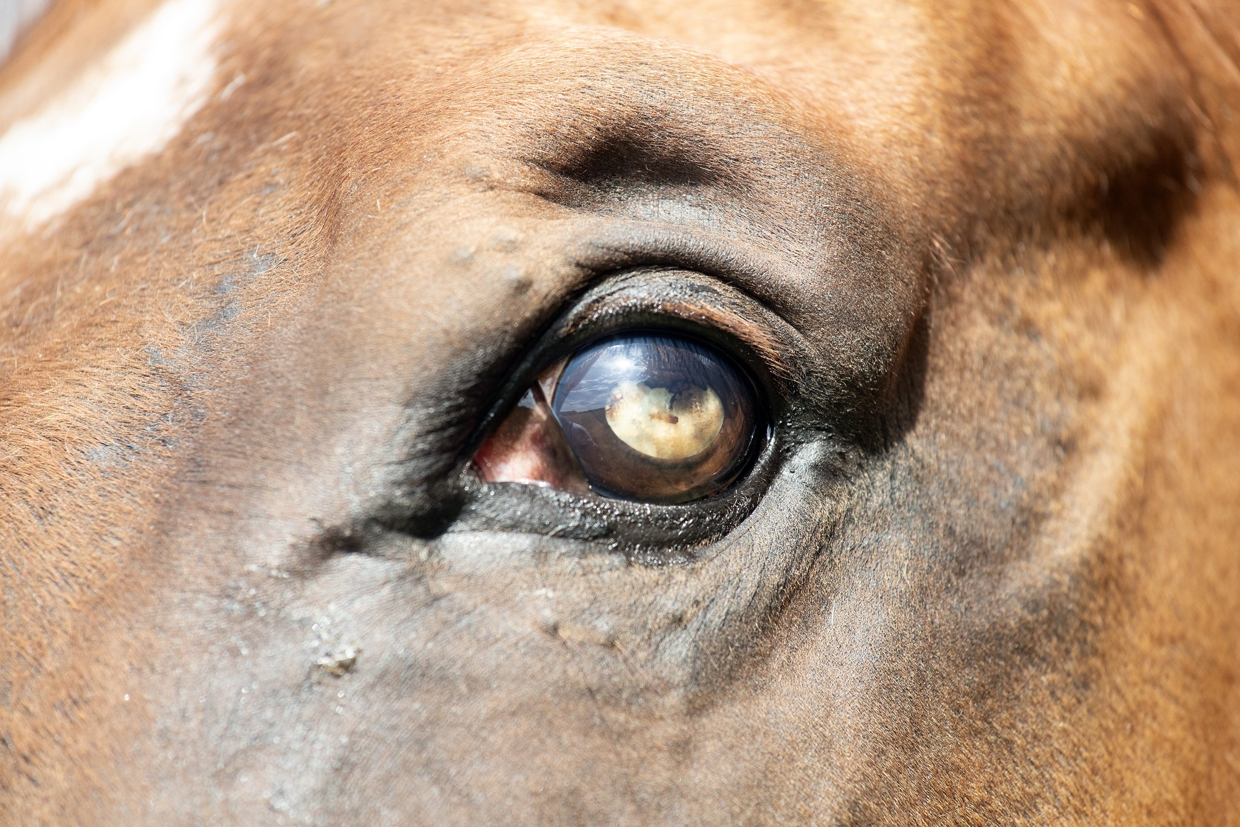
Bacterial keratitis is often seen after a corneal injury especially if an ulcer is present. The horse will demonstrate acute eye pain with serous discharge that quickly becomes mucopurulent or purulent. The clinical appearance is not usually diagnostic and cultures and scrapings should be taken from the edge of the ulcer. This procedure ensures the correct selection of treatment and pain relief.
Mycotic keratitis is uncommon in the UK but with the changing climate it may become more prominent. This type of keratitis is a result of fungal growth so tends to occur in climates supportive of this type of growth. Diagnosis is based on the history, clinical appearance and the demonstration of fungal hyphae and positive fungal culture. This disease may be a consequence of inappropriate drug therapy (such as corticosteroids) or from previous corneal trauma. Following the identification of the fungus, topical treatments can be used but may take weeks to months.
Uveal Tract
The uveal tract consists of three parts that include; The choroid which is the tissue layer filled with blood vessels; The ciliary body that is the ring of tissue containing muscles that change the shape of the lens as well as producing the clear fluid that fills the space between the cornea and the iris; The iris which is the colored part of the eye.
Persistent pupillary membranes are vascular arcades and developing tissue of the eye that fail to atrophy as the eye matures. These are very common in horses and usually have no consequence and no treatment is needed.
Cysts may arise in various parts of the uveal tract and are not uncommon in the horse. Irrespective of their origin, they may vary between pigmented to unpigmented and are smooth, round and do not invade neighboring tissue. No treatment is required except on rare occasions when they interfere with the horse’s vision.
Neoplasia of the uvea is not common but may arise and are often melanomas that are locally invasive but without cellular malignancy.

Trauma of the iris may result from direct trauma, or a secondary consequence of corneal perforation or a whiplash injury. Any uveitis that is caused by trauma can be treated medically. If there is an iris prolapse, then the iris is placed back into the anterior chamber provided they are not contaminated. Any foreign bodies must be removed and any hyphaema (bleeding in the anterior chamber between the cornea and iris) is usually left to be resorbed naturally.
Uveitis is inflammation of the uveal tract and can cause eye pain and alterations in vision. There are many causes of uveitis that include trauma, lens-associated uveitis, general viral infections (such as equine viral arteritis) and bacterial disease (such as Rhodococcus equi in foals). However, many situations of uveitis are an immune-mediated uveitis often referred to as equine recurrent uveitis (also known as moon blindness). Uveitis may present as an acute or a chronic condition.
Features of acute uveitis and chronic uveitis
This disease can be treated medically often with the use of a sub-palpebral or nasolacrimal lavage system. In addition, the patient should be placed in a quiet fly-free and dust-free environment.
Lens
The lens is a clear curved disk that sits behind the iris and in front of the vitreous of the eye, which bends light as it enters the eye to develop an image. The horse’s lens is large and minor opacities associated with embryonic remnants are common.
Cataracts are the most common lens abnormality to be encountered causing an opacity of the lens. Cataracts may be acquired from trauma or post-inflammation situations. However, cataracts may be congenital commonly seen in Arab and thoroughbred foals. These opacities can be classified in various ways:
• Age of onset – juvenile, senile or congenital
• Cause – post inflammation (uveitis) or trauma
• Location – cortical, capsular, nuclear, polar, equatorial
• Stage of development – immature, mature, hypermature
Most cataracts cause no obvious visual deficits unless they are dense and obstruct the visual axis. in which cataract surgery may be considered.
Acqueous drainage
The acqueous humor is a transparent water-like fluid similar to blood plasma but containing low protein levels. It is secreted from the ciliary body (a supporting structure of the lens) and fills both the anterior and posterior chamber of the eye.

Glaucoma is a pathological elevation of the intraocular pressure resulting in the optic nerve becoming damaged . Primary glaucoma in horses is exceptionally rare while secondary glaucoma is uncommon but may occur after anterior segmental inflammation. Often there is little to no pain but an enlarged globe and raised intraocular pressure with the lack of pupillary light reflex may be seen. Treatment may be attempted if the vision is present with various medications to reduce the intraocular pressure. If the horse is blind it may be left without treatment.
Conclusion
Good eye care is vital as the horse relies on its site to receive a great deal of information on its surrounding environment. Even with the horse holding its head forward it has remarkable peripheral vision but the horse’s vision is a little blurrier and less colorful compared to humans. In addition, both the strengths and weaknesses of the visual abilities of the horse must be seriously considered when looking at various techniques for training.
Proximal suspensory desmitis
Words - Connor Parsons DipWCF

Diagnosing proximal suspensory desmitis in the hind limb can be difficult. However, the modern diagnostic modalities available to the industry today makes it possible to isolate injuries, allowing both veterinarians and farriers to work together to achieve the best diagnosis and prognosis possible for the equine in question.
In this article, Connor Parsons reviews the anatomy and function of the suspensory ligament, causes and signs of proximal suspensory desmitis and whether there is an ideal procedure for diagnosing, treating and formulating a prognosis for the horse as part of his DipHE Farriery studies.
ANATOMY

The equine limb is complex yet effective. The suspensory ligament is made up of dense white fibrous connective tissue which suspends the fetlock and prevents hyperextension.
Originating at the proximal, plantar aspect of the third metatarsal/carpal attaching to two palmar depressions distal to the carpometacarpal and tarsometatarsal joints descending the channel formed by the 2nd, 3rd and 4th metatarsal/carpal, bifurcating two thirds of the way down the 3rd metatarsal/carpal, making a firm attachment to the palmar aspect of the proximal sesamoids, pulling the sesamoids proximally, then traveling dorsally and distally at an oblique angle to merge with the common digital extensor tendon. This forms a sling to support the fetlock joint. The ligament and its branches are strong but only slightly elastic (Devereux, 2006).
The suspensory ligament also forms a part of the hindlimb stay apparatus which is a system of ligaments, tendons and muscles that work together to allow the horse to stand and doze with minimal muscular effort. Also known as the fright and flight mechanism (Colles & Ware, 2020).

DAMAGE TO THE SUSPENSORY LIGAMENT
Suspensory ligament damage can affect horses of all breeds and ages. However, it is most common in competition horses. Proximal suspensory desmitis (PSD) is inflammation or damage of the main body at the origin of the ligament at the proximal end of the third metacarpal/metatarsal.
The suspensory ligament can be inflamed or there can be changes to the fiber pattern of the
ligament. These cases will present with lack of performance, being worse on soft surfaces. In more severe cases a core lesion (hole) can be seen on an ultrasound scan, where a number of fibers have ruptured. This type of injury will have a more sudden onset of lameness (Dyson, 1994). Injury can be solely within the ligament, involve tearing of the fibers of the ligament or be connected to avulsion fractures at the origin, involving the proximal 3rd metacarpal/tarsal (Baxter, 2020). Complete rupture is possible, however, very rare. The prognosis for a complete rupture is not favorable (Dyson, 1994).
Although the suspensory ligament has a slight elasticity to its make-up, if it is stretched it tends to heal with a loss of elasticity making it susceptible to recurrent damage (Colles & Ware, 2020).
SIGNS OF PROXIMAL SUSPENSORY DESMITIS

Proximal suspensory desmitis is a difficult condition to diagnose as the hind limb is complex and many of the functioning structures work in unison. A horse suffering with inflammation or damage to the main body of its hind suspensory can present one of three ways. It may have a unilateral lameness, a bilateral lameness or just a general decrease in performance (Dyson,1994).
CAUSES OF PROXIMAL SUSPENSORY DESMITIS OF THE HINDLIMB
Although there has been extensive research into proximal suspensory desmitis, there is no primary cause in all cases.
Proximal suspensory desmitis is a common injury in both front and hind limbs of the equine athlete. Usually bilateral in the hind limb (Dyson, 2016). All types and breeds of horses are susceptible to this type of injury. Poor conformation is a contributing factor to proximal suspensory desmitis.
Conformational defects such as straight hocks, sloping pasterns and long-toe, low-heel conformations would be at higher risk to injury. These conformational defects will all apply unnecessary pressure to the suspensory ligament. Horses that have suffered with this condition will be predisposed to a repetitive strain injury of this ligament (Devereux, 2006).
Overextension of the tarsus as a result of overextension of the fetlock has been linked to proximal lesions. The higher the severity of trauma, the higher the severity of ligamentous lesion. Working horses on deep, soft surfaces will increase the risk of this injury (Baxter, 2020). The hindlimbs are more frequently affected with this condition than the forelimbs with a much lower success rate of the horse returning back to performance prior to rest (69% hind vs 80% forelimb) (Colles & Ware, 2020).
DISCUSSION
In a study of six horses, this is an extremely small cohort of horses to be able to state an average age a horse is likely to present with this condition. This study also shows that all of the horses studied were of varying fitness levels, therefore stating that this does not affect the likelihood of injuring the hind suspensory ligament. There was only one horse in this study that was unfit and overweight. The rest were all competition fit with good muscle mass, showing that fitness doesn’t necessarily decrease the risk of this injury happening. The case history of the six horses studied did not include which discipline or level the horse was working at. This would be an interesting factor to consider when looking at which horses would be more susceptible to proximal suspensory desmitis.

Each individual case was being looked after by different veterinarians, giving a clear picture of different approaches on how to diagnose and treat this condition. Although for the purpose of a study the varying opinions will make the comparison more difficult.
All horses presented with a reduction in performance prior to veterinary contact. Only one horse was reported with a bilateral lameness behind. Flexion testing appeared to aggravate the lameness making it more prominent to see. Local analgesia has been shown to be effective in isolating the area to be investigated. Also, showing lameness on the other hind once the worse limb has been blocked out.
Using digital diagnostic modalities such as ultrasonography to diagnose this condition allows the veterinarian to study the changes in the fiber pattern of the suspensory ligament. This will allow the veterinarian to see the severity of damage caused and allow them to provide the best treatment plan possible. In this study only one horse had a lesion while the other five horses had thickening and slight changes to the fiber pattern. Horse 2 had lesions on both hind limbs however the veterinarian didn’t medicate, box rest was recommended. His prognosis was guarded.
Although radiographs of the feet don’t directly help with the diagnosis of proximal suspensory desmitis, they do allow the farrier to trim accordingly to restore the hoof back to correct hoof pastern axis and mediolateral foot balance. This will reduce lever arm forces thus reducing any unnecessary pressures on the plantar aspect of the limb.

Horses were radiographed for foot balance to aid with remedial trimming and shoeing. This will increase the equine’s prognosis allowing the farrier to have a clear picture of what is being dealt with.
All of the horses that were radiographed presented with a negative sole plane and weak heels.
The question is whether this foot conformation is because the horses are wanting to apply more pressure to the caudal aspect of the hoof in the landing phase, reducing the movement of the metacarpophalangeal articulation. This is an attempt to reduce the loading forces applied to the suspensory ligament. However, it will also cause the heels to become weak. Or, if this conformational defect has caused the suspensory ligament to become inflamed or damaged, thus causing proximal suspensory desmitis.
Proximal suspensory desmitis can be secondary to other conditions such as hock conditions or sacroiliac problems which cause the horse to adopt a different gate. Therefore causing unnecessary loading on the suspensory ligament. It is important that the primary cause is diagnosed and treated when treating proximal suspensory desmitis. This is where scintigraphy can be a useful tool to get a clear picture of the cause involved in individual cases.
Scintigraphy is an expensive diagnostic modality which carries significant health and safety risks, this must be taken into consideration when dealing with cases.
All horses studied were worse on a soft surface where it is harder for the horse to guard itself from soft tissue injuries. Horses that are worse on soft surfaces generally are suffering from soft tissue pain. However, nerve blocks will help the veterinarian pinpoint the structures involved when diagnosing lameness.
Although it is possible to have a unilateral lameness with proximal suspensory desmitis in the hind limb it is most common for the lameness to be bilateral. All of the horses in this study had a bilateral lameness, generally worse on one limb than the other. Although presenting prior to veterinary contact as lack of power or struggling to strike off on the correct canter lead.
When a veterinarian is deciding on a treatment plan, the horse is looked at carefully including its previous history as some treatments come with higher risks, although can be extremely effective for reducing inflammation. Shockwave treatment comes with minimal risks involved and is effective; however, many racing authorities require a mandatory 5 day Stand-Down period from racing following the administration of extra-corporeal shockwave therapy. Findings from this study show that the horses with the best prognosis of getting back to competitive work have undergone surgery. Understandably this is the last resort treatment as it is invasive and expensive for the client.

Only one horse from this study did not have any medical intervention and this horse had the least favorable prognosis. This would suggest that box rest alone is not generally enough if the horse is expected to get back to full athletic fitness. The most common veterinary treatment is steroidal injections into the area of interest and shockwave therapy with rest. However, the use of corticosteroids in horses in training often adopt a clear 14-day exclusion on the use of intra-articular (joint) injections before racing in line with different racing authority regulations.
Water based therapy can also be considered as part of the recovery process when bringing the horse back into work. It’s known to reduce limb oedema, stimulate nerves, and improve circulation, which speeds the healing process and provides pain relief. It also aids in joint stability, providing all-around support to the limbs.
Cold water therapy is typically prescribed when the goal is to reduce heat and inflammation. Applying cold water or ice reduces the amount of accumulating fluid to an injured area and can somewhat numb the area, causing a topical analgesic effect.

Underwater treadmills are often used for horses with tendon and ligament injuries to provide a gradual transition back into exercise and regain the range of motion. Swimming is also used to condition the horse without putting a load on the skeletal system. It is often used in the early stages of tendon and suspensory injuries due to no pressure being placed on the lower limb. Trainers who use swimming as part of their routine often find that, in addition to the cardiovascular workout, it also helps the horse relax and settle its mind.
This is not always successful and horses are then admitted for surgery. While the surgery for this condition is successful, there must be consideration taken into the fact that it is not legal to compete at certain levels once this surgery has taken place.
The study shows that the farriery treatment involved when dealing with this condition is varied, depending on which veterinarian the horse is being looked after by. However, the the author has had positive results from many different shoeing styles. The main importance of trimming and shoeing for this condition has been shown to restore the best possible hoof pastern axis through trimming, supporting the entire limb and fitting a shoe with an early breakover. This will reduce the lever arm on the metacarpophalangeal articulation, thus minimizing unnecessary pressure on the suspensory ligament.
CONCLUSION
Having such a small cohort of horses in a study makes it difficult to finish with a conclusive result. This small study however, has given a positive result in the diagnosis stages of dealing with this condition. At this stage nerve blocks are invaluable along with ultrasonography. In less obvious cases MRI is useful to gain a diagnosis and occasionally scintigraphy will be used to locate the problem. Radiography is a useful tool when dealing with PSD and checking the origin area for avulsion fractures.
This study has also shown that there is a link between a negative solar angle and proximal suspensory desmitis. However, this would need to be studied further and on a greater scale to determine why there is a link between this conformational defect and this condition.
It is paramount that correct foot balance is achieved by the farrier. To achieve this foot balance radiographs are required. This study has shown that there is no definitive way to shoe for this condition, however it has shown a positive result from an early breakover shoe, allowing the horse to relieve pressures on the caudal aspect of its hoof. Horses that had the best prognosis underwent surgery, allowing them to get back to competitive fitness.

REFERENCES
Baxter, G. M., 2020. Adams and Stashak's Lameness in Horses. 7th Edition ed. Hoboken, NJ: John Wiley & Sons.
Colles, C. & Ware, R., 2020. The Principles of Farriery. 2nd edition ed. Marlborough: J.A.Allen.
Devereux, S., 2006. The Veterinary Care Of The Horse. 2nd Edition ed. London: J.A.Allen.
Dyson, S., 1994. Proximal suspensory desmitis in the hindlimb: 42 cases. British Veterinary Journal, 150(3), pp. 279-291.
Dyson, S., 2016. American Association of Equine Practitioners. [Online] Available at: https://aaep.org/horsehealth/lowdown-high-suspensory-disease-proximal- Suspensory-desmitis [Accessed 19 11 2022].
Smith, M., 2022. Newmarket Equine Hospital. [Online] Available at: https://www.newmarketequinehospital.com/media/pm1beabc/hah349 Vet_susp_desmitis-final.pdf [Accessed 9 April 2023].
Why Embryo’s Fail and Improving Odds of Successful Horse Breeding
Words - Jackie Zions

If entering the fall, your breeding prospects have come up empty, there are considerations to ponder and actions you can take for successful breeding next year. “Don’t let those mares sit all fall and winter, with untreated conditions such as a uterine infection,” says Dr. Tracey Chenier, Theriogenologist and researcher from the Ontario Veterinary College at the University of Guelph. “Have a thorough veterinary evaluation now to help ensure her uterus is clean and she is healthy and cycling early next year, for the best chance of a positive outcome.”
Common Issues Conceiving and Potential Solutions
“The number one reason your mare didn't get pregnant in any given year may be due to uterine infection,” says Chenier. “The term we use is endometritis.” Most often caused by a bacterial infection, it is often associated with poor perineal conformation. In other words, your mare has a tilt to her vulva causing the vagina and uterus to become contaminated with bacteria every time she defecates. It is also common for these mares to wind suck, which can lead to infection and inflammation that results in a hostile environment the embryo cannot survive in. A minor surgical procedure known as a Caslick’s suture reduces the chance of contamination in most affected mares. Severely affected mares may require additional procedures to reconstruct the perineal body.

Another common form of endometritis is PBIE, or persistent breeding induced endometritis. In these cases, there is a prolonged inflammatory response to semen and contamination that occurs at breeding. Again, the mare has a hostile uterine environment in which the embryo cannot survive. To improve the chance of conception, this condition can be managed by ultrasound within 6 to 12 hours after breeding. She is checked for fluid retention and inflammation and if present, the uterus is lavaged to remove the fluid and calm the inflammation. The veterinarian may also advise administration of oxytocin to increase uterine contractions and help remove the fluid.
Another very common reason for the mare not getting pregnant is their age. “We actually consider mare’s fertility to decline as early as 12 years of age,” says Chenier, “and that surprises people that as early as 12 years, their fertility can decline significantly.” Older mares can have poorer oocyte (egg) quality. This reduces their chances of getting pregnant and can result in higher rates of mid-gestational losses.
Older mares are more susceptible to many circumstances including uterine conditions, metabolic disease, changes to the uterus, fibrosis and cysts. Fibrosis of the uterus will reduce the chances of carrying a pregnancy to term. Endometrial cysts or fluid filled sacs in the lymphatics of the uterus can block the ability of the embryo to move around and interfere with the placenta formation.
Less Common Conception Issues
“Stress, nutritional issues, and hormone deficiencies can make it difficult for an embryo to survive,” explains Chenier “but these issues are generally less common.”
The corpus luteum is the structure that forms on the ovary after the mare ovulates and its progesterone production maintains the pregnancy early on. This structure may be susceptible to effects of severe stress, illness, or inflammation in the uterus. Progesterone/altrenogest supplementation can often save these early pregnancies but the mare will have to stay on the supplements until the fetoplacental unit takes over pregnancy maintenance by 120 days. The fetoplacental unit is a crucial interface between maternal and fetal circulatory systems, providing essential nutrients and oxygen to support fetal growth and development.
Early pregnancy loss can happen from days 0–60 of gestation. To help avoid risk factors like excessive stress, ask your vet before changing or adding anything to your mare’s routine. Consult your vet before administering any vaccines or deworming products.
“Oviductal blockage is another uncommon condition,” says Chenier, “but in mares that are not conceiving and everything else is normal (no uterine infection, good stallion fertility…) it should be considered.” An effective treatment the veterinarian may suggest, involves applying the hormone prostaglandin E to the oviductal papillae, which opens the oviduct and allows that blockage to be cleared out.
Diagnostics used to investigate early embryonic loss

“A good reproductive evaluation is really important to find out the reasons why a mare either didn't get pregnant or lost a pregnancy,” says Chenier.
Veterinarians use rectal palpation, especially with ultrasound, to help detect fluid and infection. Palpation with ultrasound can detect the presence of endometrial cysts, conditions on the ovary, such as failure to ovulate and ovulatory follicles.
Cultures, gained from swabs of the uterus, are performed to detect inflammation and infection. This is helpful in cases where antibiotic use is required in order to determine what type of antibiotic to use.
Uterine biopsy is indicated in certain cases. “I recommend a biopsy in any mare that fails to get pregnant after three attempts, especially if we are not getting good answers on a swab culture and ultrasound,” says Chenier. As well as providing a prognosis, it provides a lot of information on treatment options to improve the mare’s fertility.

A biopsy can help provide better information about what's going on in the uterus and in the case of inflammation, identify the type of inflammation present.
Chronic infections are more likely to be caused by something like a dormant strep infection, and biopsy may be the only way to diagnose the fibrosis of the uterus that would be directly related to prognosis. If you found out your mare’s uterine biopsy was a Grade 3, meaning she has a lot of permanent severe changes in that uterus, her likelihood of carrying a foal to term is between zero and ten percent. This is really important information to help the breeder to decide whether they want to invest the time and money to attempt to breed a mare with a prognosis revealing these challenges.
Ultrasound is useful in identifying conditions such as endometrial cysts. Cysts can be removed by putting an endoscope in the uterus and then using either laser or electrocautery to a blade to improve a mare’s chances of pregnancy. Electrocautery involves using a heated electrode to cut or coagulate tissue during surgery. When applied to a blade, it allows for precise cutting with minimal bleeding.
“In really rare cases where all else has been ruled out, a karyotype might be considered,” says Chenier. “If it's a young maiden and everything else seems to be working, there may be a genetic reason that she's not able to get pregnant, but that would be the exception.” Karyotyping involves staining chromosomes and examining them to identify structural changes or numerical abnormalities.
Improving the odds of pregnancy
The all-important veterinary evaluation will check the mare’s general health, body condition and uterine health as well as rule out metabolic diseases like insulin resistance and Cushings.
One must ensure the broodmare’s nutritional needs are met. Calories, protein, vitamins, and minerals are all passed on to the foal while in utero. Consult your vet or an equine nutritionist to ensure your mare gets a balanced diet and to learn how her nutritional needs increase during pregnancy.

Use of light to manipulate the season is a consideration if you want to breed your mare early in the season for a January – March foal. Mares stop cycling during the winter. “I think it's helpful to expose the mare to the cold and the darkness of the fall to reset her system before you start her under lights,” say’s Chenier. She recommends lighting programs begin around December 1st with what amounts to ten-foot candles, which is equivalent to 100 lux intensity of light. In old style lighting with incandescent bulbs that was the 100 Watt bulb and the old saying was you needed to be able to read a newspaper in every corner of the stall. 16 hours of total light per day is recommended, and this includes natural light. From a practical point of view, that means if you turn your mare out at 8:00 o'clock in the morning, bring her in at four, and have the lights on in the stall until 11:00 PM, you will be providing an adequate amount of light. Chenier also describes the use of a commercially available equine light mask that is worn 24/7, like a fly mask. It is battery powered and delivers blue light to one eye on a timed basis.
“Good breeding management is always key,” emphasizes Chenier. “Negative uterine swabs before breeding ensure the mare is free of infection, limit to one cover in mares prone to infection or inflammation, correcting poor perineal conformation and then practice optimal timing.”
Not breeding at the right time is much less likely in the Thoroughbred industry, where mares are being bred by natural cover. If the mare is not in heat and not ready to ovulate, she is not likely to stand for the stallion.
If a mare is bred too early, the sperm will not live long enough. Mares can stay in heat a day or two after they have ovulated. If breeding happens too late (after ovulation), the oocytes will no longer be viable.
If the chosen stallion has fertility issues, the breeder may need to closely monitor their mare’s ovulation for the most optimal timing of breeding to improve odds of success. Chenier says, “If their sperm doesn't live very long inside the mare, we have to manage those cases differently and make sure we're breeding those mares really close to ovulation to get good fertility for those stallions.” Stallion fertility should always be considered a possibility when mares are not conceiving.
If breeding early in the season, one needs to make sure the mare is cycling properly and not just in spring transition. A vet check will confirm the mare is experiencing a real heat and ovulating for early breeding (Feb – April).

Role of the Lymphatic System
Words Peter Bollen

The role of the lymphatic system in energy metabolismIn recent years, focus has turned towards the emerging science of the lymphatic system. It is as important as the circulatory system (which includes the heart and the blood vessels), and is fundamental to the functioning of the body and central to the immune system, energy metabolism, detoxification and energy availability in the horse’s body.
A horse with a well-functioning lymphatic system can metabolize energy and clear metabolic waste more efficiently, leading to enhanced performance and faster recovery.
So how can we tap into the benefits of an effective lymphatic system? Well, the first step is in understanding what it is. We might know that nutrients and oxygen are carried in the bloodstream towards our tissues, but what about how they are actually delivered to the cells?
This is where the lymphatic system comes in. Broadly speaking, it runs in parallel to the circulatory system and creates the point of delivery to the cells and tissues. The circulatory system carries the blood via arteries and vessels, which get smaller as they reach the muscle tissue as capillaries - where the wall is only one cell thick.
This enables smaller molecules such as nutrients and oxygen to be pushed out of the capillaries and into the fluid around the cells - or lymph - while retaining larger cells like erythrocytes (red blood cells).
The lymphatic system is an extensive network of vessels and nodes that transports lymphatic fluid around the body and it has two main functions:
1. Energy metabolism - transporting nutrients and oxygen to the cells
All the cells and tissues of a horse’s body are surrounded by a watery gel-like substance called interstitial fluid. This provides a medium for dissolved oxygen and nutrients to travel across to the cells. Lymph capillaries, carrying these substances, run through the interstitial fluid. The walls of these capillaries are also only one cell thick and have overlapping junctions, which make them highly permeable and allow easy transfer of materials.
In practical terms, this means the horse’s muscles and tissues get all the energy they require to carry out all their metabolic processes. As well as being responsible for giving cells the oxygen and nutrients they need, the interstitial fluid transports salts, hormones, neurotransmitters, coenzymes, amino acids, sugars and fatty acids around the body via the lymphatic system.
“A horse with a well-functioning lymphatic system can benefit from increased energy and enhanced performance, but without feeling too fresh or fizzy”

2. Detoxification: processing waste products
During the season, horses are placed under physiological demands and their body has a lot to process. In hard exercise and in races, their body will produce a lot of waste products. Their thin-walled lymph vessels also allow interstitial fluid into them to remove the waste products of cell metabolism (such as cell debris, bacteria, dead blood cells, pathogens, toxins, lactic acid and protein molecules) from each cell. This process is just as important as carrying necessary materials to the tissues to provide energy.

These metabolic waste products are carried in the lymph away from the cells for detoxification. This is the term used for the continuous processes in the horse’s body to remove those waste products or metabolites that are naturally produced.
Detoxification takes place mainly in the liver, kidneys and intestines, so it is really important to ensure that those organs in the horse are healthy and functioning well. By supporting the liver and kidneys, you can help this breakdown process and allow the horse to better metabolize compounds that would otherwise be toxic if they remained in the body.
Gut health is also very important to the lymphatic system and detoxification, given that more than half of the lymphatic vessels of the horse are located within the gastrointestinal tract.
Peristalsis (the natural contraction and relaxation of the gut wall) drives the return of lymph to the rest of the body. Movement in the gut is stimulated by food consumption (mainly long fibers) and is crucial to lymphatic system function as there is very limited muscular contraction in the horse’s lymphatic vessels themselves. This means it relies on passive forces from movement of other systems in the horse’s body as opposed to active muscle contractions.
Ensuring sufficient feed for a horse around the clock is crucial, not only for maintaining gut health, but to maintain a healthy immune system and detoxification process as well.
Lymphatic system and immune function
In addition to lymph and lymph vessels, the horse has around 8000 lymph nodes. These bundles of lymphoid tissue and proteins act as a filter for foreign substances that travel through the lymphatic fluid and contain lymphocytes (white blood cells) that help the body fight infection and disease. This means that the lymphatic system is also key to the functioning of the immune system.
As the lymph tissue approaches each lymph node, it slows down and collects in that area. Horses have so many lymph nodes that they are extremely susceptible to lymph node blockage. Lymph nodes can actually be felt or sometimes seen at various points around the horse’s body, particularly around the head and neck or when they are swollen.
Keep on moving

Movement is so important for horses, precisely because of the fact that there are no direct muscular contractions within the walls of the lymph vessels to promote the flow of lymph. The functioning of their lymphatic system relies on the force of movement of other structures in the body, such as the movement of the skin to apply pressure to the underlying tissues and stimulate the transport of lymph through the vessels.
In the modern day, racehorses can be stabled for longer periods of time. It is very common to see leg swelling, which has a very simple explanation in terms of the immune system. This principle of continuous movement being necessary applies especially to the legs, where this elastic movement of the skin is assisted by a pump mechanism in the hoof and fetlock joint. It is thought that this is the case due to there being no muscles in the lower limbs of the horse to aid the movement of lymph from this area.
Horses therefore need to move extensively and perhaps almost constantly (up to 16 hours per day) to keep the lymph circulating around the body, and standing still for too long significantly impairs the functioning of the lymphatic system.
Practical tips for a healthy lymphatic system
1. Turn horses out as much as possible
Horses that are standing still for long periods of time are at risk of an impaired lymphatic system, which can impact the immune system, recovery time and ultimately performance. Turnout makes a happier horse and a healthier horse.
2. Cool down for at least 15 minutes after exercise
The period of time immediately after training is important for removal of metabolic waste products and keeping a horse moving after a hard training session stimulates the lymphatic system to continue to remove waste products from cells, which includes the removal of lactic acid. By cooling down properly after every session, you can significantly improve recovery times.
3. Consider feeding supplements that can directly benefit the lymphatic system

Functional herbal ingredients in innovative supplements can support lymphatic system function. Cleavers (Galium aparine) is known to bring benefits to the lymphatic system. Couch grass (Elymus repens), Nettle (Urtica dioica), and Dandelion (Taraxacum officinale) are all known to have purifying effects which can support metabolism and detoxification.
Even Lionel Messi, Usain Bolt or Iga Swiatek have picked up issues through their sporting careers, despite having elite support teams behind them. Unfortunately injuries do just happen, but with foresight, preparation and providing the best nutritional support to their horses, trainers will give themselves the greatest chance of avoiding yet another bad news phone call to an owner.
The latest Strangles research available testing for ultimate sensitivity to avoid infection
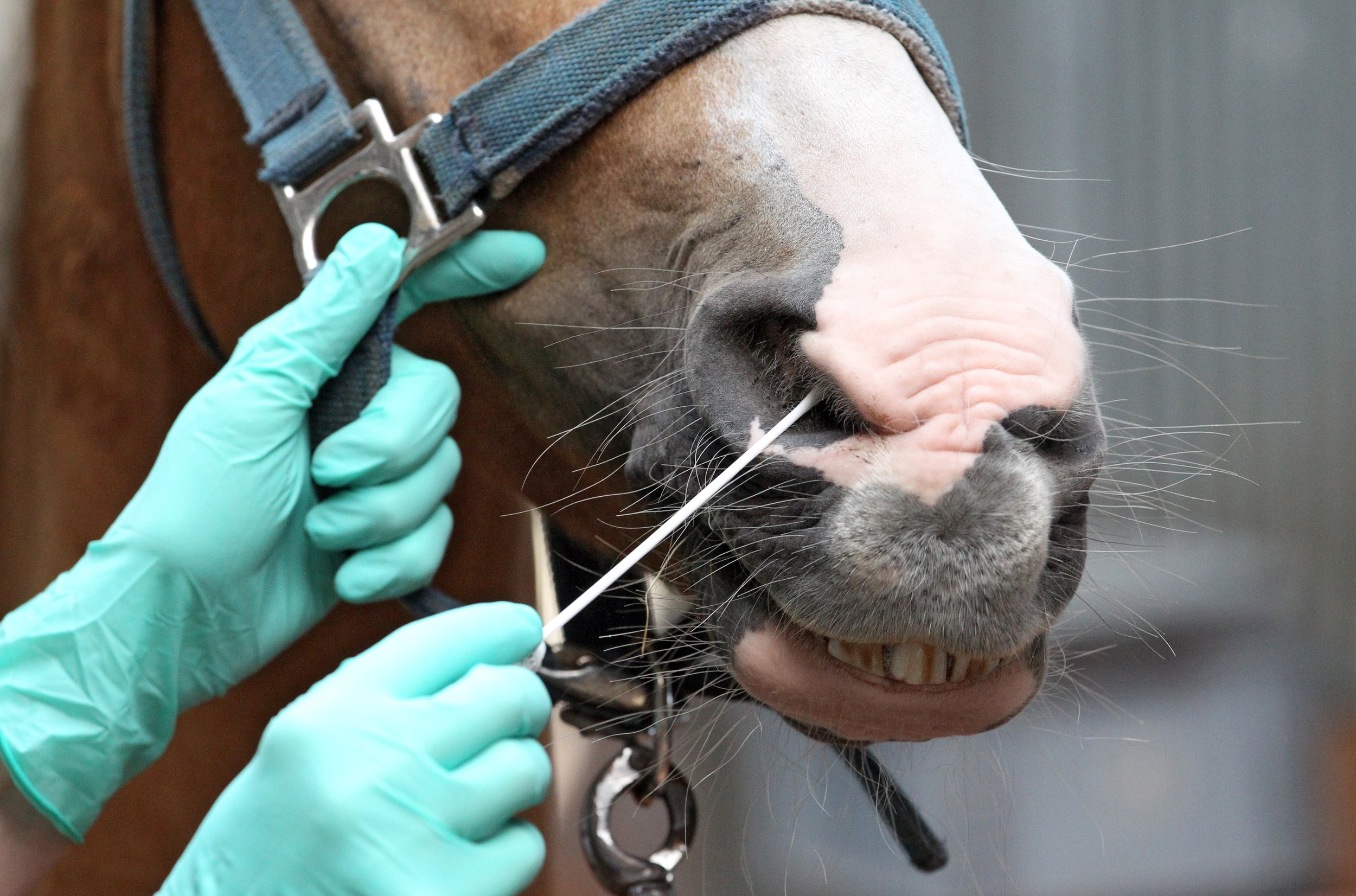
Strangles, the highly contagious upper respiratory disease caused by the bacterium, Streptococcus equi (S. equi) has been front and centre on social media lately with numerous disease alerts being posted. These alerts are triggered by positive test results for S. equi and reported by an official laboratory to the provincial or state veterinary office. Given the potential ramifications of a positive test, such as animal movement restrictions for several weeks and increased costs to horse and facility owners, a lot rides on the interpretation of these test results and the associated risk of disease spread to other horses, on and off the premises.
Testing for S. equi helps determine that a horse is free of S. equi or, in other words, not an S. equi carrier. It is usually done when the horse has recovered from clinical signs of Strangles to determine they are no longer infected and capable of transmitting S. equi, or upon request by equine facility managers, to screen a horse for carrier status prior to coming to their facility.

The two tests utilized for S. equi testing are the polymerase chain reaction (PCR) and bacterial culture. Testing utilizing bacterial culture detects living S. equi. Polymerase chain reaction (PCR) testing is much more sensitive than culture but detects DNA from both living and non-living bacteria. While the PCR sensitivity level can be useful as it can detect carrier horses that have a very low level of bacteria present in their guttural pouches, it can also detect transiently exposed/infected, asymptomatic horses, which rapidly clear the infection within a week. PCR can also flag horses that are less likely to be infectious at the time of sampling which can aid in risk management for that horse and the herd.

While these tests have their pros and cons, the relationship between S. equi PCR and bacterial culture has not been extensively studied. This is what Dr. Scott Weese of the Ontario Veterinary College and collaborators from OMAFRA and the University of Prince Edward Island set out to determine in a 2023 research study funded by Equine Guelph; ( tinyurl.com/guelph-strangles)
The relationship between quantitative real-time PCR cycle threshold and culture for detection of Streptococcus equi subspecies equi.
The 2023 study compared PCR and culture results from 158 equine respiratory tract samples submitted to an Ontario animal health laboratory for S. equi PCR testing. Of the samples that were PCR positive (CT < 40), only a minority (7.6%) were positive for S. equi on culture. That suggested that most PCR positive horses were likely a low risk for transmitting the bacterium at the time of sampling. A qPCR cycle threshold (CT ) of 34.2 was the breakpoint established, signifying that the likelihood of finding culturable S.equi above a CT of 34.2 was less likely and that the horse had a lower risk of being infectious at that point in time. These results were specific to this particular laboratory and cannot be applied to other laboratories which use their own testing procedures.
The line is not meant to be a green or red light but an indicator to aid in assessing the risk of disease transmission. Horses with PCR CT levels above 34.2, and who have developed a carrier status, can go on to produce lower CT levels (higher bacterial counts) over time and be a risk for S. equi shedding down the road. More research is needed to understand the S. equi shedding dynamics in carrier horses.

Combining culture and PCR testing is an option which comes at a higher cost to the horse owner but can be useful for an in-depth way to investigate bacterial loads and the risk of transmission at the time of sampling. While opting for ultimate sensitivity can help make sure no potentially infected horses are missed, it can start the domino effect of excessive control measures and costly interventions if not put into perspective related to the goals of testing, which may vary significantly between facilities (e.g. busy show barn, racetrack or closed herd).
Strangles has existed in horses since the 1800’s and isn’t going away anytime soon. Testing as part of a recovery plan from a Strangles outbreak is a no-brainer, but when it comes to using S. equi testing as part of a sickness prevention plan for your horse or facility, talk with your veterinarian and understand the impact a positive test result might have on your horse/herd and wallet BEFORE you start testing.
Electroarthrography to Predict Cartilage Quality
Article by Jackie Zions interviewing Dr. Adele Changoor and Dr. Judith Koenig

Researchers from the Ontario Veterinary College (OVC) and University of Toronto are developing a novel method to measure the quality of cartilage in horses using electroarthrography (EAG). EAG is a non-invasive technique that uses electrodes attached to the skin around a joint to detect electrical signals produced by the cartilage when it is loaded.
Dr. Adele Changoor, from the University of Toronto and Lunenfeld Tanenbaum Research Institute, and Ontario Veterinary College researcher Dr. Judith Koenig from the department of Clinical Studies, explain how EAG works and why it may become very useful for predicting cartilage quality and diagnosing osteoarthritis and other degenerative joints diseases in horses.
EAG is analogous to electrocardiography (ECG), which measures the electrical activity of the heart. Cartilage produces electrical signals during loading and these signals reflect its biomechanical properties, such as stiffness and permeability.
“By measuring EAG signals, we can get an idea of how healthy the cartilage is,” said Changoor.

Healthy cartilage ensures joints can move without pain and has an important role preventing wear and tear on bone.
Currently, there are no readily available tools to assess cartilage quality in horses with the exception of diagnostic arthroscopy – a minimal invasive surgery – under general anesthesia. X-rays and ultrasound are not sensitive enough to detect cartilage changes, and magnetic resonance imaging (MRI) is expensive, requires anesthesia and is often difficult to access. EAG offers a potential alternative that is fast, easy, and affordable.
“EAG is a promising tool for detecting cartilage damage early allowing intervention with treatments that can slow down or prevent further deterioration of the joint,” says Koenig “EAG could also help us monitor the effectiveness of treatments over time.”
EAG measurements were collected at the same time as the center of pressure (COP), which measures the distribution of force under the horse’s hoof when it stands or walks.
“EAG is really tied directly to cartilage biomechanical properties,” says Changoor. “We also needed to know about the joint biomechanics in order to interpret EAG properly.” A custom, portable, force mat was developed by Dr. Changoor’s graduate students that included an array of force sensors to place under the horse’s hoof when measuring EAG.
“Then we can measure how much compressive force or ground reaction force is being exerted on that joint” said Changoor. “COP, is where the ground reaction force is acting. The ground reaction force gives us the total load on the joint. COP lets us figure out where on the hoof or where on the joint surface force is being concentrated.”

COP provides information about the joint biomechanics and the horse’s balance and stability. EAG and COP testing were combined to get a comprehensive picture of the joint health and function in horses with osteoarthritis. Results were compared with MRI imaging and it was found that EAG and COP testing correlated well with MRI and could detect differences in cartilage quality between healthy and osteoarthritic joints.

In the 2023 study involving horses with osteoarthritis in the fetlock joint; the horses were treated with MSCs to decrease inflammation and stimulate tissue healing. The researchers measured EAG, COP, and MRI before and after the treatment to evaluate its impact on cartilage quality.
“We observed that MSCs improved cartilage quality in some horses and EAG and COP testing were able to capture these changes and show the responses to treatment. This suggests that EAG and COP testing could be useful for selecting treatment options for the horse,” says Dr. Koenig. “One of the biggest advantages of EAG is that it seems to correspond with our arthroscopic findings. It can perhaps evaluate the quality of the cartilage or cartilage defects, which we are at the moment only able to evaluate with arthroscopy.”
The researchers plan to conduct further studies in order to validate and refine EAG and COP testing for predicting cartilage quality in equines. They hope that these techniques will become widely available and accessible for veterinarians and horse owners in the future.
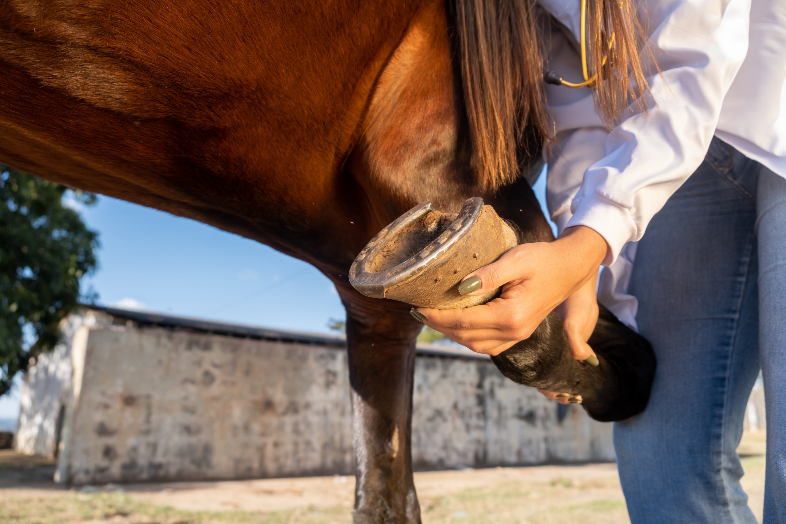
“This is an exciting and innovative research project that has the potential to improve the diagnosis and early management of osteoarthritis in horses,” says Dr. Koenig “Osteoarthritis is a major health and welfare issue for horses and their owners, and we need better tools to detect it early and treat it. EAG and COP testing could provide a simple, affordable, and accurate way to assess cartilage quality and joint function in horses.”
Many thanks go to the graduate students who worked tirelessly on the EAG study: Peter Suderman, PhD Candidate in the Department of Materials Science & Engineering at U of T, Jaylon Pascual, undergraduate co-op student finishing her fourth year in the Biomedical Engineering program at U of G, Dr Rodrigo Munevar Luque, Equine Sports Medicine Resident at OVC and PhD Candidate Biomedical Sciences at U of G, Undergraduate Research Assistants in Clinical Studies Ashley Nixon, DVM 25 (OVC) , Pjotr Roest DVM 26 (OVC), and in Biomedical Sciences Axel Koenig Parris HBA 25 (Ivey School of Business, Western University) and Rebecca Mullin BSc OVC 25.
The study was funded by the Equine Guelph Research Fund and the Natural Sciences and Engineering Research Council of Canada (NSERC).
The value of good hoof balance and how to evaluate this alongside your farrier
Article by Adam Jackson MRCVS
Introduction
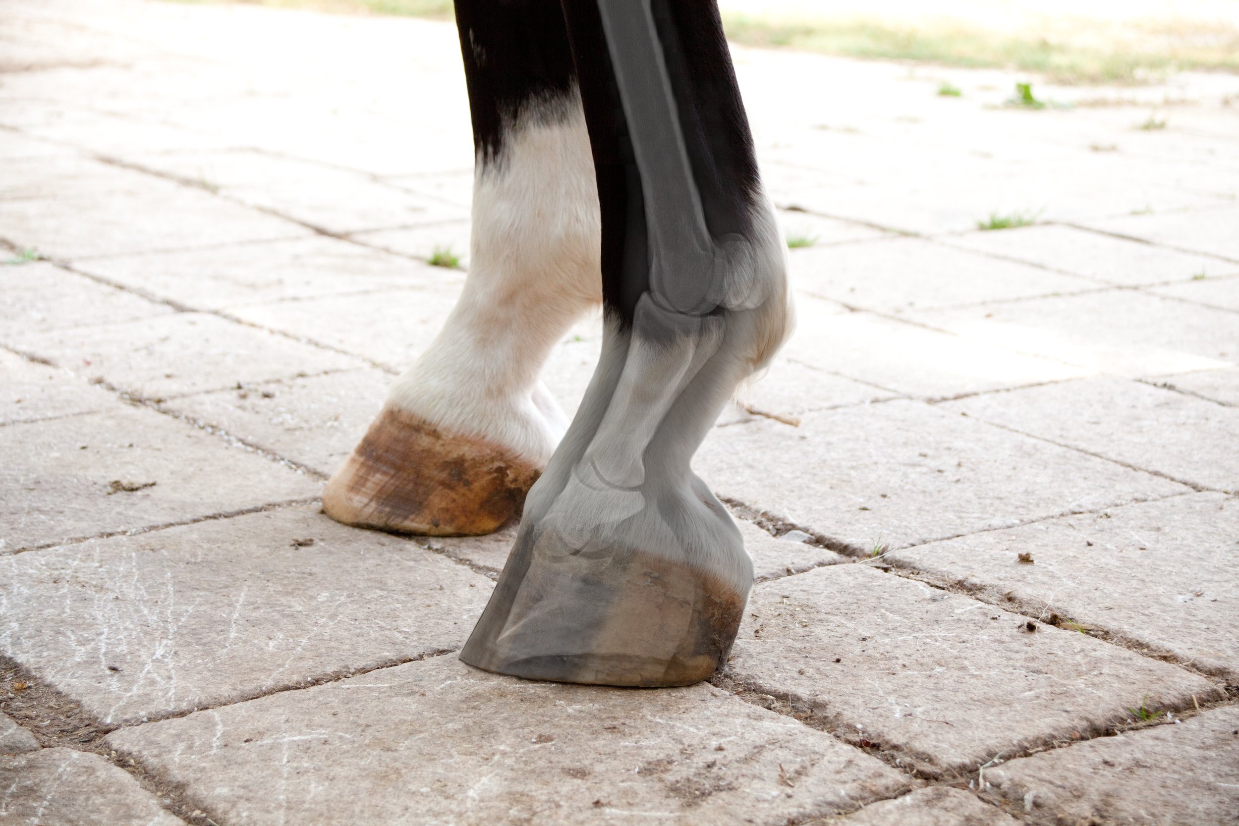
The equine foot is a unique structure and a remarkable feat of natural engineering that follows the laws of biomechanics in order to efficiently and effectively disperse concussional forces that occur during the locomotion of the horse. Hoof balance has been a term used by veterinarians and farriers to describe the ideal conformation, size and shape of the hoof relative to the limb.
Before horses were domesticated, they evolved and adapted to survive without any human intervention. With respect to their hoof maintenance, excess hoof growth was worn away due to the varied terrain in their habitat. No trimming and shoeing were required as the hoof was kept at a healthy length.
With the domestication of the horse and our continued breeding to achieve satisfactory performance and temperament, the need to manage the horse’s hoof became essential in order to ensure soundness and performance. The horse’s foot has evolved to ensure the health and soundness of the horse; therefore, every structure of the foot has an essential role and purpose. A strong working knowledge of the biology and biomechanics of the horse’s foot is essential for the veterinarian and farrier to implement appropriate farriery. It was soon concluded that a well-balanced foot, which entails symmetry in shape and size, is essential to achieve a sound and healthy horse.
Anatomy and function of the foot

The equine foot is extremely complex and consists of many parts that work simultaneously allowing the horse to be sound and cope with the various terrains and disciplines. Considering the size and weight of the horse relative to the size of the hoof, it is remarkable what nature has engineered. Being a small structure, the hooves can support so much weight and endure a great deal of force. At walk, the horse places ½ of its body weight through its limbs and 2 ½ its weight when galloping. The structure of the equine foot provides protection, weight bearing, traction, and concussional absorption. Well-balanced feet efficiently and effectively use all of the structures of the foot to disperse the forces of locomotion. In order to keep those feet healthy for a sound horse, understanding the anatomy is paramount.
The foot consists of the distal end of the second phalanx (short pastern), the distal phalanx (pedal bone, coffin bone) and the navicular bone. The distal interphalangeal joint (coffin joint) is found between the pedal and short pastern bone and includes the navicular bone with the deep digital flexor tendon supporting this joint. This coffin joint is the center of articulation over which the entire limb rotates. The navicular bone and bursa sits behind the coffin bone and is stabilized by multiple small ligaments. The navicular bone allows the deep digital flexor tendon to run smoothly and change direction in order to insert into the coffin bone. The navicular bursa is a fluid-filled sac that sits between the navicular bone and the deep digital flexor tendon.
The hoof complex can be divided into the epidermal weight-bearing structures that include the sole, frog, heel, bulbs, bars, and hoof wall and the anti-concussive structures that include the digital cushion, lamina, deep digital flexor tendons, and ungual (lateral) cartilage. The hoof wall encloses the dermal structures with its thickest part at the toe that decreases in thickness as it approaches the heel. The hoof wall is composed of viscoelastic material that allows it to deform and return its shape in order to absorb concussional forces of movement. There is enough deformation to diminish the force from the impact and load of the foot while preventing any damage to the internal structures of the foot and limb. As load is placed on the foot, there is deformation that consists of:
Expansion of the heels
Sinking of the heels
Inward movement of the dorsal wall
Biaxial compression of the dorsal wall
Depression of the coronary band
Flattening of the sole
The hoof wall, bars and their association with the sole form the heel base with the purposes of providing traction, bearing the horse’s weight while allowing the stability and flexibility for the expansion of the hoof capsule that dissipates concussional forces on foot fall. The sole is a highly keratinized structure like the hoof wall but made up of nearly 33% water so it is softer than the hoof wall and should be concave to allow the flattening of the sole on load application. The frog and heel bulbs serve a variety of special functions ranging from traction, protection, coordination, proprioception, shock absorption and the circulation of blood.
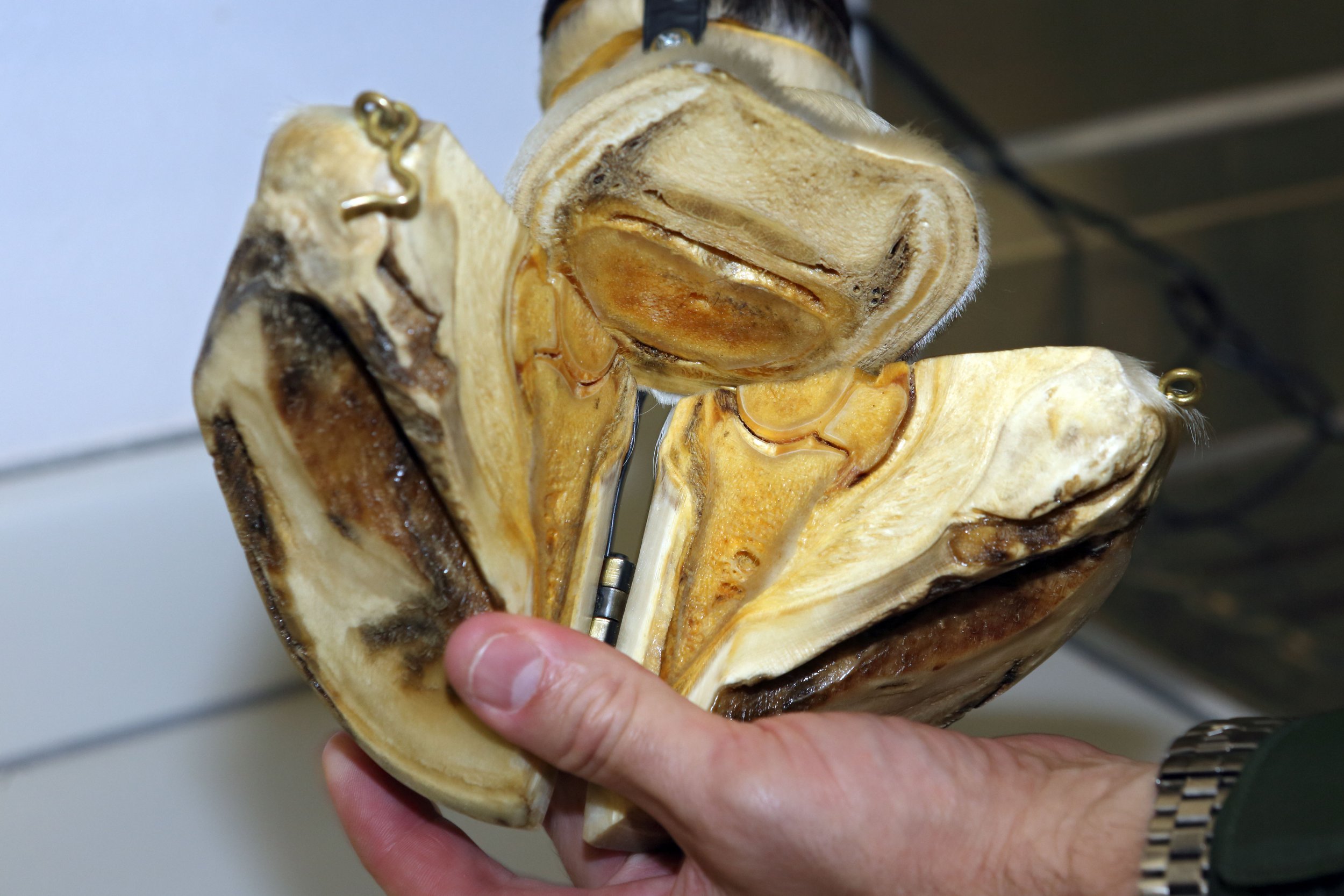
When the foot lands on the ground, the elastic, blood-filled frog helps disperse some of the force away from the bones and joints, thus, acting as a shock absorber. The venous plexus above the frog is involved in pumping blood from the foot back to the heart when the foot is loaded. In addition, there is shielding of the deep digital flexor tendon and the sensitive digital cushion (soft tissue beneath the sole that separates the frog and the heel bulb from the underlying tendons and bones). Like the heel bulbs, the frog has many sensory nerve endings allowing the horse to be aware of where his body and feet are and allows the horse to alter landing according to the condition of the ground (proprioception and coordination).
The soft tissue structures comprise and form the palmar/plantar aspect of the foot. The digital cushion lies between the lateral cartilages and above the frog and bars of the horse’s hoof. This structure is composed of collagen, fibrocartilage, adipose tissue and elastic fiber bundles. The digital cushion plays a role in shock absorption when the foot is loaded as well as a blood pumping mechanism. Interestingly, it has been found that the digital cushion composition varies across and within breeds. It is thought the variation of the composition of the digital cushion is partially dictated by a genetic predisposition. In addition, the composition of the digital cushion changes with age. As the horse ages the composition alters from elastic, fat and isolated collagen bundles to a stronger fibrocartilage. Finally, the digital cushion and connective tissue within the foot have the ability to adapt to various external stimuli such as ground contact or body weight. The lateral cartilage is a flexible sheet of fibrocartilage that suspends the pedal bone as well as acting as a spring to store and release energy. The lamina is a highly critical structure for hoof health. The lamina lies between the hoof wall and the coffin bone. There are two types of lamina known as the sensitive (dermal) lamina and insensitive (epidermal) lamina. The insensitive lamina coming in from the hoof wall connects to the sensitive lamina layer that is attached to the coffin bone and these two types of lamina interdigitate with each other to form a bond.
Hoof and Musculoskeletal System
The hoof and the musculoskeletal system are closely linked and this is particularly observed in the posture of the horse when resting or moving. Hoof shape and size and whether they are balanced directly affects the posture of the horse. Ultimately, this posture will also affect the loads placed on the skeletal system, which affects bone remodeling. With an imbalance, bone pathologies of the limbs, spine and pelvis may occur such as osteoarthritis. In addition, foot imbalances result in postural changes that lead to stress to the soft tissue structures that may lead to muscle injuries and/or tendon/ligament injuries.
Conformation and hoof balance
The terms balance and conformation are used frequently and used to describe the shape and size of the limb as a whole as well as the individual components of the limb and the spatial relations between them. Balance is the term often used to describe the foot and can be viewed as a subset of conformation.
Conformation should be considered when describing the static relations within the limb and excludes the foot. Balance should be considered when describing the dynamic and static relationship between the horse’s foot and the ground and limb as well as within the hoof itself.

These distinctions between conformation and balance are important to assess lameness and performance of the horse. Additionally, this allows the veterinarian and farrier to find optimal balance for any given conformation.
The term hoof balance does lack an intrinsic definition. The use of certain principles in order to define hoof balance, which in turn can be extended to have consistent evaluation of hoof balance as well as guide the trimming and shoeing regimens for each individual horse. In addition, these principles can be used to improve hoof capsule distortion, modify hoof conformation and alter landing patterns of the foot. These principles are:
Evaluate hoof-pastern axis
Evaluate center of articulation
The need for the heels to extend to the base of the frog
Assessing the horse’s foot balance by observing both static (geometric) balance and dynamic balance is vital. Static balance is the balance of the foot as it sits on a level, clean, hard surface. Dynamic balance is assessing the foot balance as the foot is in motion. However, horses normally do not resemble the textbook examples of perfect conformation, which creates challenges for the farriers and veterinary surgeons. The veterinarian should instigate further evaluation of the foot balance and any other ailments, in order to provide information that can be used by the farrier and veterinarian in formulating a strategy to help with the horse’s foot balance. With the farrier and veterinarian working cooperatively, the assessment of the hoof balance and shoeing of the foot should deliver a harmonious relationship between the horse’s limb, the hoof and the shoe.
Dynamic Balance

The horse should be assessed in motion as one can observe the foot landing and placement. A balanced foot when in motion should land symmetrically and flat when moving on a flat surface. When viewed from the side, the heels and toe should land concurrently (flat foot landing) or even a slight heel first landing. It is undesirable to have the toe landing first and often suggests pain localized to the heel region of the foot. When observing the horse from the front and behind, both heel bulbs should land at the same time. Sometimes, horses will land first slightly on the outside or lateral heel bulb of the foot but rarely will a horse land normally on the medial (inside) of the foot. If the horse has no conformational abnormalities or pathologies the static balance will achieve the dynamic balance.
Static Balance
Hoof –pastern axis (HPA)
The hoof pastern axis (HPA) is a helpful guideline in assessing foot balance. With the horse standing square on a hard, level surface, a line drawn through the pastern and hoof should be parallel to the dorsal hoof wall and should be straight (unbroken). The heel and toe angle should be within 5 degrees of each other. An underrun heel has been defined as the angle of the heel being 5 degrees less than the toe angle. The heel wall length should be roughly 1/3 of the dorsal wall. In addition, the cannon (metacarpus/metatarsus) bone is perpendicular to the ground and when observed from the lateral side, the HPA should be a straight line. When assessing the foot from the side, the dorsal hoof wall should be aligned with the pastern. The optimal angle of the dorsal hoof wall is often cited as being 50-54°. The length of the dorsal hoof wall is variable but guidelines have been suggested according to the weight of the horse.

It is not uncommon that the hind feet are more upright compared to the fore feet at approximately 5 degrees. A broken hoof-pastern axis is the most common hoof imbalance. There are two presentations of a broken HPA known as a broken-back HPA and a broken-forward HPA. These changes in HPA are often associated with two common hoof capsule distortions that include low or underrun heels and the upright or clubfoot, respectively.
A broken-back hoof-pastern axis occurs when the angle of the dorsal hoof wall is lower than the angle of the dorsal pastern. This presentation is commonly caused by low or underrun heel foot conformation accompanied with a long toe. This foot imbalance is common and often thought to be normal with one study finding it present in 52% of the horse population. With a low hoof angle, there is an extension of the coffin and pastern joints resulting in a delayed breakover and the heels bearing more of the horse's weight, which ultimately leads to excess stress in the deep digital flexor tendon as well as the structures around the navicular region including the bone itself.
This leads to caudal foot pain so the horse lands toe first causing subsolar bruising. In addition, this foot imbalance can contribute to chronic heel pain (bruising), quarter and heel cracks, coffin joint inflammation and caudal foot pain (navicular syndrome). The cause of underrun heels is multifactorial with a possibility of a genetic predisposition where they may have or may acquire the same foot conformation as the parents. There are also environmental factors such as excessive dryness or moisture that may lead to the imbalance.

A broken-forward hoof-pastern axis occurs at a high hoof angle with the angle of the dorsal hoof wall being higher than the dorsal pastern angle. One can distinguish between a broken-forward HPA and a clubfoot with the use of radiographs. With this foot imbalance, the heels grow long, which causes the bypassing of the soft tissue structures in the palmar/plantar area of the foot and leads to greater concussional forces on the bone. This foot imbalance promotes the landing of the toe first and leads to coffin joint flexion as well as increases heel pressure. The resulting pathologies that may occur are solar bruising, increased strain of the suspensory ligaments near the navicular bone and coffin joint inflammation.
Center of articulation
When the limb is viewed laterally, the center of articulation is determined with a vertical line drawn from the center of the lateral condyle of the short pastern to the ground. This line should bisect the middle of the foot at the widest part of the foot and demonstrates the center of articulation of the coffin joint. The widest part of the foot (colloquially known as “Ducketts Bridge”) is the one point on the sole that remains constant despite the shape and size of the foot. The distance and force on either side of the line drawn through the widest part of the foot should be equal, which provides biomechanical efficiency.
Heels extending to the base of the frog

With respect to hoof balance, another component of the foot to assess is that the heels of the hoof capsule extend to the base of the frog. The hoof capsule consists of the pedal bone occupying two-thirds of the space and one-third of the space is soft tissue structures. This area is involved in dissipating the concussional and loading forces and in order to ensure biomechanical efficiency both the bone and soft-tissue structures need to be enclosed in the hoof capsule in the same plane.
To achieve this goal the hoof wall at the heels must extend to the base of the frog. If the heels are allowed to migrate toward the center of the foot or left too long then the function of the soft tissue structures have been transferred to the bones, which is undesirable. If there is a limited amount to trim in the heels or a small amount of soft tissue mass is present in the palmar foot then some form of farriery is needed to extend the base of the frog (such as an extension of the branch of a shoe).
Medio-lateral or latero-medial balance
The medio-lateral balance is assessed by viewing the foot from the front and behind as well as from above with the foot raised. To determine if the foot has medio-lateral balance, the hoof should be bisected or a line is drawn down the middle of the pastern down to the point of the toe.
You should be able to visualize the same amount of hoof on both the left and right of that midline. In addition, one should observe the same angle to the side of the hoof wall. It is important to pick up the foot and look at the bottom. Draw a line from the middle quarter (widest part of foot) on one side to the other then draw a line from the middle of the toe to the middle sulcus of the frog.
This provides four quadrants with all quadrants being relatively the same in size (Proportions between 40/60 to 60/40 have been described as acceptable for the barefoot and are dependent on the hoof slope). The frog width should be 50-60% of its length with a wide and shallow central sulcus. The frog should be thick enough to be a part of the bearing surface of the foot. The bars should be straight and not fold to the mid frog. The sole should be concave and the intersection point of both lines should be the area of optimal biomechanical efficiency.
The less concavity means the bone is nearer to the ground, thus, bearing greater concussional force. Finally, assess the lateral and medial heel length. Look down at the heel to determine the balance in the length of both heel bulbs. Each heel bulb should be the same size and height. If there are any irregularities with the heel bulbs then sheared heels may result, which is a painful condition. Medio-lateral foot imbalance results in the uneven loading of the foot that leads to an accumulation of damage to the structures of the foot ultimately causing inflammation, pain, injury and lameness. Soles vary in thickness but a uniform sole depth of 15mm is believed to be the minimum necessary for protection.
Dorso-palmar/plantar (front to back – DP) balance
Refers to the overall hoof angle and the alignment of the hoof angle with the pastern angle when the cannon bone is perpendicular to the ground surface. When assessing the foot from the side, the dorsal hoof wall should be aligned with the pastern. The optimal angle of the dorsal hoof wall is often cited as being 50-54°. The length of the dorsal hoof wall is variable but guidelines have been suggested according to the weight of the horse.
The heel and toe angle should be within 5 degrees of each other. An underrun heel has been defined as the angle of the heel being 5 degrees less than the toe angle. The heel wall length should be roughly 1/3 of the dorsal wall.
A line dropped from the first third of the coronet should bisect the base. A vertical line that bisects the 3rd metacarpal bone should intersect the ground at the palmar aspect of the heels.
Radiographs
A useful way to assess trimming and foot balance is by having foot x-rays performed. Radiography is the only thorough and conclusive method that allows one to determine if the foot is not balanced and the bony column (HPA) is aligned.
Shoes should be removed and the foot cleaned before radiographs are executed. The horse is often placed on foot blocks to elevate the feet off the ground so that the foot can be centered in the cassette and x-ray beam.

Latero-medial view – The side view of the foot allows one to assess the dorsal and palmar aspects of the pedal bone as well as the navicular bone. The horse should be standing squarely on a flat, level surface. This projection is useful in determining the point of breakover and the hoof pastern axis should be parallel with the hoof wall. The lateral view will demonstrate the length of the toe and the alignment of the dorsal surface of the pedal bone with the hoof wall, which should be parallel. This view also allows one to determine the depth of the sole and inadequate solar depth is usually accompanied with excessive toe length (broken-back HPA). One may observe a clubfoot, broken forward.
One can distinguish between a clubfoot and a broken-forward HPA with radiographs. The broken-forward HPA the hoof angle of the heel is greater than the angle of the dorsal hoof wall. The clubfoot also demonstrates these steep/high hoof angles but additionally the alignment of the coffin, short and long pastern bones are broken forward.

Dorsopalmar/plantar views - this “front to back” view is also performed with the horse standing squarely on 2 positioning blocks. This projection allows the evaluation of medial to lateral balance and conformation of the foot with observation and measurement of the medial and lateral wall length and angle. Horses with satisfactory conformation present with a parallel joint surface of the pedal bone to the ground. The coffin joint should be even across its width. In addition, the lateral and medial coronet and the lateral and medial walls are of equal thickness and the distance from the lateral and medial solar margins to the ground are similar.
With foot imbalance, this author has observed that fore feet may have a higher lateral hoof wall, whereas, the hind feet may have a higher medial hoof wall. It is worth noting that the pelvis, stifle and hocks are adapted to move laterally allowing a slight rotating action as it moves. This action may cause uneven wear or poor trimming and shoeing may cause this limb movement to be out of line.
Trimming
Often, trimming and shoeing are based on empirical experience that includes theoretical assumptions and aesthetic decisions. The goals of trimming and shoeing are to facilitate breakover, ensure solar protection and provide heel support. Trimming is the most important aspect of farriery because it creates the base to which a shoe is fitted. Hoof conformation takes into account the function and shape of the foot in relation to the ground and lower limb both at rest and exercise. Each individual foot should have a conformation that provides protection and strength while maximizing biomechanical efficiency often viewed as foot balance.
An important question that initially needs to be addressed is whether the horse requires shoes or not. The answer does depend on what type of work the horse performs, what is the amount of workload, the conformation of the horse (especially the limbs and foot) and are there any previous or current injuries. It must be stressed that the most important aspect, whether the horse is shod or not, is that the trim ensures an appropriately balanced foot for the horse. If there is poor trimming then this may lead to uneven and increased workload on the limb leading to an increased strain of the hoof and soft tissues (i.e. ligaments, tendons) that increase the risk of injury and developing acute and chronic lameness.
The foot can be evaluated, trimmed and/or shod in a consistent, reproducible manner that considers:
Hoof-pastern axis (HPA)
The center of articulation
Heels extending to the base of the frog
Appropriate trimming and shoeing to ensure the base of the foot is under the lateral cartilage; therefore, maximizing the use of the digital cushion, can help in creating a highly effective haemodynamic mechanism. Shoeing must be done that allows full functionality of the foot so that load and concessional forces are dissipated effectively.
To implement appropriate farriery, initially observe the horse standing square on a hard service to confirm that the HPA is parallel. If The HPA is broken forward or backward then these balances should be part of the trimming plan. To determine the location of the center of rotation, palpate the dorsal and palmar aspect of the short pastern just above the coronary band and a line dropped vertically from the center of that line should correlate with the widest part of the foot.
Shoeing

When the shoe is placed on the horse, the horse is no longer standing on its feet but on the shoe; therefore, shoeing is an extension of the trim. The shoe must complement the trim and must have the same biomechanical landmarks to ensure good foot balance. It is this author’s view that the shoe should be the lightest and simplest possible. The shoe must be placed central to the widest part of the foot and the distance from the breakover point to the widest part of the foot should be equal to the distance between the widest part of the foot and the heel.
It has been shown that the use of shoes that lift the sole, frog and bars can reduce the efficient workings of the caudal foot and may lead to the prevalence of weak feet. A study by Roepstorff demonstrated there was a reduced expansion and contraction of the shod foot but improved functionality of solar and frog support. With this information, appropriate shoeing should allow increased functionality of the digital cushion, frog and bars of the foot, which improves the morphology and health of the hoof and reduces the risk of exceeding the hoof elasticity.
Disease associated with hoof imbalance
As seen in the figure above, foot imbalance can lead to multiple ailments and pathologies in the horse. It must be noted that the pathologies that may result are not necessarily exclusive for the foot but may expand to other components to the horse’s musculoskeletal system. In addition, not one but multiple pathologies may result. Diseases that may result from hoof imbalance are:

Conclusion
Foot balance is essential for your horse to lead a healthy and sound life and career. With a strong understanding of the horse anatomy and how foot imbalance can lead to lameness as well as other musculoskeletal ailments, one can work to assess and alter foot balance in order to ensure optimal performance and wellbeing of the horse. It is essential that there is a team approach involving all stakeholders as well as the veterinarian and farrier in order to achieve foot balance. With focus on foot balance, one can make a good horse into a great horse.
Gut issue biomarkers and their use in signalling dysbiosis
Article by Jackie Zions

Gastrointestinal issues (GI) are the number one cause of morbidity in horses other than old age. An unhealthy digestive system can cause poor performance, pain, discomfort, diarrhea, and a whole host of issues that can sideline your horse. It’s no wonder researchers are paying close attention to the ‘second brain’ and it’s billions of inhabitants. Ontario Veterinary College (OVC) researcher, Dr. Luis Arroyo has been studying the equine gastrointestinal systems for many years with several research projects receiving funding from Equine Guelph. Arroyo discusses what we know about equine gut health, causes of GI disorders and the extensive continuing research to understand what unstable and stable gut populations look like.
Starting with some basic anatomy Arroyo says, “The gastrointestinal tract of a horse is extremely large, and there are many things that can cause disturbances to the normal functioning or health of the gut.” A healthy gut microbiome is essential for the horse’s entire body to function optimally.
Signs of GI issues

Common signs of disorders could include abdominal pain, bloating, changes in fecal consistency (including diarrhea or constipation), excessive drooling, decrease in water consumption, lack of or poor appetite, weight loss and low body condition score.
“Some cases are more obvious to owners,” says Arroyo, “like poor performance, or acute or chronic diarrhea.”
Changes of behaviour such as becoming cranky or moody can be tell-tale signs there is unrest in the GI system. Biting at the flanks can signal abdominal pain as well as reactivity to being saddled. When the horse stops wanting to perform and athletic abilities suddenly decline, if there is no obvious lameness, GI issues are high among the considerations.
“Horses are herbivores, designed to consume a diet of forage, and to break down complex sugars within that forage.” says Arroyo. “The gut microbiota does this job and is very important for healthy digestion.” Recent research is connecting the changes in diversity of microbial communities to conditions like colic, colitis, and gastric ulcers.
Causes of GI Issues
Colic is the number one clinical condition occurring in horses. It is well-known that sudden dietary changes can be a major contributor as well as diets that are high in grain. This can create changes in the volatile fatty acids produced in the GI system, which in turn can lead to the development of gas colic. Arroyo provides the example of switching from dry hay fed in the winter, too rich, lush, spring grass as a big cause of rapid fermentation that can cause colic.

Any abrupt change, even if it’s a good quality feed to a different good quality feed, can be a source of colic. Then there is the more obvious consumption of moldy, poor, quality hay. So not only the quality but the transition/adaptation period needs to be considered when making feed changes and this goes for both changes to forage or concentrates.
A table of feed transition periods on the Equine Guelph website states an adaptation period of at least 10 – 14 days is recommended. Transition periods under seven days can increase colic risk over 22 times!
“Decrease in water consumption can be an issue, especially in countries with seasons,” says Arroyo. When water gets really cold, horses often drink less, and if it freezes, they don’t drink at all, which can lead to impaction colic. Parasite burden can also cause colic. If your horse lives in a sandy environment, like California, ingesting sand can cause impaction colic.
Non-steroidal anti-inflammatory drugs (NSAIDS) can cause colic or ulcers. NSAIDS can interfere with blood supply to the GI tract causing ulceration, for example in the mucosa of the stomach. Prolonged use can cause quite severe ulceration.
NSAIDS are not the only drugs that can contribute to GI issues. “Antibiotics - as the name says - kill many kinds of bacteria,” says Arroyo. “They are designed for that! Invariably they deplete some bacterial populations including in the intestine, and that is a problem because that may allow some other bacteria, potentially pathogenic or harmful, to overgrow, and that can cause dysbiosis.”
In a recent study, by fellow OVC researcher, Dr. Gomez and co-workers, it was determined that damage to the intestinal microbiota could occur after only 5 days of administering antibiotics to horses. Damage to the intestinal microbiota resembled dysbiosis that can potentially result in intestinal inflammation and colitis predisposing the horse to diarrhea. Judicious use of antibiotics and antimicrobials are advised.
There are infectious and non-infectious causes of colitis. Infectious examples include salmonella and then there is Neorickettsia risticii, which if ingested from contaminated sources, can cause Salmonellosis or Potomac horse fever, respectively.
“Any stress factors such as transportation, fasting or intense exercise like racing, can be a factor for developing stomach ulcers,” says Arroyo.
Current Diagnostics
Putting together a picture of the horse’s health status includes gathering clinical history from the horse owner and performing a physical examination for motility and hydration status. A biochemistry profile and complete set count can be gathered from blood testing.
Gastric ultrasound allows veterinarians to view the wall of the intestine, noting if it has thickened or distended, which could occur in cases when there is colic. They can assess appearance and find out if the intestine is displaced or if there is a twist. Gastroscopy is commonly used to find ulcers in the stomach and can reach as far as the first part of the duodenum.
GI Research

“DNA sequencing has been a breakthrough in science in terms of understanding the communities of different microorganisms living in many different niches from the skin to the lungs to the upper airways to the intestine,” says Arroyo.
It has allowed in-depth study of the population of microorganisms, providing a big picture of the different inhabitants in various areas of the GI tract, such as the lumen of the small intestine and the small and large colon. “The microorganisms vary, and they have different functions in each compartment,” says Arroyo.
DNA sequencing has allowed researchers to study microbial populations and gather information on what happens to bacterial communities when impacted by diseases like colitis. “We can see who is down, and who is up,” explains Arroyo, “and determine what populations have been depleted.” It has led to a better knowledge of which of the billions of factors are harmful to the system and which can compromise the health of the horse.
Robo-gut is one example of a fantastic system where bacterial communities are being replicated in the lab to mimic what would be found in a natural environment.
Researchers at the University of Guelph have measured metabolic profiles of the bacterial population after the addition of supplements like probiotics and prebiotics. They found they can dramatically change the metabolites that are being produced, according to what is being added to the system.
Exciting new research that could impact the future of diagnostics includes screening for biomarkers as indicators of intestinal health among equine microbiota. Dr. Arroyo is currently working with research partner, Dr. Marcio Costa, from the University of Montreal, looking for biomarkers that indicate changes in the inhabitants of the equine gut that take place during the early onset of illness.
“A biomarker is a biological molecule that you can find in different places,” explains Arroyo. “For example, you might find them in tissue, blood, urine, or different body fluids. They can signal normal or abnormal processes or could reveal a marker of a disease. For example, a biomarker can be used to see how well the body might respond to a treatment or to a disease condition.”
“The objective of a dysbiosis index is quantifying ‘X’ number of certain bacteria that are important to us,” says Arroyo. In this case, the dysbiosis derives from sequencing of the bacterial population in fecal samples.

Changes in the intestinal microbiota (dysbiosis) are present before and during the outset of diseases and after treatment with antibiotics. Arroyo cites the example of decreased Lachnospiraceae commonly observed when there is intestinal inflammation.
Bacterial biomarkers are currently being used in other species to accurately predict intestinal dysbiosis, for example in cats and dogs. One canine study quantified the number of seven different taxa of importance of the total bacterial populations. This information is entered into a mathematical algorithm that comes up with results explaining which bacteria have increased or decreased. Based on those numbers, one can use a more specific taxa to identify dysbiosis. In a feline study, it was discovered that six bacterial taxa could be accurately used to predict diarrhea in 83% of cases.
It is hoped the same results could be accomplished for horses. Developing PCR testing to screen for biomarkers could be a game changer that could potentially provide speedy, economical early diagnostics and early treatment.
So far, the most remarkable finding in the preliminary data reveals that in horses with colitis, the whole bacterial population is very depleted.
“At this stage we are in the process of increasing our numbers to find significant differences in which bacterial taxa are more important,” says Arroyo. “Soon we hope to share which bacteria taxa are more promising for predicting dysbiosis in horses with gastrointestinal disease.”
The researchers are delving into a huge biobank of samples to identify potential markers of intestinal dysbiosis in horses, utilizing PCR testing as a faster and more economical alternative to the complex DNA sequencing technologies that have been used to characterize changes in microbiota thus far. The goal is to develop simple and reliable testing that veterinarians can take right to the barn that will result in early treatment and allow closer monitoring of horses at the first onset of GI disease.
Top Tips to Protect Digestive Health

Horses are hind gut fermenters who rely on adequate amounts of fiber in the diet to maintain healthy gut function.
Make dietary changes slowly as abrupt changes disrupt the microbiota.
Avoid large grain meals as huge portions of highly fermentable diets can be quite harmful to the microbiota and can also be a source of risk for developing gastric ulcers. Opt to spread out concentrates into several smaller rations.
Prevent long periods of fasting which can also lead to ulcers. Horses are continuous-grazers, and they need to have small amounts of feed working through their digestive system to keep it functioning optimally.
Have a parasite prevention program.
Provide fresh water 24/7 to maintain good hydration and keep contents moving smoothly through the GI tract.
Keep up to date on dental appointments.
Motion is lotion – turn out and exercise are extremely important to gut function.
In closing, Arroyo states, “These top tips will help keep the horse happy and the gastrointestinal tract functioning properly.”

Lower limb anatomy and how it can be conditioned for racing
Words - Adam Jackson MRCVS

Better understanding the appropriate levels of exercise and training while the horse’s body grows and develops has been a topic of research for many years. Although it has been shown that young, growing horses are well-suited to adapt to conditioning, it is vital that continued research is performed in order to develop thoughtful and strategic training methods to promote healthy, fit and sound horses with long careers and lives.
Horses’ limbs consist of dozens of muscles, bones, tendons, ligaments, and joints that allow the horse to move as well as support its body weight. The limbs function to provide thrust and movement while absorbing impact and bearing weight. Most of the horse’s weight is supported by the fore limbs, while the propulsion of the horse is provided by the hind limbs. In addition, the horse has two apparatuses referred to as the stay apparatus and suspensory apparatus. The stay apparatus allows major joints in the limbs to lock so that the horse may rest and relax while standing. The suspensory apparatus is designed to absorb shock, carry the horse’s weight, and prevent the overextension of joints. Finally, the hooves are important structures that maintain support and traction as well as provide additional shock absorption.
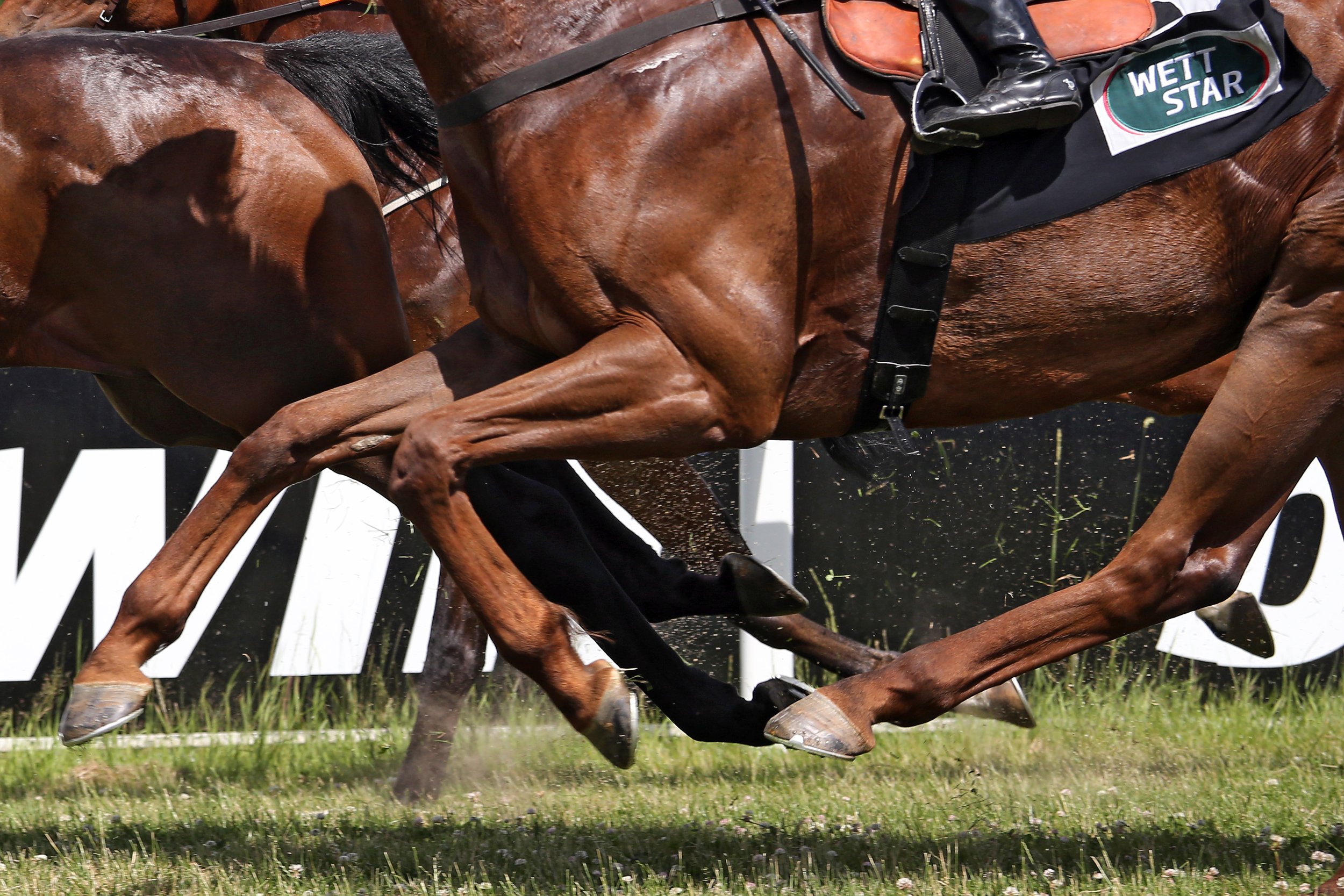
Since the cardiovascular system provides blood supply throughout the body, by responding to various stimuli, it can control the velocity and amount of blood carried through the vessels, thus, delivering oxygen, nutrients, hormones, and other important substances to cells and organs in the body. It plays a very important role in meeting the body’s demands during exercise, stress, and activity.
Exercise is used to increase the body’s ability to withstand repeated bouts of similar exercise with less impact. With a strong and healthy cardiovascular system, there is an improved ability of the musculoskeletal system receiving oxygen, thus, allowing muscles to better their capacity to use oxygen and energy. However, the adaptation period for each of these physiological systems do differ as the cardiovascular system adapts faster compared to the musculoskeletal system. This is often an overlooked consideration when developing training programmes for horses.
It is important to understand the various functions, structures, and adaptive processes of the horse’s musculoskeletal system such as bone, articular cartilage, tendons, and ligaments in order to develop appropriate training regimens.

Bone has many important roles that involve locomotion, the storage of minerals (especially calcium and phosphate), soft tissue and vital organ protection, and the support and containment of bone marrow. Bone is a specialized connective tissue, and together with cartilage forms the strong and rigid endoskeleton. The bone is continuously altering through two processes called bone modeling and bone remodeling, involving four cells referred to as osteoclasts, osteoblasts, osteocytes and bone lining cells.
Osteoblasts secrete bone matrix in the form of non-mineralized osteoid, which is then mineralized over a few weeks to form a bone matrix. Osteoclasts are involved in resorption of bone as this process occurs faster than the formation of bone. When the bone surfaces are not in the development or resorption phase, the bone surface is completely lined by a layer of flattened and elongated cells termed bone-lining cells. Osteocytes are derived from osteoblasts and are highly specialized to maintain the bone matrix. They are designed to survive hypoxic conditions and maintain biomineralization of the bone matrix. Osteocytes also control osteoblastic and osteoclastic activities allowing bone remodeling.
The function of bone modeling is to alter and maintain shape during growth. As the horse grows and develops, bone modeling occurs with the acquisition and removal of bone. While the young horse grows and develops, bone modeling allows the bone to endure strains from everyday work and exercise. The adult skeleton undergoes a minimal amount of bone modeling. Due to the presence of the high frequency of bone modeling in young horses, their skeletal strength is highly influenced by strains to their bones during exercise and daily use. With this knowledge, it has been concluded and confirmed that short-term dynamic exercise of an adolescent can lead to beneficial changes to its bone morphology.
Bone remodeling is a different process, in which old and damaged bone is renewed, which enables the bone to respond and adapt to changing functional situations. Bone remodeling is usually a coordinated relationship between bone resorption and bone formation. This process occurs throughout the horse’s life with the renewal of primary, damaged or old bone. Osteoclasts absorb old and damaged bone, and the osteoblasts form new bone and lay down new bone matrix until the earlier absorbed bone is replaced. In those animals with musculoskeletal disease or damage, there is an imbalance of osteoblast and osteoclast activity. With the knowledge that osteoblast activity to make new bone takes months whilst osteoclast activity of removing old and damaged bone only takes a few days to two weeks, bone that is being repaired is at a high risk of further injury as bone removed has not been completely replaced. Multiple studies have shown that exercise while growing can provide lifelong benefits; however, it must be done with care and knowledge. In addition, many studies have shown that exercise of a dynamic nature in moderate distances, such as that achieved in the pasture or prescribed short-distance high-speed work is beneficial to musculoskeletal development and may prevent injuries when entering race training. It has also been observed that long slow work does not increase bone strength. Below is a summary of the young horse response of the various types of exercise.

Articular cartilage is a highly specialized connective tissue found in joints with the role of providing a smooth, lubricated surface of articulation and to help transmit loads with a low amount of friction. The articular cartilage is a hyaline cartilage (flexible and strong tissue providing a smooth, slippery surface) with a dense “ExtraCellular Matrix” (ECM) consisting of specialized cells called chondrocytes, collagen and proteoglycans. These components help to retain water in the ECM that is required for the joints mechanical properties. As age increases, hydration of the matrix does decrease, resulting in stiffness. Chondrocytes are residential cells in articular cartilage that play a role in the development, maintenance, and repair of the ECM. They do respond to a variety of stimuli, including mechanical loads, growth factors, hydrostatic pressures, piezoelectric forces (formation of electric charge with force). Because of the lack of blood vessels, lymphatics, and nerves as well as being a harsh biomechanical environment, there is a limited capacity to heal and repair. In addition, chondrocytes have limited potential for replication, thus, have limited healing capacity; and chondrocytes survival depends on an optimal chemical and mechanical environment.
Maintaining joint health is vital, which requires the preservation of healthy cartilage tissue. Inactivity of joints is detrimental to articular cartilage; thus, regular movement of joints and dynamic loads is needed to provide a normal articular cartilage structure and function. Biochemical responses of the cartilage to exercise are not nearly as well known compared to bone. While the confinement of young horses stunts joint development, excessive straining of cartilage can also reduce joint development. It has been observed that pasture access was optimal for the development of joints and the confinement or excessive sprint exercise (12–32 sprints of 40 meters for 6 days a week for 5 months) causes detrimental effects on the joint and may be deemed as unnatural exercise. It is also thought that exercise is needed well before two years of age to allow cartilage thickening as well as the avoidance of confinement. It can be concluded that further studies are required with respect to level of exercise and type of exercise in order to achieve healthy cartilage tissue as there is clearly a fine line between frequency and intensity of exercise.

Tendons and ligaments are distinct but closely related tissues that have unique and important roles in musculoskeletal function and musculoskeletal disease. Tendons and ligaments are dense, fibrous connective tissues that connect muscle to bone or bone to bone, respectively. These tissues transmit mechanical forces to stabilize the skeleton and allow body movement. Tendons and ligaments consist mainly of collagen type I as well as small amounts of collagen III, IV, V, and VI. There are also various proteoglycans in tendons and ligaments that both organize and lubricate collagen fiber bundles. The elasticity of tendons and ligaments is due to the large amount of type I collagen. During locomotion, the tendon decreases energy cost to the horse by acting as a spring to store and release energy while stretching and recoiling in the stance and swing phases of each stride. Tendons and ligaments have blood vessels and nerves that allow the homeostasis and response to injury.
Tenocytes are tightly regulated by a series of growth factors and transcription factors that allow the synthesis, maintenance, and the degradation of the tendon extracellular matrix. Tendons are elastic, but tearing may occur if there is excessive loading on the tendon and the repair of collagen is a slow process. In addition, tendons have crimp morphology where the tendons buckle in a state of relaxation and act as shock absorbers. Unbuckling of the tendon occurs during loading. This crimp morphology may be disturbed if an injury occurs and also is reduced in older horses.
Due to the variation of activity of tenocytes in foals and young horses, it has been observed that both a lack of exercise and excess of exercise can impair tendon make-up and subsequent functionality. With the current data and research that has been gathered, it can be concluded that if horses take advantage of spontaneous exercise when in the paddocks (which they often do), the developing tendons may benefit and be at a lower risk of injury when racing training starts.
Conclusion
It is clear that further research is needed in order to ascertain the optimal amount and type of exercise that is needed in order to provide a strong musculoskeletal system and functional performance. However, it has been shown that prescribed exercise during the growth of the horse can increase the longevity of the horse’s health and performance. It has been observed that confinement and the lack of loading can result in weaker tissues and the loss of function of none, tendons, ligaments and articular cartilage. However, it must also be recognized that medical attempts to alleviate pain so that a horse can continue to train through an injury can greatly increase tissue damage which is detrimental to the horse’s health and career. It is far more beneficial to provide an adequate amount of time for the injury to heal, thus, putting the horse’s health and wellbeing as a top priority.
Nutritional perspective
Bone development in yearlings from the sales ring to racing — nutritional perspective
Words - Des Cronin B.Ag.Sc, M.B.A

Maintaining the equine skeleton is vital to ensure optimal development of the young growing horse, minimize risk of injury in the performance horse, and promote longevity and soundness.
The skeletal development and health of a young horse begins in utero and ensuring the broodmare receives the correct intake of key nutrients will be critical to the growth of the unborn foal. Producing high-quality milk places a significant drain on the mineral reserves of the mare. Maintaining mineral intakes during peak lactation is vital to ensure the foal receives the best nutrition to support the rapid skeletal development in the early weeks and months of growth. During this time, bone formation, body size, and muscle mass greatly increase. Risk of defective bone and related tissue formation increases with one of more of the following:
Poor diet with the incorrect balance of energy and nutrients in the daily ration
Inadequate amounts of calcium (Ca) and phosphorus (P)
A reversed Ca:P ratio
Low zinc (Zn) or copper (Cu) in the diet
Low Vitamin D
Feeding a young horse for a maximum growth rate is undesirable because bone hardening lags greatly behind bone lengthening. At 12 months old, the young horse could reach about 90 to 95 per cent of its mature height but only about 75 per cent of its mature bone mineral content.
Ideally, young horses should gain weight at a rate that their developing bones can easily support. Growing bones and connective tissues don’t have the strength to support rapid weight gain from overfeeding, especially energy. Rapid weight gain can also make other skeletal anomalies worse. In these cases the risk of developmental orthopedic disorders (DOD) and unsoundness increases.
DOD and unsoundness can also occur during uneven growth. For example, switching an underfed, slow-growing horse to a good diet that allows quick growth (compensatory growth), increases the risk of DOD. Foals between the ages of 3 and 9 months of age are at greatest risk of DOD.
Fresh forages, for example grazed grass, usually provide enough major minerals such as calcium (Ca) and phosphorus (P) for the growing horse. However, there can be significant variation in calcium and phosphorus levels in all forages but particularly preserved forages (hay and haylage). Forage analysis should always be undertaken to determine mineral composition.
For young fast-growing horses, the diet must supply the quantities of calcium and phosphorus needed for normal bone formation. In terms of Ca:P ratio, the ratio must be positive in favor of calcium. Horses are much more tolerant of high-dietary calcium than other species. For practical purposes, a good guideline would be to keep the ratio Ca:P between 1.5 to 1 and 2.5 to 1. Grains (e.g., oats) contain 10 per cent of the calcium level found in typical forages. Grains are poor sources of calcium, both in terms of the amount of calcium supplied and their effect on Ca:P ratio in the diet. Where grains are fed, supplementation will be necessary to balance the diet.
While some forages may contain adequate calcium and phosphorus, they will typically supply less than 20 per cent of the daily requirements for trace elements. Supplementation of trace elements will generally be necessary to support normal bone development.
Where concentrates are fed (especially low levels), supplementation may still be necessary to balance the overall mineral and trace element intake. Nutritional advice should be sought to ensure the horse's diet is correctly balanced.
To meet the carefully balanced requirements of key minerals, it is advisable to supplement the daily rations of growing horses and young horses entering training with an appropriate nutritional product.
Make sure that the supplement used contains the correct ratio of calcium and phosphorus, as well as other key nutrients such as vitamin D and chelated trace elements (copper, manganese, and zinc) to support normal bone development.
Supplementing branch chain amino acids in the diet ensures that growth is maintained. Lysine plays a key role when protein concentrations in the body are low. Vitamin A supports collagen formation, which is a key component of the supportive structures of joints (tendons and ligaments). Vitamin D3 is added to enhance calcium absorption.
Although growth rates slow after the age of two, they are still juvenile in their skeletal development with some growth plates, such as the shoulder and stifles, yet to completely close. Although they may look like fully grown adults, it is still important to meet nutritional requirements especially if starting training and work. With the addition of exercise and training, a young horse's nutritional needs change. The added forces from groundwork on the long bones and increased requirements of other nutrients like electrolytes need to be considered.
Finally, horses all grow and develop at different rates because of factors such as genetics. Some youngsters will need more support for longer periods of time than others, so it is important to manage accordingly.
A New Look at Lameness
Words - Jackie Zions (interviewing Dr. Koenig)

Prevention is the ideal when it comes to lameness, but practically everyone who has owned horses has dealt with a lay-up due to an unforeseen injury at some point. The following article will provide tools to sharpen your eye for detecting lameness, review prevention tips and discuss the importance of early intervention. It will also begin with a glimpse into current research endeavouring to heal tendon injuries faster, which has obvious horse welfare benefits and supports horse owners eager to return to their training programs. Dr. Judith Koenig of Ontario Veterinary College (OVC) spends half of her time as a surgeon and teacher with a strong interest in equine sports medicine and rehabilitation, and the other half as a researcher at the OVC.
Lameness is a huge focus for Koenig, whose main interest is in tissue healing. “I think over the past 20 or 30 years we have become very, very good in diagnosing the cause of lameness,” says Koenig. “In the past, we had only radiographs and ultrasound as a diagnostic tool, but by now most referral centers also have MRI available; and that allows us to diagnose joint disease or tendon disease even more. We are much better now [at] finding causes that previously may have been missed with ultrasound.”
Improvements in diagnostics have resulted in increased ability to target treatment plans. With all the different biologics on the market today, Koenig sees a shift in the management of joint disease with more people getting away from steroids as a treatment.
The following list is excerpted from Equine Guelph’s short course on lameness offered on TheHorsePortal.ca. It outlines the different diagnostics available:

Stem Cell Therapy
When asked for the latest news on research she has been involved in, Koenig proclaims, “I'm most excited about the fact that horses are responding well to stem cell treatment—better than I have seen any response to any other drug we have tried so far!”
Koenig has investigated the use of many different modalities to see if they accelerate tissue healing and has studied which cellular pathways are affected. Two recent collaborative studies have produced very exciting findings, revealing future promise for treating equine osteoarthritis with stem cell therapy.
In a safety study, Koenig and her team at the Ontario Veterinary College have shown equine pooled cryopreserved umbilical cord blood, (eCB) MSC, to be safe and effective in treatment of osteoarthritis.

“These cells are the ones harvested from umbilical cord blood at the time of foaling and then that blood is taken to the lab and the stem cells are isolated out of it,” explains Koenig. The stem cells are then put through a variety of tests to make sure they are free of infectious diseases. Once given a clean bill of health, they are expanded and frozen.
The stem cells harvested from multiple donors of equine umbilical cord blood [eCB, (kindly provided by eQcell), MSC] were compared to saline injections in research horses. “This type of cells is much more practical if you have a cell bank,” says Koenig. “You can treat more horses with it, and it’s off the shelf.” There were no systemic reactions in the safety study. Research has also shown no different reactions from sourcing from one donor or multiple donors.
In the second study, 10 million stem cells per vial were frozen for use in healing OA from fetlock chips in horses that were previously conditioned to be fit. After the fetlock chip was created, exercise commenced for six more weeks, and then osteoarthritis was evaluated by MRI for a baseline. Half the horses were treated with the pooled MSC stem cells, and the control group received saline before another month of exercise. Then MRI and lameness exams were repeated, and arthroscopy was repeated to score the cartilage and remove the chip.
Lameness was decreased and cartilage scores were improved in the group that received stem cell therapy at the time of the second look with arthroscopy.
Many diagnostics were utilized during this study. MRIs, X-rays, ultrasounds and weekly lameness evaluations all revealed signs of osteoarthritis in fetlock joints improved in the group treated with (eCB) MSCs. After six weeks of treatment, the arthroscopic score was significantly lower (better cartilage) in the MSC group compared to the control group.
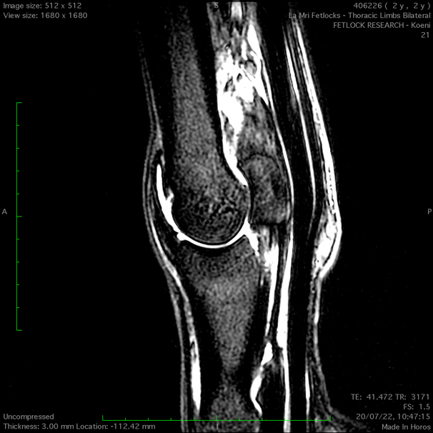
“Using the MRI, we can also see a difference that the horses treated with stem cells had less progression of osteoarthritis, which I think is awesome,” says Koenig. “They were less lame when exercised after the stem cell therapy than the horses that received saline.”
This research group also just completed a clinical trial in client-owned horses diagnosed with fetlock injuries with mild to moderate osteoarthritis changes. The horses were given either 10 million or 20 million stem cells and rechecked three weeks and six weeks after the treatment. Upon re-evaluation, the grade of lameness improved in all the horses by at least one. Only two horses presented a mild transient reaction, which dissipated after 48 hours without any need for antibiotics. The horse’s joints looked normal, with any filling in the joint reduced.
There was no difference in the 18 horses, with nine given 10 million stem cells and the other nine 20 million stem cells; so in the next clinical trial, 10 million stem cells will be used.
The research team is very happy with the results of this first-of-its-kind trial, proving that umbilical cord blood stem cells stopped the progression of osteoarthritis and that the cartilage looked better in the horses that received treatment. The future of stem cell therapy is quite promising!
Rehabilitation
Research has shown adhering to a veterinary-prescribed rehabilitation protocol results in a far better outcome than paddock turn out alone. It is beneficial for tendon healing to have a certain amount of controlled stimulation. “These horses have a much better outcome than the horses that are treated with just being turned out in a paddock for half a year,” emphasizes Koenig. “They do much better if they follow an exercise program. Of course, it is important not to overdo it.”
For example, Koenig cautions against skipping hand-walking if it has been advised. It can be so integral to stimulating healing, as proven in recent clinical trials. “The people that followed the rehab instructions together with the stem cell treatment in our last study—those horses all returned to racing,” said Koenig.
“It is super important to follow the rehab instructions when it comes to how long to rest and not to start back too early.”
Another concern when rehabilitating an injured horse would be administering any home remedies that you haven't discussed with your veterinarian. Examples included blistering an area that is actively healing or applying shockwave to mask pain and then commence exercise.
Prevention and Training Tips
While stating there are many methods and opinions when it comes to training horses, Koenig offered a few common subjects backed by research. The first being the importance of daily turnout for young developing horses.

Turnout and exercise
Many studies have looked at the quality of cartilage in young horses with ample access to turn out versus those without. It has been determined that young horses that lack exercise and are kept in a stall have very poor quality cartilage.
Horses that are started early with light exercise (like trotting short distances and a bit of hill work) and that have access to daily paddock turnout, had much better quality of cartilage. Koenig cited research from Dr. Pieter Brama and similar research groups.
Another study shows that muscle and tendon development depend greatly on low grade exercise in young horses. Evaluations at 18 months of age found that the group that had paddock turnout and a little bit of exercise such as running up and down hills had better quality cartilage, tendon and muscle.
Koenig provides a human comparison, with the example of people that recover quicker from injury when they have been active as teenagers and undergone some beneficial conditioning. The inference can be made that horses developing cardiovascular fitness at a young age stand to benefit their whole lives from the early muscle development.
Koenig says it takes six weeks to regain muscle strength after injury, but anywhere from four to six months for bone to develop strength. It needs to be repeatedly loaded, but one should not do anything too crazy! Gradual introduction of exercise is the rule of thumb.
Rest and Recovery
“Ideally they have two rest days a week, but one rest day a week as a minimum,” says Koenig. “I cannot stress enough the importance of periods of rest after strenuous work, and if you notice any type of filling in the joints after workout, you should definitely rest the horse for a couple of days and apply ice to any structures that are filled or tendons or muscles that are hard.”
Not purporting to be a trainer, Koenig does state that two speed workouts a week would be a maximum to allow for proper recovery. You will also want to make sure they have enough access to salt/electrolytes and water after training.
During a post-Covid interview, Koenig imparted important advice for bringing horses back into work methodically when they have experienced significant time off.
“You need to allow at least a six-week training period for the athletes to be slowly brought back and build up muscle mass and cardiovascular fitness,” says Koenig. “Both stamina and muscle mass need to be retrained.”
Watch video: “Lameness research - What precautions do you take to start training after time off?”
The importance was stressed to check the horse’s legs for heat and swelling before and after every ride and to always pick out the feet. A good period of walking is required in the warmup and cool down; and riders need to pay attention to soundness in the walk before commencing their work out.
Footing and Cross Training
With a European background, Koenig is no stranger to the varying track surfaces used in their training programs. Statistics suggest fewer injuries with horses that are running on turf, like they practice in the UK.
Working on hard track surfaces has been known to increase the chance of injury, but delving into footing is beyond the scope of this article.
“Cross training is very important,” says Koenig. “It is critical for the mental and proper musculoskeletal development of the athlete to have for every three training days a day off, or even better provide cross-training like trail riding on these days."
Cross-training can mitigate overtraining, giving the body and mind a mental break from intense training. It can increase motivation and also musculoskeletal strength. Varied loading from training on different terrain at different gaits means bone and muscle will be loaded differently, therefore reducing repetitive strain that can cause lameness.
Hoof care
Whether it is a horse coming back from injury, or a young horse beginning training, a proficient farrier is indispensable to ensure proper balance when trimming the feet. In fact, balancing the hoof right from the start is paramount because if they have some conformational abnormalities, like abnormal angles, they tend to load one side of their joint or bone more than the other. This predisposes them to potentially losing bone elasticity on the side they load more because the bone will lay down more calcium on that side, trying to make it stronger; but it actually makes the bone plate under the cartilage brittle.
Koenig could not overstate the importance of excellent hoof care when it comes to joint health and advises strongly to invest in a good blacksmith. Many conformational issues can be averted by having a skilled farrier right from the time they are foals. Of course, it would be remiss not to mention that prevention truly begins with nutrition. “It starts with how the broodmare is fed to prevent development of orthopedic disease,” says Koenig. Consulting with an equine nutritionist certainly plays a role in healthy bone development and keeping horses sound.

Equine Neck CT: Advancing diagnostic precision in racehorses
Words - Rachel Tucker MRCVS

Introduction
When considering neck disorders in the racehorse, we most commonly think of severe conditions such as acute neck trauma and cervical vertebral myelopathy (Wobbler Syndrome). These represent the most severe end of the scale of orthopedic and neurologic injury to the neck; and a diagnosis, or at least prognosis, is usually clear. However, neck conditions encompass a far wider range of clinical presentations.
At the milder end of the scale, signs may be subtle and easily missed, whilst still being responsible for discomfort and reduced performance. The recent ability to perform a computed tomography (CT) scan of a horse’s neck represents a major advancement in our ability to diagnose neck conditions. Timely and accurate diagnosis allows efficient and targeted treatment, the ability to plan schedules and improvement in welfare through the provision of appropriate treatment and earlier return to function.
In neurologic cases, an accurate diagnosis facilitates risk management for both the horse, their handlers and riders, while improving safety for all. As conditions and injuries of the neck are being better characterized using CT, new medical and surgical treatment options are being developed, giving the potential for improved outcomes and fewer losses from the racing industry.
This article summarizes how CT is being increasingly used by vets to diagnose conditions of the neck and how it is revealing previously unknown information and providing exciting new treatment opportunities.
Presentation

Conditions of the neck can cause a range of signs in the horse, which are wide-ranging in their presentation and variable in their severity. The manner in which these conditions present depends on which anatomical structures are affected. Issues affecting the bones, joints and/or soft tissues of the neck can all cause neck pain, which can manifest in a number of ways. Cases of neck pain can be severe, resulting in a horse with a rigid, fixed neck carriage, an unwillingness to walk and struggling to eat, perhaps due to a traumatic event. Neck fractures are thankfully uncommon but can be catastrophic.
More moderate signs might be displayed as a stiff neck, with reduced range of movement and resentment of ridden work. There may be pain on palpation of the neck and changes in the neck musculature. Increasingly, we are seeing horses with far more subtle signs, which are ultimately revealed to be due to neck pain and neck pathology. Typically, these horses might have an acceptable range of motion of their neck under most circumstances, but they suffer pain or restriction in certain scenarios, resulting in poor performance. This may be seen as tension through the neck, resisting rein contact, a reluctance to extend the neck over fences, or they may struggle on landing.
Riders might report a feeling of restriction or asymmetry in the mobility of the neck. In addition, these horses may be prone to forelimb tripping or show subtle forelimb lameness.
Any condition, which causes injury or disease to the spinal cord or nerves within the neck, also causes a specific range of neurologic signs. Compression of the spinal cord is most commonly caused by malformations or fractures of the cervical vertebrae, or enlargement of the adjacent articular process (facet) joints. This results in classic ‘Wobbler’ symptoms, which can range from subtle weakness and gait abnormalities, through to horses that are profoundly weak, ataxic and uncoordinated. This makes them prone to tripping, falling, or they may even become recumbent.
Peripheral nerve deficits are uncommon but become most relevant if they affect the nerves supplying the forelimbs, which can result in tripping, forelimb lameness, or local sensory deficits. This lameness might be evident only in certain circumstances, such as when ridden in a rein contact. This lameness is difficult to pinpoint as there will be no abnormality to find in the lame limb, indeed a negative response to nerve and joint blocks (diagnostic analgesia) will usually be part of the diagnostic process.
Horses can present with varying combinations and severities of neck pain, neurologic signs and peripheral nerve deficits, creating a wide range of manifestations of neck related disease.
Diagnosis

A diagnosis of neck pain is based on careful static and dynamic clinical examination and may be supported by seeing a positive response to treatment. Neurologic deficits are noted during a specific neurologic assessment, which includes a series of provocation tests such as asking a horse to walk over obstacles, back up, turn circles and walk up and down a hill. Confirming neck pathology as the cause of signs can be difficult. Until recently, radiography has been the mainstay imaging modality. Radiographs are useful for assessment of the cervical vertebrae and continue to play an important role in diagnosis; however the complex 3-dimensional shape of these bones, the large size of the neck and an inability to take orthogonal (right-angled) x-ray views means that this 2-dimensional imaging modality has significant limitations. High quality, well-positioned images are essential to maximize the diagnostic potential of radiographs.
A turning point in our diagnostic ability and understanding of neck dysfunction has been the recent adaptation of human CT scanners to allow imaging of the horse’s neck. A number of equine hospitals across the United Kingdom and Northern Europe now offer this imaging modality. We have been providing this service at Liphook Equine Hospital since 2017, with over 150 neck scans performed to date.
The CT procedure

A computed tomography (CT) scan combines a series of x-ray images taken from different angles around the area of interest to create a 3-dimensional volume of imaging data. This data is presented as a grayscale image which can be viewed in any plane and orientation. It provides excellent bone detail, and post processing techniques can provide information on soft tissue structures too. Additional techniques can be employed such as positive contrast myelography to provide greater detail about soft tissue structures. Myelography delineates the spinal cord using contrast medium injected into the subarachnoid space and is indicated in any case showing neurologic signs suggestive of spinal cord compression.
Neck CT is performed under a short general anesthetic. Scans without myelography typically take less than 20 minutes to complete. The entire neck is imaged, from the poll to the first thoracic vertebra. The procedure is non-invasive and low risk, with anesthetic-related complication presenting the main risk factor to the procedure. We have not encountered any significant complications in our plain CT scan caseload to date. Horses showing ataxia, weakness and incoordination (Wobbler’s), undergo CT myelography which adds around 20 minutes to the procedure. These horses are exposed to a greater level of risk due to their neurologic condition, the injection of a contrast agent and the increased chance of destabilizing a more severe lesion during the procedure.
CT is revealing more detailed information about ways that spinal cord compression can occur in Wobbler cases, about compression of spinal nerves resulting in forelimb gait deficits and precise detail about fracture configurations. It gives us detailed images of articular process joint disease, intervertebral disc disease, developmental conditions and anatomic variations. It is also revealing information about rare diseases such as vertebral abscesses or spinal neoplasia. As our caseload and confidence in the imaging modality grows, we are learning more about the value of CT in examining more subtle neck conditions. We are also bringing the benefit of a more accurate diagnosis, allowing precise targeted treatment and a better ability to provide a prognosis about outcomes—likely progression or safety factors. CT myelography allows circumferential imaging of the spinal canal and yields significantly more information than traditional x-ray myelography. As a result, we hope to enable better case selection of horses that may benefit from Wobbler surgery, with the goal of resulting in improved success rates of the surgery.
Innovations in treatment

New treatment options are emerging as a result of our more accurate diagnoses of neck pathology. Of the first 55 horses which underwent neck CT at our hospital, we were surprised to discover that 13 (24%) had some form of osteochondral fragmentation within the articular process joints of their neck. Some of these horses were young Thoroughbreds, bred to race but showing Wobbler signs. These tended to have convincing CT evidence of type 1 CVM (Wobbler Syndrome) and osteochondrosis affecting their neck. Others had fragments which were larger and more discrete, with evidence of articular process joint enlargement/arthritis but no other bony lesions. These horses were typically older and of a range of breeds and uses.
In those horses presenting with signs of neck pain but no neurologic deficits, surgical removal of these fragments was proposed. Following further consideration and cadaver training, we have begun to offer this surgery for horses that fit the appropriate criteria and have surgically accessible fragments. We have performed arthroscopic or arthroscopic-assisted fragment removal from eight articular process joints in six horses to date. No intraoperative or postoperative complications have been encountered; and five of six horses showed complete resolution of neck pain. In the sixth horse, full recovery was not anticipated due to the presence of additional neck pathology, but partial improvement occurred for two years. Fragment removal has relieved signs of neck pain and stiffness and caused improved performance in these horses.
Two procedures that are emerging to treat spinal nerve root impingement are a targeted peripheral nerve root injection and a keyhole surgical procedure to widen the intervertebral foramen. Nerve root injection is performed in the standing sedated horse under ultrasound guidance. Surgery is performed under anesthesia, using specialized minimally invasive equipment to widen the bony foramen using a burr. This surgery is in its infancy but offers an exciting treatment option.
Additionally, CT gives us the ability to better plan for fracture repair, undoubtedly improving our case selection for Wobbler surgery; it more accurately guides intra-articular injection of the articular process joints.
Summary
Computed tomography is transforming our ability to diagnose conditions of the horse’s neck. The procedure is low risk and now widely available in the UK and other parts of Europe. It is driving the innovation of novel treatment options with the goal of improving outcomes and reducing losses to conditions of the neck. Our CT findings are posing new questions about neck function, pain and neurologic disease and is an active area of ongoing research.
References:
Schulze N., Ehrle, A., Beckmann, I and Lischer, C. (2021) Arthroscopic removal of osteochondral fragments of the cervical articular process joints in three horses. Vet Surg. ;1-9.
Swagemakers J-H, Van Daele P, Mageed M. Percutaneous full endoscopic foraminotomy for treatment of cervical spinal nerve compression in horses using a uniportal approach: Feasibility study. Equine Vet J. 2023.
Tucker R, Parker RA, Meredith LE, Hughes TK, Foote AK. Surgical removal of intra-articular loose bodies from the cervical articular process joints in 5 horses. Veterinary Surgery. 2021;1-9.
Wood AD, Sinovich M, Prutton JSW, Parker RA. Ultrasonographic guidance for perineural injections of the cervical spinal nerves in horses. Veterinary Surgery. 2021; 50:816–822.
Air Quality and Air Pollution’s Impact on Your Horse’s Lungs
Article by Dr. Janet Beeler-Marfisi
There’s nothing like hearing a horse cough to set people scurrying around the barn to identify the culprit. After all, that cough could mean choke, or a respiratory virus has found its way into the barn. It could also indicate equine asthma. Yes, even those “everyday coughs” that we sometimes dismiss as "summer cough" or "hay cough" are a wake-up call to the potential for severe equine asthma.
Formerly known as heaves, broken wind, emphysema, chronic obstructive pulmonary disease (COPD), or recurrent airway obstruction (RAO), this respiratory condition is now called severe equine asthma (sEA). These names reflect how our scientific and medical understanding of this debilitating disease has changed over the years. We now consider heaves to be most comparable to severe asthma in people.

But what if your horse only coughs during or after exercise? This type of cough can mean that they have upper airway irritation (think throat and windpipe) or lower airway inflammation (think lungs) meaning inflammatory airway disease (IAD), which is now known as mild-to-moderate equine asthma (mEA). This airway disease is similar to childhood asthma, meaning that it can go away on its own. However, it is still very important to call your veterinarian out to diagnose mEA. This disease causes reduced athletic performance, and there are different subtypes of mEA that benefit from specific medical therapies. In some cases, mEA progresses to sEA.
Equine Asthma and Air Quality
What does equine asthma have to do with air quality? A lot, it turns out. Poor air quality, or air pollution, includes the barn dusts—the allergens and molds in hay and the ground-up bacteria in manure, as well as arena dusts and ammonia from urine. Also, very importantly for both people and horses, air pollution can be from gas and diesel-powered equipment. This includes equipment being driven through the barn, the truck left idling by a stall window, or the smog from even a small city that drifts nearly invisibly over the surrounding farmland. Recently, forest-fire smoke has been another serious contributor to air pollution.
Smog causes the lung inflammation associated with mEA. Therefore, it is also likely that air pollution from engines and forest fires will also trigger asthma attacks in horses with sEA. Smog and smoke contain many harmful particulates and gases, but very importantly they also contain fine particulate matter known as PM2.5. The 2.5 refers to the diameter of the particle being 2.5 microns. That’s roughly 30 times smaller than the diameter of a human hair. Because it is so small, this fine particulate is inhaled deeply into the lungs where it crosses over into the bloodstream. So, not only does PM2.5 cause lung disease, but it also causes inflammation elsewhere in the body including the heart. Worldwide, even short-term exposure is associated with an increased risk of premature death from heart disease, stroke, and lung cancer. This PM2.5 stuff is not trivial!
In horses, we know that PM2.5 causes mEA, so it’s logical that smog and forest-fire smoke exposure could exacerbate asthma in horses, but we don’t know about heart disease or risk of premature death.
Symptoms, Diagnostic Tests and Treatments

Equine asthma manifests with a spectrum of symptoms that vary in severity and the degree of debilitation they cause. Just like in people with asthma, the airways of horses with mEA and sEA are “hyperreactive.” This means that the asthmatic horse’s airways are extra sensitive to barn dusts that another horse’s lungs would just “ignore.” The asthmatic horse’s airways constrict, or become narrower, in response to these dusts. This narrowing makes it harder to get air in and out of the lungs. Think about drinking through a straw. You can drink faster with a wider straw than a skinnier one. It’s the same with air and the airways. In horses with mEA, the narrowing is mild. In horses with sEA, the constriction is extreme and is the reason why they develop the “heaves line”; they have to use their abdominal muscles to help squeeze their lungs to force the air back out of their narrow airways. They also develop flaring of their nostrils at rest to make their upper airway wider to get more air in. Horses with mEA do not develop a heaves line, but the airway narrowing and inflammation do cause reduced athletic ability.
The major signs of mEA are coughing during or just after exercise that has been going on for at least a month and decreased athletic performance. In some cases, there may also be white or watery nasal discharge particularly after exercise. Often, the signs of mEA are subtle and require a very astute owner, trainer, groom, or rider to recognize them.
Another very obvious feature of horses with sEA is their persistent hacking cough, which worsens in dusty conditions. “Hello dusty hay, arena, and track!” The cough develops because of airway hyperreactivity and because of inflammation and excess mucus in the airways. Mucus is the normal response of the lung to the presence of inhaled tiny particles or other irritants. Mucus traps these noxious substances so they can be coughed out, which protects the lung. But if an asthma-prone horse is constantly exposed to a dusty environment, it leads to chronic inflammation and mucus accumulation, and the development or worsening of asthma along with that characteristic cough.
Accurately Diagnosing Equine Asthma
Veterinarians use a combination of the information you tell them, their observation of the horse and the barn, and a careful physical and respiratory examination that often involves “rebreathing.” This is a technique where a bag is briefly placed over the horse’s nose, causing them to breathe more frequently and more deeply to make their lungs sound louder. This helps your veterinarian hear subtle changes in air movement through the lungs and amplifies the wheezes and crackles that characterize a horse experiencing a severe asthma attack. Wheezes indicate air “whistling” through constricted airways, and crackles mean airway fluid buildup. The fluid accumulation is caused by airway inflammation and contributes to the challenge of getting air into the lung.
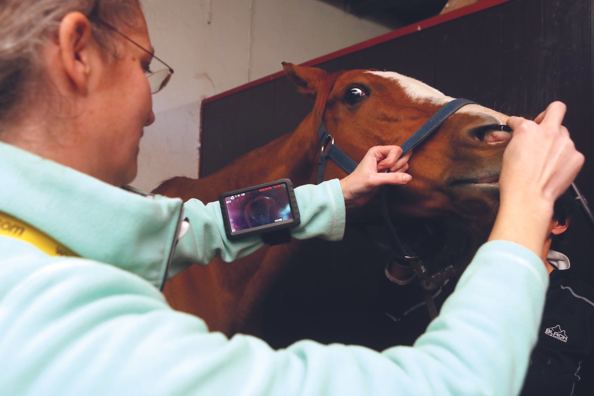
Other tests your veterinarian might use are endoscopy, bronchoalveolar lavage, and in the specialist setting, pulmonary function testing. They will also perform a complete blood count and biochemical profile assay to help rule out the presence of an infectious disease.
Endoscopy allows your veterinarian to see the mucus in the trachea and large airways of the lung. It also lets them see whether there are physical changes to the shape of the airways, which can be seen in horses with sEA.
Bronchoalveolar lavage, or “lung wash” is how your veterinarian assesses whether there is an accumulation of mucus and inflammatory cells in the smallest airways that are too deep in the lung to be seen using the endoscope. Examining lung wash fluid is a very important way to differentiate between the different types of mEA, between sEA in remission and an active asthma attack, and conditions like pneumonia or a viral lung infection.
Finally, if your veterinarian is from a specialty practice or a veterinary teaching hospital, they might also perform pulmonary function testing. This allows your veterinarian to determine if your horse’s lungs have hyperreactive airways (the hallmark of asthma), lung stiffening, and a reduced ability to breathe properly.
Results from these tests are crucial to understanding the severity and prognosis of the condition. As noted earlier, mEA can go away on its own; but medical intervention may speed healing and the return to athletic performance. With sEA, remission from an asthmatic flare is the best we can achieve. As the disease gets worse over time, eventually the affected horse may need to be euthanized.
Management, Treatment and Most Importantly—Prevention
Successful treatment of mEA and sEA flares, as well as long-term management, requires a multi-pronged approach and strict adherence to your veterinarian’s recommendations.
Rest is important because forcing your horse to exercise when they are in an asthma attack further damages the lung and impedes healing. To help avoid lung damage when smog or forest-fire smoke is high, a very useful tool is your local, online, air quality index (just search on the name of your closest city or town and “AQI”). Available worldwide, the AQI gives advice on how much activity is appropriate for people with lung and heart conditions, which are easily applied to your horse. For example, if your horse has sEA and if the AQI guidelines say that asthmatic people should limit their activity, then do the same for your horse. If the AQI says that the air quality is bad enough that even healthy people should avoid physical activity, then do the same for you AND your horse. During times of poor air quality, it is recommended to monitor the AQI forecast and plan to bring horses into the barn when the AQI is high and to turn them out once the AQI has improved.
Prevent dusty air. Think of running your finger along your tack box – whatever comes away on your finger is what your horse is breathing in. Reducing dust is critical to preventing the development of mEA and sEA, and for managing the horse in an asthmatic flare.
Logical daily practices to help reduce dust exposure:

Turn out all horses before stall cleaning
Wet down the aisle prior to sweeping
Never sweep debris into your horse’s stall
Use low-dust bedding like wood shavings or dust-extracted straw products, which should also be dampened down with water
Reduce arena, paddock, and track dust with watering and maintenance
Consider low-dust materials when selecting a footing substrate
Steam (per the machine’s instructions) or soaking hay (15–30 minutes and then draining, but never store steamed or soaked hay!)
Feed hay from the ground
Feed other low-dust feeds
Avoid hay feeding systems that allow the horse to put their nose into the middle of dry hay—this creates a “nosebag” of dust
Other critical factors include ensuring that the temperature, humidity and ventilation of your barn are seasonally optimized. Horses prefer a temperature between 10–24 ºC (50–75 ºF), ideal barn humidity is between 60–70%. Optimal air exchange in summer is 142 L/s (300 cubic feet/minute). For those regions that experience winter, air exchange of 12–19 L/s (25–40 cubic feet/minute) is ideal. In winter, needing to strip down to a single layer to do chores implies that your barn is not adequately ventilated for your horse’s optimal health. Comfortable for people is often too hot and too musty for your horse!

Medical interventions for controlling asthma are numerous. If your veterinarian chooses to perform a lung wash, they will tailor the drug therapy of your asthmatic horse to the results of the wash fluid examination. Most veterinarians will prescribe bronchodilators to alleviate airway constriction. They will also recommend aerosolized, nebulized or systemic drugs (usually a corticosteroid, an immunomodulatory drug like interferon-α, or a mast cell stabilizer like cromolyn sodium) to manage the underlying inflammation. They may also suggest nebulizing with sterile saline to help loosen airway mucus and may suggest feed additives like omega 3 fatty acids, which may have beneficial effects on airway inflammation.
New Research and Future Directions
Ongoing research is paramount to expanding our knowledge of what causes equine asthma and exploring innovative medical solutions. Scientists are actively investigating the effects of smog and barn dusts on the lungs of horses. They are also working to identify new targeted therapies, immunotherapies and other treatment modalities to improve outcomes for affected horses.
Conclusion
Both mild and severe equine asthma are caused and triggered by the same air pollutants, highlighting the need for careful barn management. The alarming rise in air pollution levels poses an additional threat to equine respiratory health. Recognizing everyday coughs as potential warning signs and implementing proper diagnostic tests, day-to-day management practices and medical therapies are crucial in combating equine asthma. By prioritizing the protection of our horse’s respiratory health and staying informed about the latest research, we can ensure the well-being of our equine companions for years to come.
Highlights
Work to prevent dust and optimize barn air exchange.
Avoid idling farm equipment and trucks around horses.
Don’t ignore a cough—call your veterinarian.
Monitor your local air quality index—it’s a free and simple way to help prevent lung damage!
Thermoregulation - Too hot to handle!
Article by Adam Jackson MRCVS

Exertional heat illness (EHI) is a complex disease where thoroughbred racehorses are at significant risk due to the fact that their workload is intensive in combination with the high rate of heat production associated with its metabolism. In order to understand how this disease manifests and to develop preventative measures and treatments, it is important to understand thermoregulation in horses.
What is thermoregulation?
With continuous alteration in the surrounding temperature, thermoregulation allows the horse to maintain its body temperature within certain limits. Thermoregulation is part of the greater process of homeostasis, which is a number of self-regulating processes the horse uses to maintain body stability in the face of changing external conditions. Homeostasis and thermoregulation are vital for the horse to maintain its internal environment to ensure its health while disruption of these processes leads to diseases.
The horse’s normal temperature range is 99–101°F (37.5–38.5°C). Hyperthermia is the condition in which the body temperature increases above normal due to heat increasing faster than the body can reduce it. Hypothermia is the opposite condition, where the body temperature decreases below normal levels as the body is losing heat faster than producing it. These conditions are due to the malfunction of thermoregulatory and homeostatic control mechanisms.
Horses are colloquially referred to as warm-blooded mammals—also known as endotherms because they maintain and regulate their core body, and this is opposite ectotherms such as reptiles. The exercising horse converts stored chemical energy into mechanical energy when contracting various muscles in its body. However, this process is relatively inefficient because it loses roughly 80% of energy released from energy stores as heat. The horse must have effective ways to dissipate this generated heat; otherwise, the raised body temperatures may be life threatening.
Transfer of body heat
There are multiple ways heat may be transferred, and this will flow from one area to another by:
1. Evaporation

The main way body heat is lost during warm temperatures is through the process of evaporation of water from the horse’s body surface. It is a combination of perspiration, sweating and panting that allows evaporation to occur.
Sweating is an inefficient process because the evaporation rate may exceed the body heat produced by the horse, resulting in the horse becoming covered and dripping sweat. This phenomenon occurs faster with humid weather (high pressure).
Insensible perspiration is the loss of water through the skin, which does not occur as perceivable sweat. Insensible perspiration takes place at an almost constant rate and is the evaporative loss from skin; but unlike sweating, the fluid loss is pure water with no solutes (salts) lost. The horse uses insensible perspiration to cool its body.
It is not common for horses to pant in order to dissipate heat; however, there is evidence that the respiratory tract of the horse can aid in evaporative heat loss through panting.
2. Conduction
Conduction is the process where heat is transferred from a hot object to a colder object, and in the case of the horse, this heat transfer is between its body and the air. However, the air has poor thermal conductivity, meaning that conduction plays a small role in thermoregulation of the horse. Conduction may help if the horse is lying in a cool area or is bathed in cool water.

The horse has the greatest temperature changes occurring at its extremities, such as its distal limbs and head. The horse can alter its blood flow by constricting or dilating its blood vessels in order to prevent heat loss or overheating, respectively.
Interestingly, the horse will lie down and draw its limbs close to its body in order to reduce its surface area and to control conduction. There also have been some adaptive changes in other equids like mules and burros, where shorter limbs, longer ears and leaner bodies increase its surface area to help in heat loss tolerance.
3. Convection
Convection is the rising motion of warmer areas of a liquid or gas and the sinking motion of cooler areas of the liquid or gas. Convection is continuously taking place between the surface of the body and the surrounding air. Free convection at the skin surface causes heat loss if the temperature is low with additional forced convective heat transfer with wind blowing across the body surface.
When faced with cold weather, a thick hair coat insulates and resists heat transfer because it traps air close to the skin; thus, preventing heat loss. Whereas, the horse has a fine hair coat in the summer to help in heat loss.
4. Radiation
Radiation is the movement of heat between objects without direct physical contact. Solar radiation is received from the sun and can be significant in hot environments, especially if the horse is exposed for long periods of time. A horse standing in bright sunlight can absorb a large amount of solar radiation that can exceed its metabolic heat production, which may cause heat stress.
How the horse regulates its body temperature
The horse must regulate its heat production and heat loss using thermoregulatory mechanisms. There are many peripheral thermoreceptors that detect changes in temperature, which leads to the production of proportional nerve impulses. These thermoregulators are located in the skin skeletal muscles, the abdomen, the spinal cord and the midbrain with the hypothalamus being instrumental in regulating the internal temperature of the horse. A coordinating center in the central nervous system receives these nerve incoming impulses and produces output signals to organs that will alter the body temperature by acting to reduce heat loss or eliminate accumulated heat.
The racehorse and thermoregulation
The main source of body heat accumulation in the racehorse is associated with muscular contraction. At the initiation of exercise, the racehorse’s metabolic heat production, arising from muscle contraction, increases abruptly. The heat production does alter the level of intensity of the work as well as the type of exercise undertaken.
During exercise, the core body temperature increases because heat is generated and the horse’s blood system distributes this heat throughout the body. Hodgson and colleagues have theorized and confirmed via treadmill studies that the racehorse has the highest rate of heat production compared to other sporting horses. In fact, the racehorse’s body temperature can rise 33°F (0.8°C) per minute, reaching 108°F (42.0°C). But what core temperature can the horse tolerate and not succumb to heat illness and mortality? The critical temperature for EHI (exertional heat illness) is not known, but studies have demonstrated that a racehorse can be found to have core temperatures between 108°F - 109° F (42–43°C) without any clinical symptoms. Currently, anecdotal evidence is only available, suggesting that a core temperature of 110°F (43.5°C) will result in manifestation of EHI with the horse demonstrating central nervous system dysfunction such as ataxia (incoordination). In addition, temperatures greater than 111°F (44°C) result in collapse.
Heat loss in horses

A horse loses heat to the environment by a combination of convection, evaporation and radiation, which is magnified during racing due to airflow across the body. However, if body heat gained through racing is not minimized by convection, then the racehorse’s body temperature is regulated entirely by evaporation of sweat. This evaporation takes place on the horse’s skin surface and respiratory tract.
The horse has highly effective sweat glands found in both haired and hairless skin, which produces sweat rates that are highest in the animal kingdom. Efficient evaporative cooling is present in the horse because its sweat has a protein called latherin, which acts as a wetting agent (surfactant); this allows the sweat to move from its skin to the hair.
Because of the horse’s highly blood-rich mucosa of its upper respiratory tract, the horse has a very efficient and effective heat exchange system. Estimates suggest this pathway dissipates 30% of generated heat by the horse during exercise. As the horse exercises, there is blood vessel dilation, which increases blood flow to the mucosa that allows more heat to be dissipated to the environment. When the respiratory tract maximizes evaporative heat loss, the horse begins to pant. Panting is a respiratory rate greater than 120 breaths per minute with the presence of dilated nostrils; and the horse adopts a rocking motion. However, if humidity is high, the ability to evaporate heat via the respiratory route and skin surface is impaired. The respiratory evaporative heat loss allows the cooling of venous blood that drains from the face and scalp. This blood may be up to 37°F (3.0°C) cooler than the core body temperature of 108°F (42.0°C). And as it enters the central circulatory system, it can significantly have a whole-body cooling effect. This system is likely an underestimated and significant means to cool the horse.
Pathophysiology of EHI in the thoroughbred
Although it is inconsistent to determine what temperature may lead to exertional heat illness (EHI), it is known that strenuous exercise, especially during heat stress conditions leads to this disease. In human medicine, this disease is recognised when nervous system dysfunction becomes apparent. There are two suggested pathways that lead to EHI, which may work independently or in combination depending on the environmental factors that are present during racing/training.
1. Heat toxicity pathway
Heat is known to detrimentally affect cells by denaturing proteins leading to irreversible damage. In general, heat causes damage to cells of the vascular system leading to widespread intravascular coagulation (blood clot formation), pathologically observed as micro thrombi (miniature blood clots) deposits in the kidneys, heart, lungs and liver. Ultimately, this leads to damaged organs and their failure.
Heat tissue damage depends on the degree of heat as well as the exposure time to this heat. Mammalian tissue has a level of thermal damage at 240 minutes at 108°F (42°C), 60 minutes at 109°F (43°C), 30 minutes at 111°F (44°C) or 15 minutes at 113°F (45°C). This heat damage must be borne in mind following a race requiring suitable and appropriate cooling methods, otherwise inadequate cooling may lead to extended periods of thermal damage causing disease.
The traditional viewpoint is that EHI is caused by strenuous exercise in extreme heat and/or humidity. However, recent studies have revealed that environmental conditions may only cause 43% of EHI cases, thus, suggesting that other factors are involved.
2. Heat sepsis pathway
In some instances. a horse suffering from EHI may present with symptoms and clinical signs similar to sepsis like that seen in an acute bacterial infection.

A bacterial infection leading to sepsis causes an extreme body response and a life threatening medical emergency. Sepsis triggers a chain reaction throughout the body particularly affecting the lungs, urinary tract, skin and gastrointestinal tract.
Strenuous exercise in combination with adverse environmental conditions may lead to sepsis without the presence of a bacterial infection— also known as an endotoxemic pathway—causing poor oxygen supply to the mucosal gastrointestinal barrier. Ultimately, the integrity of the gastrointestinal tract is compromised, allowing endotoxins to enter the blood system and resulting in exercise-induced gastrointestinal syndrome (EIGS).
However, researchers have observed that EHI in racehorses is unpredictable as EHI may develop in horses following exercise despite “safe” environmental conditions. Even with adequate cooling and resuscitative therapies, tissue damage that occurs demonstrates that thermoregulatory and inflammatory pathways may vary, and hyperthermia may be the trigger but may not necessarily be driving the condition.
Diagnosis of EHI
The diagnosis of EHI is based on the malfunctioning of the central nervous system.
Initially, hyperthermia reduces the blood flow to the cerebrum of the brain, leading to a decrease of oxygen to that area—also known as ischemia. As a result, the clinical signs are:
Extreme restlessness
Confusion
Substantial headache
If this hyperthermia continues, then the blood-brain barrier (an immunological barrier between circulating blood that may contain microorganisms like bacteria and viruses to the central nervous system) begins to leak plasma proteins, resulting in cerebral oedema (build up of fluid causing affected organ to become swollen). If treatment is not initiated at this point, then neuronal injury will result especially in the cerebellum.
EHI follows and involves serious CNS dysfunction. The clinical signs associated with EHI are:
Delirium
Horses unaware of their surroundings
The final stage of EHI occurs when the swollen oedematous brain compresses vital tissue causing cellular damage. The clinical signs of end-stage EHI are:
Collapse
Unconsciousness
Coma
Death
Definition of EHI
EHI most commonly occurs immediately after a race when the horse is panting, sweating profusely and may be dripping with sweat. The most reliable indication of EHI is clinical signs associated with the dysfunction of the central nervous system in the presence of hyperthermia. Researchers have provided descriptions of levels of CNS dysfunction, ranging from level 1 to level 4.
Level 1 – The earliest recognizable signs of CNS dysfunction
The horse becomes restless, agitated and irritable. There is often head nodding or head shaking. The horse is difficult to restrain and will not stand still. Therapeutic intervention such as cooling can resolve these clinical signs, but if the horse is inadequately cooled then the disease can escalate.
Level 2 – Obvious neurological dysfunction
Often misdiagnosed as colic symptoms, the horse becomes further agitated and irritable with the horse kicking out without any particular stimulus present. This stage is dangerous to all handlers involved as the horse’s behavior is unpredictable.
Level 3 – Bizarre neurological signs
At this stage, the horse has an altered mentation appearing vacant, glassy-eyed and “spaced-out”. In addition, there is extreme disorientation with a head tilt and leaning to one side with varying levels of ataxia (wobbly). It has been observed that horses may walk forward, stop, rear and throw themselves backwards. It is a very dangerous stage, as horses are known to run at fences, obstacles and people. Horses may also present as having a hind limb lameness appearing as a fractured leg with hopping on the good limb. These clinical signs may resolve with treatment intervention.
Level 4 – Severe CNS dysfunction
There is severe CNS dysfunction at this stage of EHI with extreme ataxia, disorientation and lack of unawareness of its surroundings. The horse will continuously stagger and repeatedly fall down and get up while possibly colliding with people or objects with a plunging action. Unsurprising, the horse is at risk of severe and significant injury. Eventual collapse with the loss of consciousness and even death may arise.
Treatment of EHI
In order to achieve success in the treatment of EHI, it is imperative that there is early detection, rapid assessment and aggressive cooling. The shorter the period is between recognising the condition and treatment, the greater the chance of a successful outcome. In particular settings such as racecourses or on particularly hot and humid days, events must be properly equipped with easily accessible veterinary care and cooling devices. It is highly effective if a trained worker inspects every horse in order to identify those horses at risk or exhibiting symptoms.
If EHI is recognised, veterinary intervention will be paramount in the recovery to prevent further illness and suppress symptoms. It will be important to note any withdrawal periods of any non-steroidal anti-inflammatories (NSAIDs) and analgesics before returning to racing. There are a number of effective ways to cool the horse with easily accessible resources.
Whole body cooling systems
Cooling the horse with ice-cold water is an effective way to draw heat from the underlying tissues. In addition, cooling the skin redistributes cooled blood back to the central circulatory system thus reducing thermal strain with the cooling of core body temperature.

The system that works best for horses due to its size is spray cooling heat transfer. It is ideal to have two operators to spray either side of the horse. It is recommended to begin at the head and neck followed by the chest and forelimbs then the body, hind limbs and between the legs. Spray nozzles are recommended to provide an even coverage of the skin surface.
Dousing is another technique in which horses are placed in stalls and showered continuously until the condition resolves. Pouring buckets over the entire body of the horse is not recommended as most of the water falls to the ground, thus, not efficient at cooling the horse.
Because most horses suffer from EHI immediately after the race, the appropriate location for inspection, cooling systems and veterinary care should be in the dismounting yard and tie-up stalls. There must be an adequate supply of ice to ensure ice-cold water treatment.
When treating a horse with EHI, there must be continuous and uninterrupted cooling until the CNS dysfunction has disappeared.

When the skin surface temperature decreases to 86°F (30°C), cutaneous skin vessels begin to disappear; CNS function returns to normal, and there is the normalization of behavior. Cooling can be stopped, and the horse can be walked once CNS abnormalities have resolved. It must remain closely monitored for a further 30 minutes in a well-ventilated and shaded region. It is important that they are not unattended.
Scraping sweat off of the horse must only be done if the conditions are humid with no airflow. However, if it is hot and there is good airflow, scraping is unnecessary because the sweat will evaporate.
Cooling collars
During strenuous exercise, there is a combination of heat production in the brain, reduced cerebral blood flow, creating cerebral ischaemia as well as the brain being perfused with hot blood. It is believed that cooling the carotid artery that aids in blood perfusion of the brain might be a strategy to cool the brain. A large collar is placed on either side and around the full length of the horse’s neck and is cooled by crushed ice providing a heat sink around the carotid artery; and it is able to pump cooled blood into the brain.
Another possible benefit of this device is the cooling of the jugular veins, which lie adjacent to the carotid arteries. The cooled blood in the jugular veins enter the heart and is pumped to the rest of the body, hence, potentially cooling the whole body. In addition, it is thought that the cooling of the carotid artery causes it to dilate, allowing greater blood flow into the brain.
Provision of shaded areas
Shaded areas with surfaces that reflect heat, dry fans providing air flow and strategically placed hoses to provide cool water is an important welfare initiative at racecourses in order to minimize risk of EHI and treat when necessary.
Conclusion
The most effective treatment of EHI is the early detection of the disease as well as post-race infrastructure that allows monitoring of horses in cooling conditions, while providing easily accessible treatment modalities when they are needed.
Evaluating the horse’s central nervous system dysfunction is essential to recognise both the disease as well as monitoring the progression of the disease. CNS dysfunction allows one to define the severity of the condition.
Understanding the pathophysiology of EHI is essential. It is important to recognise that it is a complex condition where both the inflammatory and thermoregulatory pathways work in combination. With a better understanding of these pathways, more effective treatment for this disease may be found.

Why bucked shins are so prevalent in the racehorse
Article by Adam Jackson MRCVS

One of the most common causes of lost days to training and racing in racehorses is dorsal metacarpal disease (DMD), often referred to as “bucked shins” or “sore shins.”
Often a frustration to trainers and owners, this problem rears its ugly head at the time of highest expectations, such as arising the last day of work before a horse’s first race, right after a horse’s first victory or after a horse was purchased at a two-year-old sale.
This disease presents with heat, pain with or without inflammation (swelling) on the dorsal (front) surface or the dorsomedial (front inside) surface of the third metacarpal bone (cannon), which is referred to as acute periostitis. With rest and reduced exercise, the condition can improve, but catastrophic fractures of the cannon may occur at the site of previous DMD episodes. A good understanding of this disease and strategies of prevention are vital in order to improve the welfare of the horse and reduce the potential expenses to all shareholders.
Introduction

The cannon bone is an important structure in the weight bearing and absorbing shock. As the horse moves, the bone bends a little and then returns to its original shape like an elastic band, which is often referred to as elastic deformation. In addition, it has been observed that horses that work slowly have tension on the front of the cannon bone; in other words, the bone is stressed by a stretching force rather than a compressing force. However, at higher speeds, these forces change from stretching to compressing forces.
Repeated bending forces (stress cycle) on the cannon bone causes dorsal metatarsal disease. When the horse is young, it has a thin bone cortex. As the horse grows and is repeatedly subjected to these forces, the bones remodel and the cortex thickens, making it stronger. However, if the bending forces exceed the bone’s ability to remodel, then this leads to stress fatigue and bone damage.
The occurrence of bucked shins is most common when horses are developing, typically at two to three years old as training becomes more intensive. But it must be noted that if the horse is not bone fit, any aged racehorse is susceptible to these diseases when they begin training. Roughly at the age of five years, when a horse is fit, they are at a low risk of this disease. Within the first six months of training, DMD may present in one or both front limbs. If the condition does occur in both front limbs and the horse is being trained on a circular track, then it is likely the inside leg is where it will occur first. In other words, if the training tends to be in a counterclockwise training circuit, then there are greater forces on the left limb than the right, thus the left is more likely to develop the disease before the right limb.
Risk Factors of DMD


Age: DMD occurs most commonly in two to three-year-olds often within their first six months of training. It is rarely seen in horses with a mature skeleton (age four and over). However, this disease has been seen in five-year-olds especially if they have been stalled for a long amount of time after weaning and not racing until that age.
Gender: It is believed that the gender of the horse does not alter its risk to DMD.
Breed: Most common in Thoroughbreds but may be seen in both Standardbreds and Quarter horses.
Genetics: The risk of DMD is influenced by genetics as variation in limb bone geometry (inherited) behaves differently to force/strains on the bone. In addition, the longer the cannon bone, the greater the load is at flexion of the dorsal cortex of the bone, making it more susceptible to DMD.
Training and racing surfaces: The different types of training and racing surface alter the risk to DMD because there are variations in the force applied to limbs as well as the acceleration rates of hoof impact. Furthermore, the impact of these forces is increased with greater speed. Dirt tracks tend to be the hardest surface, whereas synthetic tracks reduce hoof and limb impact and loading force. However, it is important to remember that the hardness all of these surfaces can be altered by a number of other factors such as:

Training and racing surfaces
Different surface materials
Changes in weather, temperature and humidity
Surface maintenance (i.e., soaking, harrowing)
Changes in horse body weight
Age of surface, wear and tear of surface
Human opinion of condition of track
6. Training: The length of time for bones to respond to different training practices is unknown. Although further research is required, it is suggested that fast work should be avoided in the early stages of training as it is thought that high-speed exercise introduced too quickly (within one month) was detrimental to bone health.
7. Direction of training: Track direction varies globally. Thoroughbreds tend to lead with the inside forelimb around turns then switch to the outer forelimb on the straight. It has been suggested that due to greater forces on the leading limb on the turn, that limb is more at risk of bucked shins. However, more research is required to make accurate conclusions.
8. Speed: Current research is contradictory. Some research indicates a reduction in the risk of DMD if the horse is trained at high speeds with every extra mile worked, and canter work increases the risk. However, other research suggests that short periods of work (< 1 month) at high speed increases the risk of DMD.
9. Camber: In the U.S., tracks are usually flat in contrast with European tracks, which tend to vary in their design and often include slopes, twists, turns, uphill sections, and cambers, with turf being the prevalent surface. In addition, races may be run straight, clockwise or counterclockwise. Although it is known that this variation in track characteristics alters the horse’s gait, thus altering forces on the forelimbs, further research is needed to understand if these variations increase the risk of DMD.
How does DMD develop?
Bucked shin is the formation of tiny stress fractures on the front or inside of the cannon bone of the horse’s front legs. DMD occurs when the stress on the legs with high-speed training exceeds the bone’s ability to adapt to those stresses.
Bone is a dynamic tissue that is constantly adapting its structure. Once the bone is formed in immature animals, the bone grows and changes shape by a process called modeling. Bone remodeling is different to modeling in that its function is to renew the skeleton and involves both bone resorption and formation to occur at the same location in a sequential manner.
With high-speed training, there is high-strain fatigue, which causes excessive compression of the bone. During this compression, there is an insufficient amount of bone remodeling at the point of stress. At this site, this new bone is much weaker; thus, it is susceptible to inflammation, and pain and may lead to fractures.
Treatment of dorsal metacarpal disease
Treatment of DMD is designed to alleviate pain and inflammation while allowing the remodeling process of the bone to catch up with the damage that has been caused from stress cycling.

The core of the treatment is rest and providing pain relief, followed by a slow and gradual increase in exercise levels.
Fractures of the bone cortex can be treated with surgery using lag screw fixation and osteostixis. Osteostixis is the drilling of many holes around the site of fracture in order to promote bone healing. Lag screw fixation is the drilling of a screw across the fracture line to compress and stabilize the bone. However, fracture recurrence is common with both techniques and requires five to six months out of training.

There are additional treatments that may be used to complement core treatments. Extracorporeal shock wave therapy (ESWT) is commonly used for treatment and involves a highly concentrated, powerful acoustic (sound) energy source being applied to the site of injury. The rationale is that ESWT increases blood flow, increases growth of new blood vessels and increases the production of natural healing factors in the treated area. The research findings are limited on its effectiveness but anecdotally among the veterinary profession, it seems to work on bucked shins and stress fractures.
In North America, horses are not permitted to race or breeze for 30 days following treatment as per the Horseracing Integrity and Safety Authority’s (HISA) rulings. In Europe, horses must not have had shock wave therapy on the day of racing, or on any of the five days before the race day in which the horse is declared to run.
With all treatment options, there must be a careful and considered discussion with the veterinarian and all stakeholders on the desired outcome while bearing in mind the important factor of the horse’s welfare and wellbeing.
What about bisphosphonates?
Some clinicians are using a combination of shockwave and bisphosphonates (TildrenTM, OsPhosTM) to treat DMD. Bisphosphonates were first seen in human medicine and used for osteoporosis. Bones are constantly remodeling in a process that removes old bone cells and deposits new ones. Bisphosphonates help prevent bones from losing calcium and other minerals by slowing or stopping that natural process that dissolves bone tissue, thus, helping bones remain strong and intact. Veterinary surgeons report mixed results with these therapies, and long-term use of bisphosphonates is expensive and has serious consequences. Bisphosphonates are toxic to the gastrointestinal and renal systems, thus, potentially causing colic and kidney disease. Their safety has not been evaluated for the use in horses younger than four years old nor in pregnant and lactating mares.
RULES ARE CHANGING - Bisphosphonates

HISA’s Anti-Doping and Medication Control (ADMC) Program came into effect on March 27 and with it, new regulations regarding the presence and use of bisphosphonates.
The Horseracing Integrity & Welfare Unit (HIWU) states “The ADMC Program regulations categorize bisphosphonates as a banned substance, meaning that they are prohibited from being administered to, or present in, covered horses at any time. Covered horses that test positive for bisphosphonates under the ADMC Program are subject to lifetime ineligibility, and associated covered persons may incur an Anti-Doping Rule Violation.”
“HIWU will not pursue disciplinary action against Covered Horses or their associated covered person(s) for the presence of bisphosphonates if the covered person(s) can provide documentation (e.g., medical records or a positive test result) to HIWU of the administration or presence of bisphosphonates prior to the implementation date of the ADMC Program.”
In Europe, bisphosphonates are not to be administered to a racehorse under the age of three years and six months as determined by its recorded date of birth, on the day of the race or on any of the 30 days before the day of the race in which the horse is declared to run as per The International Federation of Horseracing Authorities rulings.
Training Regimens
With DMD, it must be remembered that it is an appropriate response for new bone formation when the cannon endures cyclic stress and injury. This injury cannot be ignored but addressed to reduce the risk of serious consequences. Exercise is the root of the problem; therefore, the solution is to alter the patterns of exercise.
Dr. David Nunamaker, DVM, of the University of Pennsylvania, has developed a training program, which is believed to reduce the risk of DMD. The rationale when developing this modified training program is that horses are not born with the right bone structure for racing. The bones are to develop and adapt to racing. By providing training programs that mimic racing, the bones can adapt to the forces that are applied during racing, thus, reducing the risk of developing bucked shins.
When initiating this training regimen, it is assumed that young horses are broken to ride in autumn and able to gallop a mile by January so that training can start.
Stage 1 (5-week duration) – Horses finish the gallops two times a week with the last 1/8th of the mile (last 200 meters of 1600 meters) completed in an open gallop in 15 seconds.
Stage 2 (5-week duration) – Twice a week, open gallops for ¼ of a mile (400 meters of 1600 meters) in 30 seconds including a one-mile (1600 meters) gallop.
Stage 3 (7-week duration) – The addition of speed work once per week. Breezing (moderate speed) for ¼ mile (400 meters) and daily gallops lengthened to 1¼ miles twice per week for four weeks. In the following three weeks, the ¼ mile breeze is continued with a strong gallop out for another furlong (roughly 40 seconds total for a breeze).

Conclusion
The findings of exercise research are often varied and contradictory due to many research variables, making comparisons and conclusions difficult. In addition, most of the research of musculoskeletal issues in racehorses uses racing data, but most injuries occur during training.
Because more research is needed, there remain conflicting views of the effects of racing on horses before skeletal maturity and the most effective and safe way to introduce speed exercise. At present, the data suggests that distance and speed should be implemented gradually and should include high-speed work at full racing speed.
The racing industry must continue to work cooperatively to address the welfare concerns associated with horses experiencing DMD.