Outlook for stem cell therapy - its role in tendon regeneration - different treatments for horse tendon injuries

Outlook for Stem Cell Therapy: Role in Tendon Regeneration(1943/2000 words)Tendon injuries occur very commonly in racing thoroughbreds and account for 46% of all limb injuries. The superficial digital flexor tendon (SDFT) is the most at risk of injury due to the large strains that are placed upon it at the gallop. Studies have reported that the SDFT experiences strains of up to 11-16% in a galloping a thoroughbred, which is very close to the 12-21% strain that causes the SDFT to completely rupture in a laboratory setting.An acute tendon injury leads to rupture of the collagen fibres and total disruption of the well organised tendon tissue (Figure 1). There are three phases to tendon healing: an inflammatory phase that lasts for around one week, where new blood vessels bring in large numbers of inflammatory blood cells to the damaged site—a proliferative phase that lasts for a few weeks, where the tendon cells rapidly multiply and start making new collagen to replace the damaged tissue; and a remodelling phase that can last for many months, where the new collagen fibres are arranged into the correct alignment and the newly made structural components are re-organised.Figure 1. A) The healthy tendon consists predominantly of collagen fibres (light pink), which are uniformly arranged with tendon cells (blue) evenly interspersed and relatively few blood vessels (arrows). B) After an injury the collagen fibres rupture, the tissue becomes much more vascular, promoting the arrival of inflammatory blood cells. The tendon cells themselves also multiply to start the process of rebuilding the damaged structure.After a tendon injury occurs, horses need time off work with a period of box rest. Controlled exercise is then introduced, which is built up slowly to allow a very gradual return to work. This controlled exercise is an important element of the rehabilitation process, as evidence suggests that exposing the tendon to small amounts of strain has positive effects on the remodelling phase of tendon healing. However, depending on the severity of the initial injury, it can take up to a year before a horse can return to racing. Furthermore, when tendon injuries heal, they repair by forming scar tissue instead of regenerating the normal tendon tissue. Scar tissue does not have the same strength and elasticity as the original tendon tissue, and this makes the tendon susceptible to re-injury when the horse returns to work. The rate of re-injury depends on the extent of the initial injury and the competition level that the horse returns to, but re-injury rates of up to 67% have been reported in racing thoroughbreds. The long periods of rest and the high chance of re-injury therefore combine to make tendon injuries the most common veterinary reason for retirement in racehorses. New treatments for tendon injuries aim to reduce scar tissue formation and increase healthy tissue regeneration, thereby lowering the risk of horses having a re-injury and improving their chance of successfully returning to racing.Over the past 15 years, the use of stem cells to improve tendon regeneration has been investigated. Stem cells are cells which have the remarkable ability to replicate themselves and turn into other cell types. Stem cells exist from the early stages of development all the way through to adulthood. In some tissues (e.g., skin), where cells are lost during regular turnover, stem cells have crucial roles in normal tissue maintenance. However, in most adult tissues, including the tendon, adult stem cells and the tendon cells themselves are not able to fully regenerate the tissue in response to an injury. In contrast, experimental studies have shown that injuries to fetal tissues including the tendon, are capable of undergoing total regeneration in the absence of any scarring. At the Animal Health Trust in Newmarket, we have an ongoing research project to identify the differences between adult and fetal tendon cells and this is beginning to shed light on why adult cells lead to tendon repair through scarring, but fetal cells can produce tendon regeneration. Understanding the processes involved in fetal tendon regeneration and adult tendon repair might enable new cell based and/or therapeutic treatments to be developed to improve tendon regeneration in adult horses.In many tissues, including fat and bone marrow, there is a population of stem cells known as mesenchymal stem cells (MSCs). These cells can turn into cells such as bone, cartilage and tendon in the laboratory, suggesting that they might improve tendon tissue regeneration after an injury. MSC-based therapies are now widely available for the treatment of horse tendon injuries. However, research has demonstrated that after injection into the injured tendon, MSCs do not turn into tendon cells. Instead, MSCs produce factors to reduce inflammation and encourage better repair by the tissue’s own cells. So rather than being the builders of new tendon tissue, MSCs act as the foreman to direct tissue repair by other cell types. Although there is some positive data to support the clinical application of MSCs to treat tendon injuries in horses, placebo controlled clinical trial data is lacking. Currently, every horse is treated with its own MSCs. This involves taking a tissue biopsy (most often bone marrow or adipose tissue), growing the cells for 2-4 weeks in the laboratory and then injecting them into the site of injury. This means the horse must undergo an extra clinical procedure. There is inherent variation in the product, and the cells cannot be injected immediately after an injury when they may be the most beneficial.To allow the prompt treatment of a tendon injury and to improve the ability to standardise the product, allogeneic cells must be used. This means isolating the cells from donor horses and using them to treat unrelated horses. Experimental and clinical studies in horses, mice and humans suggest that this is safe to do with MSCs, and recently an allogeneic MSC product was approved for use in the EU for the treatment of joint inflammation in horses. These cells are isolated from the circulating blood of disease-screened donor horses and are partially turned into cartilage cells in the laboratory. They are then available “off the shelf” to treat unrelated animals. Allogeneic MSC products for tendon injuries are not yet available, but this would provide a significant step forward as it would allow horses to be treated immediately following an injury. However, MSCs exhibit poor survival and retention in the injured tendon and improvements to their persistence in the injury site, and with a better understanding of how they aid tissue regeneration, they are required to enable better optimised therapies in the future.Our research has previously derived stem cells from very early horse embryos (termed embryonic stem cells, ESCs. Figure 2). ESCs can grow in the laboratory indefinitely and turn into any cell type of the body. These properties make them exciting candidates to provide unlimited numbers of cells to treat a wide range of tissue injuries and diseases. Our experimental work in horses has shown that, in contrast to MSCs, ESCs demonstrate high survival rates in the injured tendon and successfully turn into tendon cells. This suggests that ESCs can directly contribute to tissue regeneration.Figure 2. A) A day 7 horse embryo used for the isolation of ESCs. Embryos at this stage of development have reached the mare’s uterus and can be flushed out non-invasively. B) “Colonies” of ESCs can grow forever in the laboratory.To understand if ESCs can be used to aid tendon regeneration, they must be shown to be both safe and effective. In a clinical setting, ESC-derived tendon cells would be implanted into horses that were unrelated to the original horse embryo from which the ESCs were derived. The recipient horse may therefore recognise the cells as “foreign” and raise an immune response against them. Using laboratory models, we have shown that ESCs which have been turned into tendon cells do not appear recognisable by the immune cells of unrelated horses. This may be due to the very early developmental stage that ESCs originate from, and it suggests that they would be safe to transplant into unrelated horses.To determine if ESCs would be effective and improve tendon regeneration, without the use of experimental animals, we have established a laboratory system to make “artificial” 3D tendons (Figure 3). ESC-tendon cells can produce artificial 3D tendons just as efficiently as adult and fetal cells, and this system allows us to make detailed comparisons between the different cell types. The 3D cellular environment more closely resembles the tendons present in the adult horse, thus providing a more physiological relevant experimental model system. This system has demonstrated that ESC-tendon cells more closely resemble fetal tendon cells than adult tendon cells. This may make them more likely to initiate the regenerative healing process that occurs in fetal tendons, rather than the scarring process that occurs in adult tendons. However, this will only truly be ascertained by performing placebo-controlled clinical trials and following up treated horses over time to determine if an ESC-treatment increases the number of horses that return to work and/or reduces the number of horses that suffer from re-injury.Figure 3. Artificial 3D tendons grown in the laboratory are used to study different sources of tendon cells and help us work out how safe and effective an ESC-based therapy will be. A) Artificial 3D tendons are 1.5 cm in length. B) a highly magnified view of a section through an artificial tendon showing well-organised collagen fibres in green and tendon cells in blue.ESC-tendon cells have other unique properties that may enable them to produce better tendon tissue regeneration. For example, in the early stages following a tendon injury there is a significant increase in inflammation. Inflammation is likely to contribute to the poor tissue regeneration that occurs because it has profound negative consequences for adult tendon cells. We have demonstrated that adult tendon cells cannot produce artificial tendons efficiently when exposed to inflammation (Figure 4). In contrast, we found that ESC-tendon cells behaved normally when exposed to inflammation due to a lack of certain receptors for inflammatory signals on their surface. This means that tendon cells derived from ESCs may provide a useful source of cells for clinical transplantation into the injured tendon, as they are unlikely to suffer any negative effects from being placed into an inflamed environment. Furthermore, it opens up the possibility of further studies to understand more about how ESC-tendon cells protect themselves from different inflammatory signals, allowing for the development of new drug treatments that could be used to protect adult tendon cells following a tendon injury. Protecting tendon cells from inflammation could help to improve the regeneration of healthy tendon tissue, thereby reducing the risk of re-injury and allowing more horses to remain in active work.Figure 4. Adult tendon cells exposed to inflammation can no longer make well organised artificial tendons. However, ESC-tendon cells do not have the receptors for some of these inflammation signals and so produce well organised artificial tendons even in the presence of inflammation.Many treatments for horse tendon injuries have been tested over the years, and to date none of them have resulted in significant improvements in re-injury rates compared to the standard use of box rest and controlled exercise alone. Stem cell therapies could allow us to shift the balance between tendon repair and regeneration, ultimately reducing the risk of re-injury and allowing more horses to return to successful racing careers. There is scope to improve the current MSC-based therapies and research to harness the potential of ESCs for tendon regeneration is ongoing, but we hope that it will have a significant impact on horse welfare in the future.

By Dr Debbie Guest

Tendon injuries occur very commonly in racing thoroughbreds and account for 46% of all limb injuries. The superficial digital flexor tendon (SDFT) is the most at risk of injury due to the large strains that are placed upon it at the gallop. Studies have reported that the SDFT experiences strains of up to 11-16% in a galloping a thoroughbred, which is very close to the 12-21% strain that causes the SDFT to completely rupture in a laboratory setting.  

Screenshot 2020-10-24 at 13.05.48.png

An acute tendon injury leads to rupture of the collagen fibres and total disruption of the well organised tendon tissue (Figure 1). There are three phases to tendon healing: an inflammatory phase that lasts for around one week, where new blood vessels bring in large numbers of inflammatory blood cells to the damaged site—a proliferative phase that lasts for a few weeks, where the tendon cells rapidly multiply and start making new collagen to replace the damaged tissue; and a remodelling phase that can last for many months, where the new collagen fibres are arranged into the correct alignment and the newly made structural components are re-organised.

Figure 1. A) The healthy tendon consists predominantly of collagen fibres (light pink), which are uniformly arranged with tendon cells (blue) evenly interspersed and relatively few blood vessels (arrows). B) After an injury the collagen fibres rupture, the tissue becomes much more vascular, promoting the arrival of inflammatory blood cells. The tendon cells themselves also multiply to start the process of rebuilding the damaged structure.

Figure 1. A) The healthy tendon consists predominantly of collagen fibres (light pink), which are uniformly arranged with tendon cells (blue) evenly interspersed and relatively few blood vessels (arrows). B) After an injury the collagen fibres rupture, the tissue becomes much more vascular, promoting the arrival of inflammatory blood cells. The tendon cells themselves also multiply to start the process of rebuilding the damaged structure.

After a tendon injury occurs, horses need time off work with a period of box rest. Controlled exercise is then introduced, which is built up slowly to allow a very gradual return to work. This controlled exercise is an important element of the rehabilitation process, as evidence suggests that exposing the tendon to small amounts of strain has positive effects on the remodelling phase of tendon healing. However, depending on the severity of the initial injury, it can take up to a year before a horse can return to racing. Furthermore, when tendon injuries heal, they repair by forming scar tissue instead of regenerating the normal tendon tissue. Scar tissue does not have the same strength and elasticity as the original tendon tissue, and this makes the tendon susceptible to re-injury when the horse returns to work. The rate of re-injury depends on the extent of the initial injury and the competition level that the horse returns to, but re-injury rates of up to 67% have been reported in racing thoroughbreds. The long periods of rest and the high chance of re-injury therefore combine to make tendon injuries the most common veterinary reason for retirement in racehorses. New treatments for tendon injuries aim to reduce scar tissue formation and increase healthy tissue regeneration, thereby lowering the risk of horses having a re-injury and improving their chance of successfully returning to racing.


Over the past 15 years, the use of stem cells to improve tendon regeneration has been investigated. Stem cells are cells which have the remarkable ability to replicate themselves and turn into other cell types. Stem cells exist from the early stages of development all the way through to adulthood. In some tissues (e.g., skin), where cells are lost during regular turnover, stem cells have crucial roles in normal tissue maintenance. However, in most adult tissues, including the tendon, adult stem cells and the tendon cells themselves are not able to fully regenerate the tissue in response to an injury. In contrast, experimental studies have shown that injuries to fetal tissues including the tendon, are capable of undergoing total regeneration in the absence of any scarring. At the Animal Health Trust in Newmarket, we have an ongoing research project to identify the differences between adult and fetal tendon cells and this is beginning to shed light on why adult cells lead to tendon repair through scarring, but fetal cells can produce tendon regeneration. Understanding the processes involved in fetal tendon regeneration and adult tendon repair might enable new cell based and/or therapeutic treatments to be developed to improve tendon regeneration in adult horses.


In many tissues, including fat and bone marrow, there is a population of stem cells known as mesenchymal stem cells (MSCs). These cells can turn into cells such as bone, cartilage and tendon in the laboratory, suggesting that they might improve tendon tissue regeneration after an injury. MSC-based therapies are now widely available for the treatment of horse tendon injuries. However, research has demonstrated that after injection into the injured tendon, MSCs do not turn into tendon cells. Instead, MSCs produce factors to reduce inflammation and encourage better repair by the tissue’s own cells. So rather than being the builders of new tendon tissue, MSCs act as the foreman to direct tissue repair by other cell types. Although there is some positive data to support the clinical application of MSCs to treat tendon injuries in horses, placebo controlled clinical trial data is lacking. Currently, every horse is treated with its own MSCs. This involves taking a tissue biopsy (most often bone marrow or adipose tissue), growing the cells for 2-4 weeks in the laboratory and then injecting them into the site of injury. This means the horse must undergo an extra clinical procedure. There is inherent variation in the product, and the cells cannot be injected immediately after an injury when they may be the most beneficial. 


To allow the prompt treatment of a tendon injury and to improve the ability to standardise the product, allogeneic cells must be used. This means isolating the cells from donor horses and using them to treat unrelated horses. Experimental and clinical studies in horses, mice and humans suggest that this is safe to do with MSCs, and recently an allogeneic MSC product was approved for use in the EU for the treatment of joint inflammation in horses. These cells are isolated from the circulating blood of disease-screened donor horses and are partially turned into cartilage cells in the laboratory. They are then available “off the shelf” to treat unrelated animals. Allogeneic MSC products for tendon injuries are not yet available, but this would provide a significant step forward as it would allow horses to be treated immediately following an injury. However, MSCs exhibit poor survival and retention in the injured tendon and improvements to their persistence in the injury site, and with a better understanding of how they aid tissue regeneration, they are required to enable better optimised therapies in the future.


Our research has previously derived stem cells from very early horse embryos (termed embryonic stem cells, ESCs. Figure 2). ESCs can grow in the laboratory indefinitely and turn into any cell type of the body. These properties make them exciting candidates to provide unlimited numbers of cells to treat a wide range of tissue injuries and diseases. Our experimental work in horses has shown that, in contrast to MSCs, ESCs demonstrate high survival rates in the injured tendon and successfully turn into tendon cells. This suggests that ESCs can directly contribute to tissue regeneration.

Figure 2. A) A day 7 horse embryo used for the isolation of ESCs. Embryos at this stage of development have reached the mare’s uterus and can be flushed out non-invasively. B) “Colonies” of ESCs can grow forever in the laboratory.

Figure 2. A) A day 7 horse embryo used for the isolation of ESCs. Embryos at this stage of development have reached the mare’s uterus and can be flushed out non-invasively. B) “Colonies” of ESCs can grow forever in the laboratory.

To understand if ESCs can be used to aid tendon regeneration, they must be shown to be both safe and effective. In a clinical setting, ESC-derived tendon cells would be implanted into horses that were unrelated to the original horse embryo from which the ESCs were derived. The recipient horse may therefore recognise the cells as “foreign” and raise an immune response against them. Using laboratory models, we have shown that ESCs which have been turned into tendon cells do not appear recognisable by the immune cells of unrelated horses. This may be due to the very early developmental stage that ESCs originate from, and it suggests that they would be safe to transplant into unrelated horses. 

To determine if ESCs would be effective and improve tendon regeneration, without the use of experimental animals, we have established a laboratory system to make “artificial” 3D tendons (Figure 3).

Figure 3. Artificial 3D tendons grown in the laboratory are used to study different sources of tendon cells and help us work out how safe and effective an ESC-based therapy will be. A) Artificial 3D tendons are 1.5 cm in length. B) a highly magnified view of a section through an artificial tendon showing well-organised collagen fibres in green and tendon cells in blue.

Figure 3. Artificial 3D tendons grown in the laboratory are used to study different sources of tendon cells and help us work out how safe and effective an ESC-based therapy will be. A) Artificial 3D tendons are 1.5 cm in length. B) a highly magnified view of a section through an artificial tendon showing well-organised collagen fibres in green and tendon cells in blue.

ESC-tendon cells can produce artificial 3D tendons just as efficiently as adult and fetal cells, and this system allows us to make detailed comparisons between the different cell types. …

CLICK HERE to return to issue contents

ISSUE 58 (PRINT)

$6.95

ISSUE 58 (DIGITAL)

$3.99

WHY NOT SUBSCRIBE?

DON'T MISS OUT AND SUBSCRIBE TO RECEIVE THE NEXT FOUR ISSUES!

Four issue subscription - ONLY $24.95

IF YOU LIKE THIS ARTICLE

WHY NOT SUBSCRIBE - OR ORDER THE CONTENT FROM THIS ISSUE IN PRINT?

Don’t test your luck - equine stomach ulcer diagnosis deserves a proven treatment

BIVI_288_CMYK-Logo.png

In association with

Ulceration of the stomach is a common health problem among athletic horses around the world. The reported prevalence of ulcers in racing Thoroughbred or Standardbred horses is high, commonly quoted between 55% and 90%.1 The economic impact of this disease is difficult to calculate because the impact on athletic performance has not been accurately determined. However, there are well-defined costs attributable to diagnosis, medication and the labor required for treatment.

When it comes to cost associated with ulcer medication, it is often said that “the most expensive treatments are the ones that don’t work.” Proton pump inhibitors, of which omeprazole is considered the best studied in horses, make up one of the most commonly used classes of drugs for equine gastric ulcers. But while there are several omeprazole products on the market, are they all created equal? The simple answer is no.

Animal drugs are approved by the U.S. Food and Drug Administration (FDA) for use in specific species to treat particular conditions. To be legally marketed, animal drugs, with few exceptions, must meet the FDA’s stringent safety and efficacy standards. However, many animal drugs are manufactured and sold by compounding pharmacies, and as such are not FDA-approved brand-name drugs.

Compounded drugs can serve an important medical need for patients in certain circumstances, but they do not have the same safety, quality and effectiveness assurances as FDA-approved drugs. In addition, the FDA does not review the manufacturing methods used to make them or the accuracy of their labeling. Therefore, poor compounding practices can result in serious drug quality problems — such as bacterial or fungal contamination, or drugs that have either too much or too little active ingredient(s) — which can lead to serious patient injury and death. The American Association of Equine Practitioners (AAEP) advises: “Limit the use of compounded drugs to unique needs in specific patients.”2

There have been various headlines, articles, and research studies over the years regarding improperly compounded medications and their associated risks to horse health and welfare, and their financial implications. When it comes to stability and efficacy, compounded omeprazole products often don’t measure up. Compounded and illegally manufactured omeprazole products often have safety, efficacy, and production quality control problems like incomplete filling of syringes, air pockets, and variations in homogeneity (see Figure 1.).3

Figure 1. Radiographs of compounded and illegal omeprazole products shows inadequate syringe fill and air pockets in the products. From Wallace M. Radiographic Evaluation of Compounded and Illegal Over-the-counter Omeprazole Products (Abstract E47). ACVIM 2017

Figure 1. Radiographs of compounded and illegal omeprazole products shows inadequate syringe fill and air pockets in the products. From Wallace M. Radiographic Evaluation of Compounded and Illegal Over-the-counter Omeprazole Products (Abstract E47). ACVIM 2017

While it is vital to take every step to protect your horse's gastric health, it is a good idea to look closely when considering compounded omeprazole products. In laboratory testing, some compounded omeprazole products contained as little as 6% of their labeled value (see Figure 2.).4

In a 60-day in-vitro study comparing five compounded pastes to Gastrogard® (omeprazole), compounded formulations started as low as 63% of labeled concentration on day 0 and fell to as low as 17% on day 60.5 GASTROGARD concentrations remained stable and at label concentration throughout the study.

Figure 2. Active omeprazole vs. label claim in 10 compounded products

Figure 2. Active omeprazole vs. label claim in 10 compounded products

In another study involving 32 adult racehorses in active training, results suggested that while administration of Gastrogard® (omeprazole) was effective in promoting healing of gastric ulcers in these horses, administration of the compounded omeprazole suspension was ineffective.6

GASTROGARD is one of the most widely used prescription medications and the only FDA-approved oral formulation of omeprazole available in the United States for horses.

The omeprazole in GASTROGARD is specially protected allowing it to reach the site of absorption in the small intestine so that it can suppress gastric acid production to a level that allows ulcers to heal.7

In addition to premarket review, FDA-approved animal drugs are subject to requirements once they are on the market. For instance, manufacturers must submit adverse event reports — including reports of product defects — and provide information to the FDA related to safety, effectiveness, and manufacturing quality throughout the product’s lifetime. These reports allow the FDA to continue to monitor the safety and effectiveness of the drug after approval. Unlike manufacturers of FDA-approved animal drugs, compounders are not required to report adverse events and product defects.

What are the advantages of FDA-approved drugs?8

• Safety and efficacy based on thorough scientific review

• Meet quality, purity and potency specifications

• Continual monitoring ensures product performance

• Consistent manufacturing standards

Furthermore, FDA believes it is critically important to disclose risk information of prescription drugs appropriately and effectively to healthcare professionals and consumers. Important safety information of the drug can be found in its entirety on the package label and/or package insert. GASTROGARD is labeled for use in horses and foals 4 weeks of age and older. The safety of GASTROGARD paste has not been determined in pregnant or lactating mares. GASTROGARD is not to be used in horses intended for human consumption. 

Knowledge is power when it comes to deciding whether to use compounded versus FDA-approved drugs. Do not hesitate to ask your veterinarian about the medications being used in your horses. At the end of the day, your horse’s health and welfare is worth becoming as educated as possible about gastric health, and how to best manage it.

IMPORTANT SAFETY INFORMATION: GASTROGARD safety has not been determined in pregnant or lactating mares. For use in horses and foals 4 weeks of age and older. Keep this and all drugs out of the reach of children. In case of ingestion, contact a physician. Caution: Federal (USA) law restricts this drug to use by or on the order of a licensed veterinarian.

For prescribing information, click here.

References: 

1 Sykes BW, Hewetson M, Hepburn RJ, Luthersson N, Tamzali Y. European College of Equine Internal Medicine Consensus Statement — Equine Gastric Ulcer Syndrome in Adult Horses. J Vet Intern Med. 2015 Sep-Oct;29(5):1288-99.

2 Equine Veterinary Compounding Guidelines. 2005. Available at: http://www.aaep.org/pdfs/drug_compounding_guidelines.pdf. Accessed Dec. 17, 2009.

3 Wallace M. Radiographic Evaluation of Compounded and Illegal Over-the-counter Omeprazole Products (Abstract E47). ACVIM 2017. Available at: https://images.saymedia-content.com/.image/cs_srgb/MTQ4NzczNDgxOTU2MjU1NDEx/44465-acvim_poster_fa.pdf. Accessed May 28, 2020. 

4 Data on file.

5 Stanley SD, Knych HK. Comparison of Pharmaceutical Equivalence for Commercially Available Preparations of Omeprazole. AAEP Proceedings. 2011;57:63.

6 Nieto JE, Spier S, Pipers FS, Stanley S, Aleman MR, Smith DC, Snyder JR. Comparison of Paste and Suspension Formulations of Omeprazole in the Healing of Gastric Ulcers in Racehorses in Active Training. J Am Vet Med Assoc. 2002 Oct 15;221(8):1139-43.

7 GASTROGARD product label.

8 Animal Health Institute and American Veterinary Medical Association and American Veterinary Distributors Association. Veterinary Compounding. Available at: http://www.aaep.org/siteadmin/modules/page_editor/images/files/AHI%20Compounding.pdf. Accessed Feb. 9, 2012.

IF YOU LIKE THIS ARTICLE

WHY NOT SUBSCRIBE - OR ORDER THE CONTENT FROM THIS ISSUE IN PRINT?

Minimizing serious fractures of the racehorse fetlock

Minimising serious fractures of the racehorse fetlockLink to EVJ article:https://beva.onlinelibrary.wiley.com/doi/10.1111/evj.13273VA Colgate, PHL Ramzan and CM Marr.In March 2020, a symposium was held in Newmarket, UK, aiming to devise measures which could be used internationally to reduce the risk of catastrophic fracture associated with the fetlock joint. The meeting was supported by the Gerald Leigh Charitable Trust, the Beaufort Cottage Charitable Trust and the Jockey Club with additional contributions from a number of industry stakeholders. On the first day a panel of international experts made up of academic professors, Chris Whitton (Melbourne, Australia), Sue Stover (Davis, California), Chris Kawcak (Colorado), Tim Parkin (Glasgow) and Peter Muir (Wisconsin); experienced racehorse clinicians, Ryan Carpenter (Santa Anita) and Peter Ramzan (Newmarket); imaging experts, Sarah Powell (Newmarket) and Mathieu Spriet (Davis, California); and vets with experience in racing regulatory bodies, Scott Palmer (New York) and Chris Riggs (Hong Kong) joined forces to discuss risk assessment protocols, particularly those based on imaging features which might indicate increased risk of imminent fracture. This was followed by a wider discussion with a diverse invited audience of veterinary and industry stakeholders on how our current knowledge of fracture pathophysiology and risk factors for injury could be used to target risk assessment protocols. A report of the workshop outcomes was recently published in Equine Veterinary Journal.<< EVJ logo near here>>The importance of risk reductionWith the ethics of the racing industry now in the public spotlight, there is recognition that together veterinary and horseracing professionals must strive to realise an improvement in equine injury rates. Intervention through risk profiling programmes, primarily based on training and racing metrics, has a proven track record; and the success of a racing risk management program in New York gives evidence that intervention can and will be successful.The fetlock of the thoroughbred racehorse is subjected to very great loads during fast work and racing, and over the course of a training career this can result in cumulative changes in the bone underlying the articular cartilage (‘subchondral’ bone) that causes lameness and may in some circumstances lead to fracture. Fracture propagation involving the bones of the fetlock (cannon, pastern or proximal sesamoid bones) during fast work or racing can have catastrophic consequences, and while serious musculoskeletal injuries are a rare event when measured against race starts, there are obviously welfare and public interest imperatives to reduce the risk to racehorses even further. The dilemma that faces researchers and clinicians is that ‘fatigue’ injuries of the subchondral bone at some sites within the fetlock can be tolerated by many racehorses in training while others develop pathology that tips over into serious fracture. Differentiating horses at imminent risk of raceday fracture from those that are ‘safe’ to run has not proven particularly easy based on clinical grounds to date, and advances in diagnostic imaging offer great promise.Profiling to inform risk assessmentRisk profiling examines the nature and levels of threat faced by an individual and seeks to define the likelihood of adverse events occurring. Catastrophic fracture is usually the end result of repetitive loading, but currently there are no techniques that can accurately determine that a bone is becoming fatigued until some degree of structural failure has actually occurred. However, diagnostic imaging has clear potential to provide information about pathological changes which indicate the early stages of structural damage.Previous research has identified a plethora of epidemiological factors associated with increased risk of serious catastrophic musculoskeletal injury on the racetrack. These can be distilled into race, horse and management-related risk factors that could be combined in statistical models to enable identification of individual horses that may be at increased risk of injury.In North America, the Equine Injury Database compiles fatal and non-fatal injury information for thoroughbred racing in North America. Since 2009, equine fatalities are down 23%; and important risk factors for injury have been identified, and this work has driven ongoing improvement.The problem with all statistics-based models created so far for prediction of racehorse injury is that they have limited predictive ability due to the low prevalence of racetrack catastrophic events. If an event is very rare, and a predictive tool is not entirely accurate, many horses will be incorrectly flagged up as at increased risk. At the Newmarket Fetlock workshop, Prof Tim Parkin shared his work on a model which was based on data from over 2 million race starts and almost 4 million workout starts. Despite the large amount of data used to formulate the model, Tim Parkin suggested that if we had to choose between two horses starting in a race, this model would only correctly identify the horse about to sustain a fracture 65% of the time. Furthermore, the low prevalence of catastrophic injury means it will always be difficult to predict, regardless of which diagnostic procedure is employed.Where do the solutions lie?One possible strategy to overcome the inherent challenge of predicting a rare event involves serial testing. Essentially with this approach, a sequence of tests is carried out to refine sub-populations of interest and thus improve the predictive ability of the specific tests applied. An additional consideration in the design of any such practical profiling system would have to be the ability to speedily come to a decision. For example, starting with a model based on racing and training metrics such as number of starts and length of lay-off periods, as well as information about the risk associated with any particular track or racing jurisdiction, entries could be screened to separate those that are not considered to be at increased risk of injury from a smaller sub-group of horses that warrant further evaluation and will progress to Phase 2. The second phase of screening would be something relatively simple. Although not yet available, there is hope that blood tests for bone biomarkers or genetic profiles could be used to further distil horses into a second sub-group. This second sub-group might then be subjected to more detailed veterinary examination, and from that a third sub-group, involving a very small and manageable number of horses flagged as potentially at increased risk, would undergo advanced imaging. The results of such diagnostic imaging would then allow vets to make evidence-based decisions on whether or not there is sufficient concern to prompt withdrawal of an individual from a specific race from a health and welfare perspective. Of course there are other considerations which limit the feasibility of such a system, including availability of diagnostic equipment and whether or not imaging can be quickly and safely performed without use of sedation or other drugs, which are prohibited near to a race start.Diagnostic techniques for fetlock injury risk profilingCurrently there is no clear consensus on the interpretation of images from all diagnostic imaging modalities, and important areas of uncertainty exist. Although a range of imaging modalities are available, each has its own strengths and weaknesses, and advances in technology currently outstrip our accumulation of published evidence on which to base interpretation of the images obtained.<< box 1 near here>>Interpretation is easy when the imaging modality shows an unequivocal fracture such as a short fissure in a cannon bone. Here the decision is simple: the horse has a fracture and must stop exercising. Many cases, however, demonstrate less clearly defined changes that may be associated with bone fatigue injury.Currently radiography remains the most important imaging modality in fetlock bone risk assessment. With wide availability and the knowledge gained by more advanced imaging techniques refining the most appropriate projections to use; radiography represents a relatively untapped resource that through education of primary care vets could immediately have a profound impact on injury mitigation. The most suitable projection with which to detect prodromal condylar fracture pathology in the equine distal limb is the flexed dorsopalmar (forelimb) or plantarodorsal (hindlimb) projection. On this projection, focal radiolucency in the parasagittal groove, whether well or poorly defined, with or without increased radio-opacity in the surrounding bone, should be considered representative of fracture pathology unless evidence from other diagnostic imaging modalities demonstrates otherwise.<< fig 1 near here>>Computed Tomography (CT) excels at identification of structural changes and is better than radiography at showing very small fissures in the bone. However, additional research is needed to determine specific criteria for interpretation of the significance of small lesions in the parasagittal groove with respect to imminent risk of serious injury. There are good indications that fissure lesion size and proximal sesamoid bone volumetric measurements have the potential to be useful criteria for prediction of condylar and proximal sesamoid bone fractures respectively. With technological advancement, it is likely that CT will be more widely used in quantitative risk analysis in the future.<< fig 2 near here>>Magnetic Resonance Imaging (MRI) has the ability to detect alterations in the fluid content of bones, which allows assessment of acute, active changes. Indeed standing, low-field MRI has been shown to be capable of detecting bone abnormalities not readily identifiable on radiography and has been successfully used for injury mitigation in racehorse practice for some time. However, when used for evaluation of cartilage and subchondral bone lesions, there is a relatively high likelihood of false positive results.Positron Emission Tomography (PET) is a relatively new technique in the veterinary field which relies on similar principles to scintigraphy and provides information on how bone is functioning, enabling it to differentiate between active and inactive structural damage. Early results suggest PET is extremely sensitive, but as with MRI and CT, there is an urgent need to determine the relevance of imaging abnormalities detected in the identification and prediction of individuals at increased risk of serious fetlock injury.Lessons to be learnt from human sports medicineA presentation on the programmes carried out on elite human athletes from Dr Rod Jaques, Director of Medical Services at the English Institute of Sport (EIS), put into sharp focus both the progress equine racecourse veterinary safety assessments have made but also the direction future efforts must take. In elite sports overseen by the EIS, there is a predetermined pathway from diagnosis of any medical condition to management of the condition identified and return of the athlete to competition. The entire pathway is implemented by independent bodies to ensure protocols are followed and athletes fully informed of the consequences of abnormal findings prior to participation.Whilst veterinary assessment and regulatory pathways are in place in many racing jurisdictions globally, transparency about the process and standardisation across countries is lacking. For optimal assessment and accurate identification of horses which are and are not fit to run, there is a need for participation and respect amongst all stakeholders, underpinned by effective education and communication between parties so that trust is built. The workshop participants agreed that primary care vets should be encouraged to share pertinent veterinary history, where deemed necessary, and within the limits of client confidentiality. This maximises information available to racecourse veterinary assessment teams and assists them in making decisions in the interests of equine welfare. Equally, owners, trainers and other stakeholders must understand their obligation to comply with the risk assessment process if they wish to enter a horse in a race. They must also respect the decisions made by regulatory vets and appreciate that these decisions are formulated based on the information and findings available at a specific point in time. Confidence in the pre-race risk assessment process will increase with greater transparency, improved communication and evidence-based decision making.Workshop outcomesIt is clear that further research is needed to enhance knowledge in areas that will advance catastrophic fracture prevention through identification of horses with high immediate risk. The workshop identified several key areas where action is needed:The workshop members have contacted veterinary associations internationally to provide training resources to help improve standards in radiography.More effort is needed to educate horsemen on how serious fatigue injury develops progressively. Identification of early signs will provide the opportunity for prevention of further progression through appropriate modification of athletic activity.In light of the current lag between technological advancements in diagnostic imaging and knowledge of the significance of lesions identified, there is a need to share anonymised medical data as a research tool.Finally, it is clear that advanced diagnostic imaging in particular is a fast-moving field, and periodic revision of recommendations will be required in the future.

By VA Colgate, PHL Ramzan & CM Marr

Minimizing serious fractures of the racehorse fetlock

In March 2020, a symposium was held in Newmarket, UK, aiming to devise measures which could be used internationally to reduce the risk of catastrophic fracture associated with the fetlock joint. The meeting was supported by the Gerald Leigh Charitable Trust, the Beaufort Cottage Charitable Trust and the Jockey Club with additional contributions from a number of industry stakeholders. On the first day a panel of international experts made up of academic professors, Chris Whitton (Melbourne, Australia), Sue Stover (Davis, California), Chris Kawcak (Colorado), Tim Parkin (Glasgow) and Peter Muir (Wisconsin); experienced racehorse clinicians, Ryan Carpenter (Santa Anita) and Peter Ramzan (Newmarket); imaging experts, Sarah Powell (Newmarket) and Mathieu Spriet (Davis, California); and vets with experience in racing regulatory bodies, Scott Palmer (New York) and Chris Riggs (Hong Kong) joined forces to discuss risk assessment protocols, particularly those based on imaging features which might indicate increased risk of imminent fracture. This was followed by a wider discussion with a diverse invited audience of veterinary and industry stakeholders on how our current knowledge of fracture pathophysiology and risk factors for injury could be used to target risk assessment protocols. A report of the workshop outcomes was recently published in Equine Veterinary Journal.

The importance of risk reduction

With the ethics of the racing industry now in the public spotlight, there is recognition that together veterinary and horseracing professionals must strive to realise an improvement in equine injury rates. Intervention through risk profiling programmes, primarily based on training and racing metrics, has a proven track record; and the success of a racing risk management program in New York gives evidence that intervention can and will be successful. 

The fetlock of the thoroughbred racehorse is subjected to very great loads during fast work and racing, and over the course of a training career this can result in cumulative changes in the bone underlying the articular cartilage (‘subchondral’ bone) that causes lameness and may in some circumstances lead to fracture. Fracture propagation involving the bones of the fetlock (cannon, pastern or proximal sesamoid bones) during fast work or racing can have catastrophic consequences, and while serious musculoskeletal injuries are a rare event when measured against race starts, there are obviously welfare and public interest imperatives to reduce the risk to racehorses even further. The dilemma that faces researchers and clinicians is that ‘fatigue’ injuries of the subchondral bone at some sites within the fetlock can be tolerated by many racehorses in training while others develop pathology that tips over into serious fracture. Differentiating horses at imminent risk of raceday fracture from those that are ‘safe’ to run has not proven particularly easy based on clinical grounds to date, and advances in diagnostic imaging offer great promise.

Profiling to inform risk assessment

Risk profiling examines the nature and levels of threat faced by an individual and seeks to define the likelihood of adverse events occurring. Catastrophic fracture is usually the end result of repetitive loading, but currently there are no techniques that can accurately determine that a bone is becoming fatigued until some degree of structural failure has actually occurred. However, diagnostic imaging has clear potential to provide information about pathological changes which indicate the early stages of structural damage. 

Previous research has identified a plethora of epidemiological factors associated with increased risk of serious catastrophic musculoskeletal injury on the racetrack. These can be distilled into race, horse and management-related risk factors that could be combined in statistical models to enable identification of individual horses that may be at increased risk of injury. 

In North America, the Equine Injury Database compiles fatal and non-fatal injury information for thoroughbred racing in North America. Since 2009, equine fatalities are down 23%; and important risk factors for injury have been identified, and this work has driven ongoing improvement.

The problem with all statistics-based models created so far for prediction of racehorse injury is that they have limited predictive ability due to the low prevalence of racetrack catastrophic events. If an event is very rare, and a predictive tool is not entirely accurate, many horses will be incorrectly flagged up as at increased risk. At the Newmarket Fetlock workshop, Prof Tim Parkin shared his work on a model which was based on data from over 2 million race starts and almost 4 million workout starts. Despite the large amount of data used to formulate the model, Tim Parkin suggested that if we had to choose between two horses starting in a race, this model would only correctly identify the horse about to sustain a fracture 65% of the time. Furthermore, the low prevalence of catastrophic injury means it will always be difficult to predict, regardless of which diagnostic procedure is employed. 

Where do the solutions lie?

A radiograph showing a racing thoroughbred’s fetlock joint. The arrow points to a linear radiolucency in the parasagittal groove of the lower cannon bone—a finding that is frequently detectable before progression to serious injury.

A radiograph showing a racing thoroughbred’s fetlock joint. The arrow points to a linear radiolucency in the parasagittal groove of the lower cannon bone—a finding that is frequently detectable before progression to serious injury.

One possible strategy to overcome the inherent challenge of predicting a rare event involves serial testing. Essentially with this approach, a sequence of tests is carried out to refine sub-populations of interest and thus improve the predictive ability of the specific tests applied. An additional consideration in the design of any such practical profiling system would have to be the ability to speedily come to a decision. For example, starting with a model based on racing and training metrics such as number of starts and length of lay-off periods, as well as information about the risk associated with any particular track or racing jurisdiction, entries could be screened to separate those that are not considered to be at increased risk of injury from a smaller sub-group of horses that warrant further evaluation and will progress to Phase 2. The second phase of screening would be something relatively simple. Although not yet available, there is hope that blood tests for bone biomarkers or genetic profiles could be used to further distil horses into a second sub-group. This second sub-group might then be subjected to more detailed veterinary examination, and from that a third sub-group, involving a very small and manageable number of horses flagged as potentially at increased risk, would undergo advanced imaging. The results of such diagnostic imaging would then allow vets to make evidence-based decisions on whether or not there is sufficient concern to prompt withdrawal of an individual from a specific race from a health and welfare perspective. Of course there are other considerations which limit the feasibility of such a system, including availability of diagnostic equipment and whether or not imaging can be quickly and safely performed without use of sedation or other drugs, which are prohibited near to a race start. 

Diagnostic techniques for fetlock injury risk profiling

Currently there is no clear consensus on the interpretation of images from all diagnostic imaging modalities, and important areas of uncertainty exist. Although a range of imaging modalities are available, each has its own strengths and weaknesses, and advances in technology currently outstrip our accumulation of published evidence on which to base interpretation of the images obtained.  

Interpretation is easy when the imaging modality shows an unequivocal fracture such as a short fissure in a cannon bone. Here the decision is simple: the horse has a fracture and must stop exercising. Many cases, however, demonstrate less clearly defined changes that may be associated with bone fatigue injury. 

Currently radiography remains the most important imaging modality in fetlock bone risk assessment. With wide availability and the knowledge gained by more advanced imaging techniques refining the most appropriate projections to use; radiography represents a relatively untapped resource that through education of primary care vets could immediately have a profound impact on injury mitigation. The most suitable projection with which to detect prodromal condylar fracture pathology in the equine distal limb is the flexed dorsopalmar (forelimb) or plantarodorsal (hindlimb) projection. On this projection, focal radiolucency in the parasagittal groove, whether well or poorly defined, with or without increased radio-opacity in the surrounding bone, should be considered representative of fracture pathology unless evidence from other diagnostic imaging modalities demonstrates otherwise. 

Computed Tomography (CT) excels at identification of structural changes and is better than radiography at showing very small fissures in the bone. However, additional research is needed to determine specific criteria for interpretation of the significance of small lesions in the parasagittal groove with respect to imminent risk of serious injury. There are good indications that fissure lesion size and proximal sesamoid bone volumetric measurements have the potential to be useful criteria for prediction of condylar and proximal sesamoid bone fractures respectively. With technological advancement, it is likely that CT will be more widely used in quantitative risk analysis in the future. 

Magnetic Resonance Imaging (MRI) has the ability to detect alterations in the fluid content of bones, which allows assessment of acute, active changes. Indeed standing, low-field MRI has been shown to be capable of detecting bone abnormalities not readily identifiable on radiography and has been successfully used for injury mitigation in racehorse practice for some time. However, when used for evaluation of cartilage and subchondral bone lesions, there is a relatively high likelihood of false positive results.  

PET is the most recent advance in diagnostic imaging. It is being developed in California and, when combined with CT, provides information on bone activity and structure. In these three images of the same fetlock, from different aspects, the orange spots indicate increased activity in the proximal sesamoid bone, which is a potential precursor to more serious injury.Image courtesy of Dr M. Spriet, University of California, Davis.

PET is the most recent advance in diagnostic imaging. It is being developed in California and, when combined with CT, provides information on bone activity and structure. In these three images of the same fetlock, from different aspects, the orange spots indicate increased activity in the proximal sesamoid bone, which is a potential precursor to more serious injury.

Image courtesy of Dr M. Spriet, University of California, Davis.

CLICK HERE to return to issue contents

ISSUE 57 (PRINT)

$6.95

ISSUE 57 (DIGITAL)

$3.99

WHY NOT SUBSCRIBE?

DON'T MISS OUT AND SUBSCRIBE TO RECEIVE THE NEXT FOUR ISSUES!

Four issue subscription - ONLY $24.95


Bleeders - facts- fiction - future - treatment - of exercise-induced haemorrhages

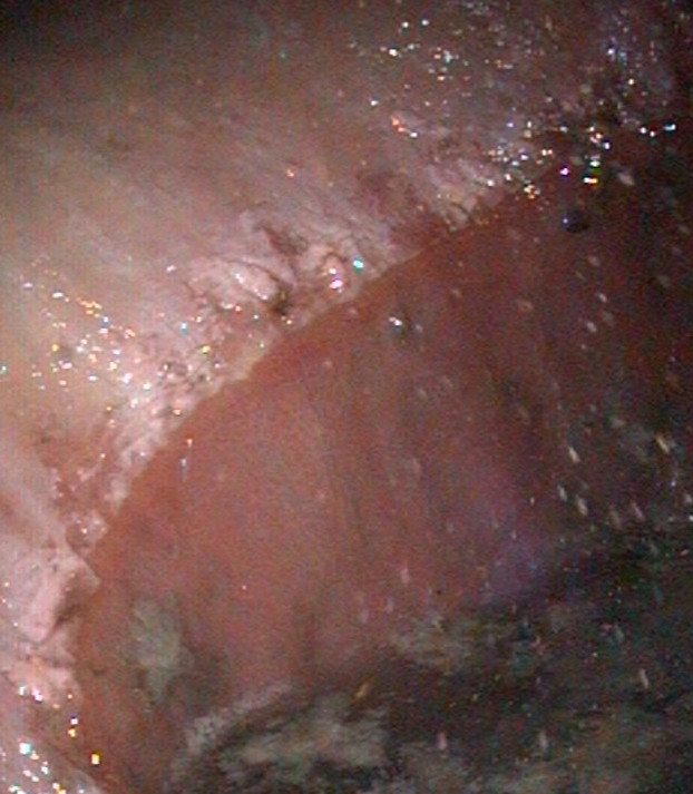
By Dr. David MarlinWe are now approaching half a century since Bob Cook pioneered the use of the flexible fibreoptic endoscope, which allowed examination of the respiratory tract in the conscious horse. One of the important outcomes of this technique was that it opened the door to the study of “bleeding” or exercise-induced pulmonary hemorrhage (EIPH).But nearly 50 years on the irony is perhaps that whilst we have become good at describing the prevalence of EIPH and some of the factors that appear to increase the severity of EIPH within individual horses, we still lack a clear understanding of the condition and how to manage it.I use the term manage rather than treat or prevent as our knowledge of EIPH must show us that EIPH cannot be stopped entirely; it is a consequence of intense exercise. The other irony is that in the past 50 years, by far the majority of research into the management of EIPH has focused on the use of the diuretic furosemide. Whilst we have good evidence from controlled studies that furosemide reduces the severity of EIPH on a single occasion, we still lack good evidence to suggest that furosemide is effective when used repeatedly during training and or racing; and there is also evidence to the contrary. Let’s review some basic facts about EIPH, which should not be contentious.• EIPH is the appearance of blood in the airways associated with exercise.• EIPH occurs as a result of moderate to intense exercise. In fact, EIPH has been found after trotting when deep lung wash (bronchoalveolar lavage or BAL) is done after exercise.• EIPH most often involves the smallest blood vessels (capillaries) but can sometimes and less commonly be due to the rupture of larger blood vessels.• The smallest blood vessels are extremely thin. Around 1/100th the thickness of a human hair. But this extremely thin membrane is also what allows racehorses such as Thoroughbreds, Standardbreds and Arabs to use oxygen at such a high rate and is a major reason for their athleticism.• EIPH is a progressive condition. The chance of seeing blood in the trachea after exercise increases with time in racing.• EIPH is variable over time, even when horses are scoped after the same type of work.• If you ‘scope a horse after three gallops in a row, you can expect to see blood in the trachea on at least one occasion.• EIPH damage to the lungs starts at the back and top, and over time moves forward and down and is approximately symmetrical.• Following EIPH the lung becomes fibrotic (as scar tissue), stiffer and does not work as well. The iron from the blood is combined with protein and stored permanently in the lung tissue where it can cause inflammation.• High blood pressure within the lung is a contributing factor in EIPH. Horses with higher blood pressure appear to suffer worse EIPH.• There is also evidence that upper airway resistance and breathing pattern can play a role in EIPH.• Airway inflammation and poor air quality may increase the severity of EIPH within individual horses.• Increasing severity of EIPH appears to have an increasing negative effect on performance.• Visible bleeding (epistaxis) has a very clear and marked negative effect on performance. In order to make progress in the management of EIPH (i.e., to minimize the severity of EIPH in each individual), there are certain steps that trainers can take based on the information we have to date. These include:• Ensuring good air quality in stables • Regular respiratory examination and treatment of airway inflammation • Reduced intensity of training during periods of treatment for moderate to severe airway inflammation• Extended periods of rest and light work with a slower return to work for horses following viral infection• Addressing anything that increases upper airway resistance (e.g., roaring, gurgling)• Avoiding intense work in cold weather• Avoiding extremes of going• Limiting number of training days in race preparation and increasing interval between races.FUTURE OPPORTUNITIES IN UNDERSTANDING AND MANAGING EIPHWe have to accept EIPH as a normal consequence of intense exercise in horses. Our aim should be to reduce the severity to a minimum in each individual horse. However, there are areas in which we still need a much greater scientific understanding.What actually causes the capillaries to leak or rupture?If you ask any vet, scientist or informed trainer what is the cause of EIPH, they will give the phrase “pulmonary capillary stress-failure”.But this is simply a description of what happens—NOT an explanation or a mechanism. EIPH and pulmonary capillary stress-failure are both descriptions of what’s happening. We know high blood pressure makes the capillaries stiff. But what makes them actually rupture? A balloon filled with water may be distended and under a lot of stress. But a pin prick will actually make it burst. The pin is the cause.Assessing EIPHAt present the most common way to assess the severity of EIPH in horses in training and racing is by ‘scoping 30-40 minutes after exercise and scoring the amount of blood in the trachea. This is a crude method, and when we see a horse that has a score of 1 after one gallop and a 3 after the next gallop, we don’t know whether this is due to differences in how quickly the blood has moved from the periphery of the lung into the trachea or due to a true difference in the amount of bleeding. We know our ‘scoping scores vary from gallop to gallop; we just don’t know why.BAL (deep lung wash) is not the answer either. It will pick up blood when there is none to be seen in the trachea (i.e., it’s a more sensitive technique), but with BAL we are looking atrelatively small areas of the lung. What we need is a technique that will allow us to image the whole lung and map the blood that is in the airways and not in the blood vessels so we can assess volume and distribution of hemorrhage.

By Dr. David Marlin

We are now approaching half a century since Bob Cook pioneered the use of the flexible fibreoptic endoscope, which allowed examination of the respiratory tract in the conscious horse. One of the important outcomes of this technique was that it opened the door to the study of “bleeding” or exercise-induced pulmonary hemorrhage (EIPH).

But nearly 50 years on the irony is perhaps that whilst we have become good at describing the prevalence of EIPH and some of the factors that appear to increase the severity of EIPH within individual horses, we still lack a clear understanding of the condition and how to manage it.

I use the term manage rather than treat or prevent as our knowledge of EIPH must show us that EIPH cannot be stopped entirely; it is a consequence of intense exercise. The other irony is that in the past 50 years, by far the majority of research into the management of EIPH has focused on the use of the diuretic furosemide. Whilst we have good evidence from controlled studies that furosemide reduces the severity of EIPH on a single occasion, we still lack good evidence to suggest that furosemide is effective when used repeatedly during training and or racing; and there is also evidence to the contrary. Let’s review some basic facts about EIPH, which should not be contentious.

• EIPH is the appearance of blood in the airways associated with exercise.

• EIPH occurs as a result of moderate to intense exercise. In fact, EIPH has been found after trotting when deep lung wash (bronchoalveolar lavage or BAL) is done after exercise.

• EIPH most often involves the smallest blood vessels (capillaries) but can sometimes and less commonly be due to the rupture of larger blood vessels.

• The smallest blood vessels are extremely thin. Around 1/100th the thickness of a human hair. But this extremely thin membrane is also what allows racehorses such as Thoroughbreds, Standardbreds and Arabs to use oxygen at such a high rate and is a major reason for their athleticism.

• EIPH is a progressive condition. The chance of seeing blood in the trachea after exercise increases with time in racing.

• EIPH is variable over time, even when horses are scoped after the same type of work.

• If you ‘scope a horse after three gallops in a row, you can expect to see blood in the trachea on at least one occasion.

• EIPH damage to the lungs starts at the back and top, and over time moves forward and down and is approximately symmetrical.

• Following EIPH the lung becomes fibrotic (as scar tissue), stiffer and does not work as well. The iron from the blood is combined with protein and stored permanently in the lung tissue where it can cause inflammation.

• High blood pressure within the lung is a contributing factor in EIPH. Horses with higher blood pressure appear to suffer worse EIPH.

• There is also evidence that upper airway resistance and breathing pattern can play a role in EIPH.

• Airway inflammation and poor air quality may increase the severity of EIPH within individual horses.

• Increasing severity of EIPH appears to have an increasing negative effect on performance.

• Visible bleeding (epistaxis) has a very clear and marked negative effect on performance. In order to make progress in the management of EIPH (i.e., to minimize the severity of EIPH in each individual), there are certain steps that trainers can take based on the information we have to date. These include:

• Ensuring good air quality in stables • Regular respiratory examination and treatment of airway inflammation • Reduced intensity of training during periods of treatment for moderate to severe airway inflammation

• Extended periods of rest and light work with a slower return to work for horses following viral infection

• Addressing anything that increases upper airway resistance (e.g., roaring, gurgling)

• Avoiding intense work in cold weather

• Avoiding extremes of going

endoscopy.jpg

• Limiting number of training days in race preparation and increasing interval between races.

FUTURE OPPORTUNITIES IN UNDERSTANDING AND MANAGING EIPH

We have to accept EIPH as a normal consequence of intense exercise in horses. Our aim should be to reduce the severity to a minimum in each individual horse. However, there are areas in which we still need a much greater scientific understanding.

What actually causes the capillaries to leak or rupture?

If you ask any vet, scientist or informed trainer what is the cause of EIPH, they will give the phrase “pulmonary capillary stress-failure”. But this is simply a description of what happens—NOT an explanation or a mechanism. EIPH and pulmonary capillary stress-failure are both descriptions of what’s happening. We know high blood pressure makes the capillaries stiff. But what makes them actually rupture? A balloon filled with water may be distended and under a lot of stress. But a pin prick will actually make it burst. The pin is the cause.

Assessing EIPH

At present the most common way to assess the severity of EIPH in horses in training and racing is by ‘scoping 30-40 minutes after exercise and scoring the amount of blood in the trachea. This is a crude method, and when we see a horse that has a score of 1 after one gallop and a 3 after the next gallop, we don’t know whether this is due to differences in how quickly the blood has moved from the periphery of the lung into the trachea or due to a true difference in the amount of bleeding. We know our ‘scoping scores vary from gallop to gallop; we just don’t know why.BAL (deep lung wash) is not the answer either. It will pick up blood when there is none to be seen in the trachea (i.e., it’s a more sensitive technique), but with BAL we are looking atrelatively small areas of the lung. What we need is a technique that will allow us to image the whole lung and map the blood that is in the airways and not in the blood vessels so we can assess volume and distribution of hemorrhage.

Furosemide is not the answer

Screenshot 2019-09-19 at 17.04.30.png
Fig 3e.jpg

A number of well-conducted and well-written scientific studies have shown conclusively that furosemide is effective in reducing the severity of EIPH in individual horses when used ONE time! We lack convincing studies that prove furosemide works as well when used one to two times a week for two to three months. In fact, several studies suggest that furosemide becomes less effective with regular use, such as the return to previous performance of horses after initial racing and improved performance on furosemide. In human medicine, repetitive administration of furosemide induces short-term (braking phenomenon, acute diuretic resistance) and longterm (chronic diuretic resistance) tolerance (i.e., if you give the same dose repeatedly, the body becomes tolerant and you get less and less urine production). A study in horses from Michigan State University in 2017 showed horses develop tolerance to furosemide. Why, when we have had nearly 50 years of research into EIPH with more published studies devoted to furosemide than any other aspect, do we still not know if furosemide is effective when used on a regular basis?

Is EIPH really blood?…

CLICK HERE to return to issue content

BUY THIS ISSUE IN PRINT OR DOWNLOAD

ISSUE 56 (PRINT)

$6.95


ISSUE 56 (DIGITAL)

$3.99


WHY NOT SUBSCRIBE?

DON'T MISS OUT AND SUBSCRIBE TO RECEIVE THE NEXT FOUR ISSUES!

Four issue subscription - PRINT & ONLINE - ONLY $24.95


IF YOU LIKE THIS ARTICLE

WHY NOT SUBSCRIBE - OR ORDER THE CONTENT FROM THIS ISSUE IN PRINT?

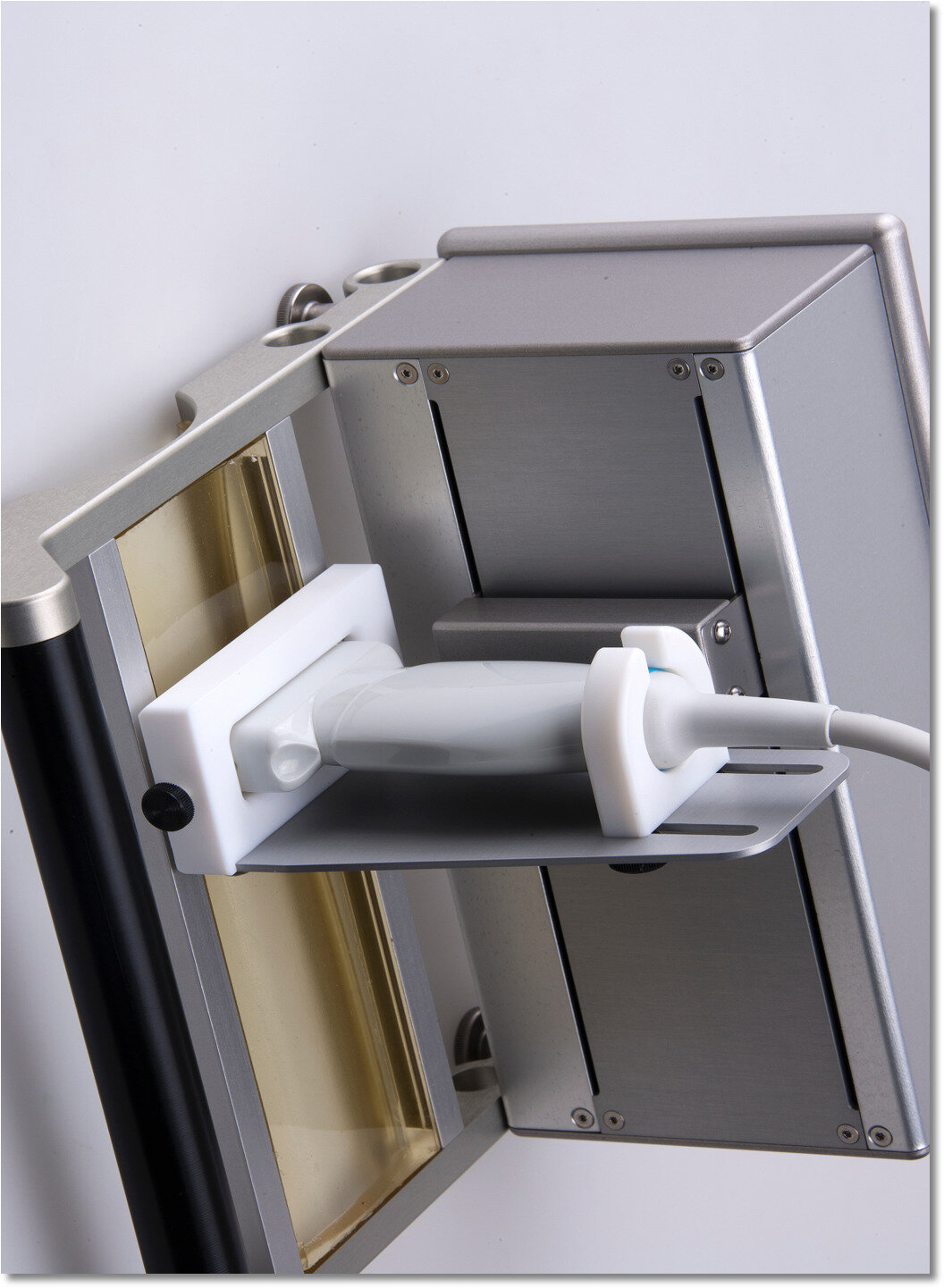
Bowed tendons - different treatment options - new ultrasound technology - ultrasound tissue characterization

Overstrain injuries to the superficial digital flexor tendon (SDFT) are among the most common musculoskeletal injuries for all athletic equine disciplines but account for a significant amount of wastage in the Thoroughbred (TB) racehorse.Treatment options for such ‘bowed tendons’ are many and varied, but all have a couple of things in common: time out of training; expense and no guarantee of success.It makes sense then, that prevention of injury should always be the goal, and failing that, a method to optimally guide rehabilitation is needed.Unfortunately, limitations of current imaging diagnostics have restricted their use for accurately monitoring the tendon.A new ultrasound technology, however, called ultrasound tissue characterization, may get us one step closer to achieving the goals of injury prevention and optimal rehabilitation.What would the ideal tendon imaging modality allow us to do?Monitor the effects of exercise on the tendonEarly detection of overstrain injuriesBe able to stage the lesion, i.e., determine the level of degenerative change within the tendon structureFine-tune therapyGuide rehabilitationWhy are tendon injuries so tricky?A normal healthy tendon is made from aligned organized tendon bundles. (Figure 1) Deterioration of this structure ranges on a spectrum from complete disruption (core lesion) to more minor changes, but all affect the ability of the tendon to function optimally.Degenerative changes within the tendon matrix are not uniform—meaning that not all overstrain injuries to the SDFT are represented by the same level of deterioration or structural change, so there is not a one-size-fits-all pathology or diagnosis, and therefore there cannot be a cure-all treatment.Most tendon injuries have a sneaky onset with tendon degeneration developing initially without clinical signs, so problems start without you or your horse even knowing about them. Often by the time you realize there is a problem, tendon matrix degradation has already begun.Staging the structural integrity of the tendon or classifying the extent of structural deterioration present is, therefore, imperative—not only for optimal therapy selection and appropriate rehabilitation guidance but also if prevention of injury is ever to be achieved.Why isn’t conventional ultrasound enough?Unfortunately, although conventional ultrasound has historically been used to evaluate equine tendon, limitations have restricted its ability to accurately monitor tendon structure, predict injury or guide rehabilitation.Clinical improvement is usually not accurately correlated with changes in imaging status using conventional ultrasound, especially in the later stages of healing with conventional ultrasound not demonstrating enough sensitivity to determine the type of tendon tissue under investigation.So, while regular ultrasound can easily demonstrate the presence of a core lesion when it first appears, by about two months post injury, its capacity to provide information regarding the health of the tendon is limited. Because of its inability to interpret the integrity of the underlying tendon structure accurately, along with inconsistencies in imaging, reliance on operator skills and the inherent lack of ability of a 2D conventional ultrasound image to fully decipher a 3D tendon structure, its ability to reliably evaluate and monitor the SDFT following the initial acute period is severely restricted. What is ultrasound tissue characterization?Ultrasound tissue characterization is a relatively new technique intended to alleviate some of the problems encountered with conventional ultrasound by improving objective tendon characterization. It does this by providing a 3D reconstruction of the tendon and by classifying and then quantifying tendon tissue into one of four color-coded echo types based on the integrity of the tendon structure.It can assess in detail the structural integrity of the tendon; it can discriminate a variety of pathological states and is sensitive enough to detect the effect of changing loads on the tendon within days.What do the colors mean? (Figure 2)Green (type 1 echoes) are normal, well-aligned and organized tendon bundles, and at least 85-90% of this echo type should be found in a healthy tendon (SDFT). Blue (type 2 echoes) are areas of wavy or swollen tendon bundles. They can represent remodeling and adapting tendon or inferior repair. Red (type 3 echoes) represents fibrillar tissue (the smaller basic unit or building block of tendon). This echo type can represent partial rupture of the tendon where they reflect breakdown of normal structure or they can represent initial healing as the tendon begins to rebuild. Black (type 4 echoes) are areas of cells or fluid and represent core lesions where no normal tendon tissue exists.How is ultrasound tissue characterization currently used?The aim of ultrasound tissue characterization is not to replace conventional ultrasound but on the contrary, it is recommended to perform an evaluation with both conventional B mode ultrasound and ultrasound tissue characterization to achieve a complete picture of tendon health.Currently it is used successfully in elite human athletes such as NBA and soccer players to monitor the health of their tendons (Achilles tendon and patellar tendons) and to guide exercise regimens post injury.In the equine field, it is used in elite sport horses as part of routine maintenance evaluations to direct exercise, to monitor tendon health and guide rehabilitation following an injury.How does it work?It consists of a standard linear ultrasound probe mounted onto a motorized tracking device (Figure 3). Due to the sensitivity of this equipment, the limbs should be clipped in order to obtain good quality images.The probe moves non-invasively and automatically down the tendon from top to bottom over a 12-cm scanning distance (Figure 4): As it does so, transverse images are captured at regular distances and stored in real time in a high-capacity laptop for processing. Images are automatically recorded every 0.2 mm to generate a 3D tendon volume made up of 600 images. This precise spatial ‘stacking’ of images is simply not possible to achieve with conventional ultrasound and is fundamental to the ultrasound tissue characterization technology. Image acquisition takes approximately 45 seconds. (Figure 5)This tendon volume can subsequently be used for visualization of the tendon in 3D, for tissue characterization (to determine the structural composition of the tendon) and for quantification of tendon matrix integrity. (Figure 6)The color-coded echo types provide objective information regarding the integrity of the tendon matrix and reflect the underlying tendon health. Ultrasound tissue characterization can discriminate between healthy normal tendon, adaptive/remodeling tendon and injured/healing tendon—often in cases where conventional ultrasound appears unremarkable in appearance. (Figure 7)The key to this technology is to perform successive evaluations. This allows comparison of differences in tendon structure between scans. Such consecutive examinations, along with clinical data and history, allow veterinarians to determine if a tendon is static, adaptive, healing or degenerating; and this information enables changes in training intensity to be made accordingly. (Figure 8)ResearchPublished research has reported correlation of ultrasound tissue characterization echo types with histological studies, meaning they correspond to postmortem findings.Numerous peer-reviewed research studies exist, documenting the ability of ultrasound tissue characterization to evaluate and monitor tendons both in human and equine athletes.Research has reported ultrasound tissue characterization to be highly reproducible with the ability to detect subtle changes in tendon structure in response to exercise loads in both human and equine athletes—something not possible utilizing conventional imaging modalities.Two studies specific to the TB racehorse exist both demonstrate the ability of ultrasound tissue characterization to monitor changes in tendon structure.How can it be used in the racehorse?RehabilitationIt is widely accepted that complete removal of load on tendons post injury is deleterious for tendon health, and the complete removal of exercise is only advocated in the very acute inflammatory phase following a tendon injury. Appropriately progressive loading of the tendon is desired to stimulate remodeling and healing, and this is where ultrasound tissue characterization is proving to be most useful.Typically, an exercise regimen post injury follows a generic format using clinical signs and conventional ultrasound as the only methods of assessment. But because most early tendon degradation is silent and conventional ultrasound struggles to decipher the integrity of the tendon unless a lesion is present, it has been traditionally difficult to precisely guide exercise regimens during rehabilitation.By providing real-time information regarding the integrity of the tendon matrix, ultrasound tissue characterization, in contrast, allows veterinarians to take advantage of the limited-time window of opportunity that exists for appropriate tendon remodeling after injury. By mapping the ultrastructure of the healing tendon and its remodeling response to exercise at each step in the rehabilitation regimen, it allows optimization of the most vital tool we have in our rehabilitation arsenal: exercise.While ultrasound tissue characterization technology is groundbreaking in its ability to non-invasively evaluate tendon structure and aid in tendon rehabilitation, it must be remembered that once a tendon is injured it will always be inferior to an uninjured tendon. Scar tissue will always be scar tissue. So, while green echoes are the goal (normal and aligned tendon bundles) and represent success for a rehabilitating tendon, they still just represent scar tissue—albeit aligned appropriately and in the best state to combat the strains of training and racing. This technology doesn’t remove the risk of reinjury in the bowed tendon, and it doesn’t provide information regarding its biochemical makeup. It simply tells us if the tendon is structurally normal; and by doing so, it improves our ability to monitor and guide healing. It provides veterinarians the best opportunity, currently, to adjust and tailor exercise regimens for the specific needs of the individual tendon and horse, allowing for informed decisions regarding the tendon’s capacity for performance. (Figure 9)The Future: Injury Prediction?While the current scientific literature seems to support the use of ultrasound tissue characterization to guide rehabilitation and monitor the effects of changing loads during training on the tendon, numerous anecdotal accounts from clinical practice. And both human and equine also report the ability of ultrasound tissue characterization to warn of impending injury. Although human research is currently ongoing to definitively confirm this, further equine research is needed to determine the specifics of any predictive capabilities it may have in the TB racehorse. For now, however, the evidence suggests that ultrasound tissue characterization can reliably and accurately be used to help guide rehabilitation of injured tendons—in both humans and horses—often resulting in a more successful return from injury.Referencesvan Schie HT, Bakker EM, Jonker AM and van Weeren PR. Efficacy of computerized discrimination between structure-related and non-structure-related echoes in ultrasonographic images for the quantitative evaluation of the structural integrity of superficial digital flexor tendons in horses. Am J Vet Res 2001; 62(7): 1159-1166.van Schie HT, Bakker EM, Cherdchutham W, Jonker AM, van de Lest CH, van Weeren PR. Monitoring of the repair process of surgically created lesions in equine superficial digital flexor tendons by use of computerized ultrasonography. Am J Vet Res. 2009; 70(1): 37-48.Docking SI, Rosengarten SD, Cook J. Achilles tendon structure improves on UTC imaging over a 5-month pre-season in elite Australian football players. Scand J Med Sci Sports. 2015.Docking SI. Tendon structure changes after maximal exercise in the Thoroughbred horse: use of ultrasound tissue characterization to detect in vivo tendon response. Vet J. 2012 Dec;194(3):338-342.S. Plevin, J. McLellan, H. van Schie, T. Parkin. Ultrasound tissue characterization of the superficial digital flexor tendons in juvenile Thoroughbred racehorses during early race training. Equine Veterinary Journal.Jarrod Antflick. Management of Tendinopathies with Ultrasound Tissue Characterization. SportEX Medicine 2014;61(July):26-30.

By Sarah Plevin

Overstrain injuries to the superficial digital flexor tendon (SDFT) are among the most common musculoskeletal injuries for all athletic equine disciplines but account for a significant amount of wastage in the Thoroughbred (TB) racehorse.

Treatment options for such ‘bowed tendons’ are many and varied, but all have a couple of things in common: time out of training; expense and no guarantee of success.

It makes sense then, that prevention of injury should always be the goal, and failing that, a method to optimally guide rehabilitation is needed.

Unfortunately, limitations of current imaging diagnostics have restricted their use for accurately monitoring the tendon.

A new ultrasound technology, however, called ultrasound tissue characterization, may get us one step closer to achieving the goals of injury prevention and optimal rehabilitation.

What would the ideal tendon imaging modality allow us to do?

  • Monitor the effects of exercise on the tendon

  • Early detection of overstrain injuries

  • Be able to stage the lesion, i.e., determine the level of degenerative change within the tendon structure

  • Fine-tune therapy

  • Guide rehabilitation

Why are tendon injuries so tricky?

Figure 1: Functionally normal healthy aligned tendon bundles.

Figure 1: Functionally normal healthy aligned tendon bundles.

  • A normal healthy tendon is made from aligned organized tendon bundles. (Figure 1) Deterioration of this structure ranges on a spectrum from complete disruption (core lesion) to more minor changes, but all affect the ability of the tendon to function optimally.

  • Degenerative changes within the tendon matrix are not uniform—meaning that not all overstrain injuries to the SDFT are represented by the same level of deterioration or structural change, so there is not a one-size-fits-all pathology or diagnosis, and therefore there cannot be a cure-all treatment.

  • Most tendon injuries have a sneaky onset with tendon degeneration developing initially without clinical signs, so problems start without you or your horse even knowing about them. Often by the time you realize there is a problem, tendon matrix degradation has already begun.

  • Staging the structural integrity of the tendon or classifying the extent of structural deterioration present is, therefore, imperative—not only for optimal therapy selection and appropriate rehabilitation guidance but also if prevention of injury is ever to be achieved.


Why isn’t conventional ultrasound enough?

  • Unfortunately, although conventional ultrasound has historically been used to evaluate equine tendon, limitations have restricted its ability to accurately monitor tendon structure, predict injury or guide rehabilitation. 

  • Clinical improvement is usually not accurately correlated with changes in imaging status using conventional ultrasound, especially in the later stages of healing with conventional ultrasound not demonstrating enough sensitivity to determine the type of tendon tissue under investigation. 

  • So, while regular ultrasound can easily demonstrate the presence of a core lesion when it first appears, by about two months post injury, its capacity to provide information regarding the health of the tendon is limited. Because of its inability to interpret the integrity of the underlying tendon structure accurately, along with inconsistencies in imaging, reliance on operator skills and the inherent lack of ability of a 2D conventional ultrasound image to fully decipher a 3D tendon structure, its ability to reliably evaluate and monitor the SDFT following the initial acute period is severely restricted. 

What is ultrasound tissue characterization?

Ultrasound tissue characterization is a relatively new technique intended to alleviate some of the problems encountered with conventional ultrasound by improving objective tendon characterization. It does this by providing a 3D reconstruction of the tendon and by classifying and then quantifying tendon tissue into one of four color-coded echo types based on the integrity of the tendon structure.

It can assess in detail the structural integrity of the tendon; it can discriminate a variety of pathological states and is sensitive enough to detect the effect of changing loads on the tendon within days.

What do the colors mean? (Figure 2)

Figure 2: Color-coded ultrasound tissue characterization echo types represent the stability of echo pattern over contiguous images related to tendon matrix integrity.

Figure 2: Color-coded ultrasound tissue characterization echo types represent the stability of echo pattern over contiguous images related to tendon matrix integrity.

Green (type 1 echoes) are normal, well-aligned and organized tendon bundles, and at least 85-90% of this echo type should be found in a healthy tendon (SDFT). Blue (type 2 echoes) are areas of wavy or swollen tendon bundles. They can represent remodeling and adapting tendon or inferior repair. Red (type 3 echoes) represents fibrillar tissue (the smaller basic unit or building block of tendon). This echo type can represent partial rupture of the tendon where they reflect breakdown of normal structure or they can represent initial healing as the tendon begins to rebuild. Black (type 4 echoes) are areas of cells or fluid and represent core lesions where no normal tendon tissue exists. 

How is ultrasound tissue characterization currently used?

The aim of ultrasound tissue characterization is not to replace conventional ultrasound but on the contrary, it is recommended to perform an evaluation with both conventional B mode ultrasound and ultrasound tissue characterization to achieve a complete picture of tendon health.

Currently it is used successfully in elite human athletes such as NBA and soccer players to monitor the health of their tendons (Achilles tendon and patellar tendons) and to guide exercise regimens post injury.

Figure 3: Ultrasound tissue characterization tracker frame with attached ultrasound probe.

Figure 3: Ultrasound tissue characterization tracker frame with attached ultrasound probe.

In the equine field, it is used in elite sport horses as part of routine maintenance evaluations to direct exercise, to monitor tendon health and guide rehabilitation following an injury.  

How does it work?

It consists of a standard linear ultrasound probe mounted onto a motorized tracking device (Figure 3). Due to the sensitivity of this equipment, the limbs should be clipped in order to obtain good quality images.

The probe moves non-invasively and automatically down the tendon from top to bottom over a 12-cm scanning distance (see Introphoto) …

CLICK HERE to return to issue contents

BUY THIS ISSUE IN PRINT OR DOWNLOAD

ISSUE 56 (PRINT)

$6.95


ISSUE 56 (DIGITAL)

$3.99


WHY NOT SUBSCRIBE?

DON'T MISS OUT AND SUBSCRIBE TO RECEIVE THE NEXT FOUR ISSUES!

Four issue subscription - PRINT & ONLINE - ONLY $24.95


PET scanning - reduces catastrophic fractures - latest advance in equine imaging - designed to image horse legs

By Mathieu Spriet, Associate Professor, University of California, Davis

PET: the latest advance in equine imagingMathieu Spriet, Associate Professor, University of California, Davis<< EVJ new logo near here>>Santa Anita Park, the iconic Southern California racetrack, currently under public and political pressure due to a high number of horse fatalities during the 2019 season, announced in December 2019 the installation of a PET scanner specifically designed to image horse legs. It is hoped that this one-of-a-kind scanner will provide information about bone changes in racehorses to help prevent catastrophic breakdowns.What is PET?PET stands for positron emission tomography. Although this advanced form of imaging only recently became available for horses, the principles behind PET imaging have been commonly used at racetracks for many years. PET is a nuclear medicine imaging technique, similar to scintigraphy, which is more commonly known as “bone scan”. For nuclear imaging techniques, a small dose of radioactive tracer is injected to the horse, and the location of the tracer is identified with a camera in order to create an image. The tracers used for racehorse imaging are molecules that will attach to sites on high bone turnover, which typically occurs in areas of bone subject to high stress. Both scintigraphic and PET scans detect “hot spots” that indicate—although a conventional X-ray might not show anything abnormal in a bone—there are microscopic changes that may develop into more severe injuries.Development of PET in CaliforniaThe big innovation with the PET scan is that it provides 3D information, whereas the traditional bone scan only acquires 2D images. The PET scan also has a higher spatial resolution, which means it is able to detect smaller changes and provide a better localisation of the abnormal sites. PET’s technological challenge is that to acquire the 3D data in horses, it is necessary to use a ring of detectors that fully encircles the leg.The first ever equine PET scan was performed at the School of Veterinary Medicine at the University of California in 2015. At the time, a scanner designed to image the human brain was used (PiPET, Brain-Biosciences, Inc.). This scanner consists of a horizontal cylinder with an opening of 22cm in diameter. Although the dimensions are convenient to image the horse leg, the configuration required the horse be anesthetised in order to fit the equipment around the limb.<< Fig 1 near here>>The initial studies performed on anesthetised horses with the original scanner demonstrated the value of the technique. A first study, published in Equine Veterinary Journal, demonstrated that PET showed damage in the equine navicular bone when all other imaging techniques, including bone scan, MRI and CT did not recognise any abnormality.<< Figure 2>> near hereA pilot study looking at the racehorse fetlock, also published in Equine Veterinary Journal, showed that PET detects hot spots in areas known to be involved in catastrophic fractures. This confirmed the value of PET for racehorse imaging, but the requirement for anesthesia remained a major barrier to introducing the technology at the racetrack. To overcome this, LONGMILE Veterinary Imaging, a division of Brain-Biosciences Inc, in collaboration with the University of California Davis, designed a scanner which could image standing horses. To do this, the technology had to be adapted so that the ring of detectors could be opened and positioned around the limb.With the support from the Grayson Jockey Club Research Foundation, the Southern California Equine Foundation and the Stronach Group, this unique scanner became a reality and, after the completion of an initial validation study in Davis, the scanner was installed at Santa Anita Park in December 2019.PET at the racetrackThe new PET scanner has been used to image the equine limb from the foot to the knee. The current main focus at the racetrack is fetlock imaging, as the majority of catastrophic breakdown in racehorses affects this area. The UC Davis pilot study highlighted the value of PET for detecting abnormalities in the proximal sesamoid bones—the two small bones at the back of the cannon bone—that are commonly involved in catastrophic fractures. Previous necropsy research on horses which suffered breakdowns has shown that changes can be present in the bones prior to the development of major injuries. The goal of the Californian PET project is to detect these warning signs in order to avoid training and racing horses at high risk for catastrophic breakdown.<<Figure 3 near here>>Alternative imaging techniquesOther imaging techniques are available for examining equine bone. Scintigraphic bone scans are doing an excellent job at detecting stress fractures of the humerus or tibia, and this has helped markedly decrease catastrophic injuries in these areas. Bone scan is also used for fetlocks; but “hot fetlocks” are common on bone scan, and the lower resolution 2D images often do not allow to truly determine whether horses are at high risk of fractures or have normal bone adaptation to training.MRI is used for fetlock imaging too, and MRI scanners designed for imaging standing horses have been available for over 15 years. Several large racing centers are equipped with such scanners, and MRI excels in particular at detecting changes in the cannon bone that precede condylar fractures. MRI can detect areas of bone densification, or even accumulation of fluid in the bone, typically indicative of microtrauma that can weaken the bone.Computed tomography (CT) has also recently been used for standing imaging of the fetlock. At the moment, there are a few centers equipped with a CT scanner allowing standing fetlock imaging, but they are only available at, for example, New Bolton Center, Pennsylvania - USA, and the University of Melbourne, Australia. CT uses X-rays to create 3D images. Similar to MRI, CT can detect areas of bone densification or areas of bone loss.PET’s advantagesThe big advantage of PET is what is called “sensitivity”—the ability to detect early and subtle findings. This is because PET detects changes at the molecular level before structural changes have occurred. MRI and CT rely on changes in the density and shape of the structures they are imaging; i.e., structural change must have occurred before these techniques can identify that the bone is abnormal. MRI and CT might miss early information that a PET scan can detect; but they provide complementary information, and these techniques will be important to further characterise abnormalities found on PET. For these reasons, PET and MRI or CT can be combined: a PET image is “fused” on an MRI or a CT, combining the sensitivity of PET with the anatomical detail of the other imaging tool.<< Figure 5 near here>>As PET is a newly available modality at the racetrack, there is still a lot to learn. The goal of the first year at Santa Anita is to image as many horses as possible and compare with the PET information with bone scan or MRI information. The pilot study at Davis and the initial cases at Santa Anita tend to show that it is normal to see some bone activity in specific areas of the fetlock, e.g., the palmar condyles; but the presence of hot spots in other areas, for example in the middle of the sesamoid bones, is an abnormal finding that could be an indicator of higher risk of fracture.Other roles for PETIn addition to its use in racehorses, PET has been used in over a 100 sport horses at UC Davis in the last three years. All these scans have been performed with horses under anesthesia and combined to a CT. The main reason to perform a PET scan is either when other imaging modalities do not find a reason to explain a lameness or to better understand changes seen with other modalities. PET is a “functional” technique; this means that a hot spot indicates an area where an injury is active. MRI can meet difficulties distinguishing between scar tissue and active injury, but PET is the ideal modality for this. The majority of the work done in sport horses has used the same bone tracer as in racehorses. The most common injuries found with this tracer in sport horses result in navicular disease and early arthritis (joint disease).PET is not restricted to imaging; with an alternative tracer, it can be used to look at injuries in the soft tissues. This is something that is not possible with scintigraphy, and the soft tissue tracer has been used successfully to identify tendon injuries—distinguishing between active and inactive tendon lesions. Another important area of interest where the soft tissue tracer has been used is for the assessment of laminitis. This disease is extremely complex, and PET is bringing new information about laminitis, which hopefully will help find new ways to fight this serious life-threatening disease.PET in the futureThe development of equine PET is the biggest step forward in horse imaging since the introduction of equine MRI over 20 years ago. The development of the standing system has considerably facilitated the use of the technique. PET is currently at the forefront of the solutions proposed to improve racehorse safety, but PET will also help with other important health issues in horses.

Santa Anita Park, the iconic Southern California racetrack, currently under public and political pressure due to a high number of horse fatalities during the 2019 season, announced in December 2019 the installation of a PET scanner specifically designed to image horse legs. It is hoped that this one-of-a-kind scanner will provide information about bone changes in racehorses to help prevent catastrophic breakdowns.

What is PET?

Figure 1: The first equine PET was performed in 2015 at the University of California Davis on a research horse laid down with anesthesia. The scanner used was a PET prototype designed for the human brain (piPET, Brain- Biosciences Inc., Rockville, MD).

Figure 1: The first equine PET was performed in 2015 at the University of California Davis on a research horse laid down with anesthesia. The scanner used was a PET prototype designed for the human brain (piPET, Brain- Biosciences Inc., Rockville, MD).

PET stands for positron emission tomography. Although this advanced form of imaging only recently became available for horses, the principles behind PET imaging have been commonly used at racetracks for many years. PET is a nuclear medicine imaging technique, similar to scintigraphy, which is more commonly known as “bone scan”. For nuclear imaging techniques, a small dose of radioactive tracer is injected to the horse, and the location of the tracer is identified with a camera in order to create an image. The tracers used for racehorse imaging are molecules that will attach to sites on high bone turnover, which typically occurs in areas of bone subject to high stress. Both scintigraphic and PET scans detect “hot spots” that indicate—although a conventional X-ray might not show anything abnormal in a bone—there are microscopic changes that may develop into more severe injuries.

Development of PET in California

Santa Anita_ 6N2A9803.jpg

The big innovation with the PET scan is that it provides 3D information, whereas the traditional bone scan only acquires 2D images. The PET scan also has a higher spatial resolution, which means it is able to detect smaller changes and provide a better localisation of the abnormal sites. PET’s technological challenge is that to acquire the 3D data in horses, it is necessary to use a ring of detectors that fully encircles the leg. 

The first ever equine PET scan was performed at the School of Veterinary Medicine at the University of California in 2015. At the time, a scanner designed to image the human brain was used (PiPET, Brain-Biosciences, Inc.). This scanner consists of a horizontal cylinder with an opening of 22cm in diameter. Although the dimensions are convenient to image the horse leg, the configuration required the horse be anesthetised in order to fit the equipment around the limb. 

The initial studies performed on anesthetised horses with the original scanner demonstrated the value of the technique. A first study, published in Equine Veterinary Journal, demonstrated that PET showed damage in the equine navicular bone when all other imaging techniques, including bone scan, MRI and CT did not recognise any abnormality.

A pilot study looking at the racehorse fetlock, also published in Equine Veterinary Journal,  showed that PET detects hot spots in areas known to be involved in catastrophic fractures.

Figure 2: These are images from the first horse image with PET. From left to right, PET, CT, MRI, and bone scan. The top row shows the left front foot that has a severe navicular bone injury. This is shown by the yellow area on the PET image and abnormalities are also seen with CT, MRI and bone scan. The bottom row is the right front foot from the same horse, the PET shows a small yellow area that indicates that the navicular bone is also abnormal. The other imaging techniques however did not recognize any abnormalities.

Figure 2: These are images from the first horse image with PET. From left to right, PET, CT, MRI, and bone scan. The top row shows the left front foot that has a severe navicular bone injury. This is shown by the yellow area on the PET image and abnormalities are also seen with CT, MRI and bone scan. The bottom row is the right front foot from the same horse, the PET shows a small yellow area that indicates that the navicular bone is also abnormal. The other imaging techniques however did not recognize any abnormalities.

This confirmed the value of PET for racehorse imaging, but the requirement for anesthesia remained a major barrier to introducing the technology at the racetrack. To overcome this, LONGMILE Veterinary Imaging, a division of Brain-Biosciences Inc, in collaboration with the University of California Davis, designed a scanner which could image standing horses. To do this, the technology had to be adapted so that the ring of detectors could be opened and positioned around the limb. 

With the support from the Grayson Jockey Club Research Foundation, the Southern California Equine Foundation and the Stronach Group, this unique scanner became a reality and, after the completion of an initial validation study in Davis, the scanner was installed at Santa Anita Park in December 2019.

Figure 3: The two images on the left are bone scan images from a 4-year-old Thoroughbred racehorse. The images on the right are 3D projection of PET images of the same fetlock. The bone scan revealed an abnormality at the bottom of the cannon bone. The PET scan confirmed this abnormality and helped better localize it. In addition, several other abnormalities were found on the PET scan in the sesamoid bones.

Figure 3: The two images on the left are bone scan images from a 4-year-old Thoroughbred racehorse. The images on the right are 3D projection of PET images of the same fetlock. The bone scan revealed an abnormality at the bottom of the cannon bone. The PET scan confirmed this abnormality and helped better localize it. In addition, several other abnormalities were found on the PET scan in the sesamoid bones.

PET at the racetrack

The new PET scanner has been used to image the equine limb from the foot to the knee. The current main focus at the racetrack is fetlock imaging, as the majority of catastrophic breakdown in racehorses affects this area. The UC Davis pilot study highlighted the value of PET for detecting abnormalities in the proximal sesamoid bones—the two small bones at the back of the cannon bone—that are commonly involved in catastrophic fractures. Previous necropsy research on horses which suffered breakdowns has shown that changes can be present in the bones prior to the development of major injuries. The goal of the Californian PET project is to detect these warning signs in order to avoid training and racing horses at high risk for catastrophic breakdown.

Alternative imaging techniques

Other imaging techniques are available for examining equine bone. Scintigraphic bone scans are doing an excellent job at detecting stress fractures of the humerus or tibia, and this has helped markedly decrease catastrophic injuries in these areas. Bone scan is also used for fetlocks; but “hot fetlocks” are common on bone scan, and the lower resolution 2D images often do not allow to truly determine whether horses are at high risk of fractures or have normal bone adaptation to training.

Figure 4: The MILE-PET scanner (LONGMILE Veterinary imaging, Rockville, MD) is the first PET scanner specifically designed to image standing horses. An openable ring of detectors allows easy positioning and safe scanning.

Figure 4: The MILE-PET scanner (LONGMILE Veterinary imaging, Rockville, MD) is the first PET scanner specifically designed to image standing horses. An openable ring of detectors allows easy positioning and safe scanning.

MRI is used for fetlock imaging too, and MRI scanners designed for imaging standing horses have been available for over 15 years. Several large racing centers are equipped with such scanners, and MRI excels in particular at detecting changes in the cannon bone that precede condylar fractures. MRI can detect areas of bone densification, or even accumulation of fluid in the bone, typically indicative of microtrauma that can weaken the bone.

Computed tomography (CT) has also recently been used for standing imaging of the fetlock. At the moment, there are a few centers equipped with a CT scanner allowing standing fetlock imaging, but they are only available at, for example, New Bolton Center, Pennsylvania - USA, and the University of Melbourne, Australia. CT uses X-rays to create 3D images. Similar to MRI, CT can detect areas of bone densification or areas of bone loss. …

CLICK HERE to return to issue contents

BUY THIS ISSUE IN PRINT OR DOWNLOAD

ISSUE 56 (PRINT)

$6.95


ISSUE 56 (DIGITAL)

$3.99


WHY NOT SUBSCRIBE?

DON'T MISS OUT AND SUBSCRIBE TO RECEIVE THE NEXT FOUR ISSUES!

Four issue subscription - PRINT & ONLINE - ONLY $24.95


Colic – effects of inflammation

By Dr. Zofia Lisowski

Overview of colic

Colic is a term used to describe the display of abdominal pain in a horse. It is the most common emergency in horses with four to ten out of every 100 horses likely to experience at least one episode of colic each year. It is also the single most common cause of equine mortality. In the U.S., one study showed that Thoroughbreds were more likely to develop colic1 than other breeds. It is of great welfare concern to horse owners, and with the estimated costs associated with colic in the U.S. exceeding $115 million per year2 and the average cost of a horse undergoing colic surgery requiring a resection being approximately $8,3703, it is also a significant economic issue for horse owners. 

Horses with abdominal pain show a wide range of clinical signs, ranging from flank watching and pawing the ground in mild cases, to rolling and being unable to remain standing for any significant period of time in more severe cases. There are numerous (over 50) specific causes of colic. In general, colic occurs as a result of disruption to the normal function of the gastrointestinal tract. This may be attributable to mechanical causes such as an obstruction (constipation), distension (excess gas) or a volvulus (twisted gut). It may also have a functional cause, whereby the intestine doesn’t work as normal in the absence of an associated mechanical problem; for example, equine grass sickness is associated with a functional derangement of intestinal motility due to loss of nerves within the intestine. 

Management of colic depends on the cause and can necessitate either a medical or surgical approach. Most horses with colic will either improve spontaneously or with simple medical treatment alone; however, a significant proportion may need more intensive medical treatment or surgery. Fortunately, due to improvements in surgical techniques and post-operative management, outcomes of colic surgery have improved over the past few decades with up to 85% of horses surviving to discharge. Crucially for the equine Thoroughbred racehorse population, several studies focused on racehorses that had undergone colic surgery and survived to discharge, reporting that 63-73% returned to racing. Furthermore, surgical treatment did not appear to negatively impact athletic performance. A similar finding was also seen in the general sport horse population.

Despite significant advancement in colic surgery per se, complications following surgery can have a significant impact on post-operative survival and return to athletic function. Common post-operative complications include:

  1. Complications at the site of the incision (surgical wound)

Infection: Infections at the surgical incision site are relatively common. Antibiotics are usually administered before surgery and after surgery. Infections are not normally severe but can increase treatment costs. Horses that develop infections are at greater risk of developing an incisional hernia.  

Hernia: Incisional hernias occur when the abdominal wall muscles fail to heal, leaving a “gap.” Hernia size can vary from just a few centimeters, up to the full length of the incision. Most hernias will not require further treatment; but in more severe cases, further surgery may be required to repair the hernia.

  1. Complications within the abdomen

Hemoperitoneum: A rare complication where there is blood within the abdomen from bleeding at the surgical site.

Anastomosis complications: The anastomosis site is where two opposing ends of intestine that have been opened are sutured back together again. It is important that at this site no leakage of intestinal contents occurs. Leakage or breakdown at this site can lead to peritonitis, which is inflammation or infection within the abdominal cavity and is a potentially life-threatening complication. 

Adhesions: Scar tissue can form within the abdomen following abdominal surgery. Occasionally this may cause further colic episodes.

Further colic episodes

Further colic episodes can occur following surgery. These can occur days to months following discharge.

Endotoxemia

In some rare cases, horses may develop sepsis in response to toxins released by damaged intestines.

Diarrhea 

This is a rare complication. It can develop as a result of infections with C. difficile or Salmonella. As a consequence, some horses may need to be treated in isolation to ensure infection doesn’t spread to other horses or humans.

Post-operative ileus 

Post-operative ileus is one of the potential post-operative complications that can lead to a significant increase in hospital stay duration, increased treatment costs and is also associated with reduced survival rates. Post-operative ileus is a condition that affects the muscle function in the intestinal wall. The intestine is a long tube-like structure that has a muscular wall throughout its entire length from the esophagus to the anus. The function of this muscle is to contract in waves to mix and move food along the length of the intestinal tract, within which digestion occurs and nutrients are absorbed, terminating in the excretion of waste material as feces. In post-operative ileus these contractions stop and thus intestinal contents are not moved throughout the intestinal tract. In most cases, it is transient and lasts for up to 48 hours following surgery; however, in some cases it can last longer. A build-up of fluid develops within the intestine as a result of the lack of propulsion. This stretches the intestines and stomach, resulting in pain and the horse’s inability to eat. Unlike humans, the horse is unable to vomit; consequently, this excess fluid must be removed from the stomach by other means, otherwise there is a risk of the stomach rupturing with fatal consequences. Post-operative ileus may occur in up to 60% of horses undergoing abdominal surgery and mortality rates as high as 86% have been reported. Horses in which the small intestine manipulated is extensively manipulated during surgery and those that require removal of segments of intestine are at higher risk. Despite the significant risk of post-operative ileus following colic surgery in horses, there is a lack of studies investigating the mechanisms underpinning this condition in horses; consequently, the precise cause of this condition in horses is not fully known. 

What causes the intestine to stop functioning? …

BUY THIS ISSUE IN PRINT OR DOWNLOAD

ISSUE 55 (PRINT)

$6.95


ISSUE 55 (DIGITAL)

$3.99

WHY NOT SUBSCRIBE?

DON'T MISS OUT AND SUBSCRIBE TO RECEIVE THE NEXT FOUR ISSUES!

Four issue subscription - PRINT & ONLINE - ONLY $24.95


IF YOU LIKE THIS ARTICLE

WHY NOT SUBSCRIBE - OR ORDER THE CONTENT FROM THIS ISSUE IN PRINT?

Indiana's New "Biological Samples" Testing Law: Integrity Assured Or Invasive Overreach?

By Peter J. Sacopulos

From House Bill To Horse Law

On May 1, 2019, Governor Eric Holcomb signed Indiana House Bill 1196 into law. The statute, which took effect on July 1 of this year, directs the Indiana Horse Racing Commission (IHRC) to adopt a variety of new rules and procedures governing horse racing within the state. 

Governor Holcomb and Indiana State Representative Bob Cherry, who introduced HB 1196 to the legislature, are Republicans. However, the bill enjoyed broad bipartisan support—a rarity in current American politics. In fact, the final version sailed through both chambers, receiving not a single “nay” vote in the House and a mere three “nays” in the Senate. 

HB 1196 is something of an equine regulatory smorgasbord. Examples of its provisions include officially changing references to the IHRC’s “secretary” to “executive director,” altering the way breed advisory committee members are appointed, specifying that certain funds be directed to the Indiana-sired horses program, and the creation of new privacy protections to guard the personal information required on license applications.

Items like these, as well as several others included in HB 1196, are unlikely to cause ripples within the racing community. However, the new law also includes provisions designed to enhance and expand the Commission’s ability to detect, police and reduce the use of banned substances. And while this is undoubtedly a worthwhile cause, a two-word term used in the drug-testing language of HB 1196 has the potential to negatively impact horses and trainers for years to come, with consequences that may spread well beyond the borders of Indiana. 


Two Words, Too Broad?

The two-word term is “biological sample.” It is legally defined in the statue as follows:

“Biological sample” refers to any fluid, tissue or other substance obtained from a horse through an internal or external means to test for foreign substances, natural substances at abnormal levels, and prohibited medications. The term includes blood, urine, saliva, hair, muscle tissue collected at a necropsy, semen, and other substances appropriate for testing as determined by the commission. 

This definition goes well beyond the longstanding blood, saliva, urine, and more recently, hair, samples routinely collected from Thoroughbred competitors for analysis. It is also disturbingly open-ended. Indeed, the phrase: “…and other substances appropriate for testing as determined by the commission…”  is a definition that is essentially wide open, providing the IHRC staff the power to define or redefine a “biological sample.”

While there was discussion and temporary agreement to limit the use of biological samples to necropsy purposes, that limitation was removed from the final version of the bill that was signed into law and became effective July 1, 2019. Therefore, the Commission Staff is authorized and may elect to take muscle tissue, and other biological samples, from live animals as it deems and determines necessary and appropriate. This rule and its definition of biological sample establishes a new frontier of testing. 


Is This Risk Really Necessary?

One of the primary concerns and positions advanced in opposition to allowing biological samples to be taken from live animals is the risk of injury.

Taking saliva and hair samples from a Thoroughbred is painless and easy. And anyone who has ever been around horses knows that they are more than happy to provide all the urine you could ever want! Drawing blood from a horse is only slightly more difficult and rarely involves the use of a local anesthetic. 

However, taking “…any fluid, tissue or other substance… through an internal or external means…” is another matter entirely. It opens the door to far more invasive collection techniques that carry far greater risks for horses than blood, saliva, urine or hair sampling. To be clear, I am referring to biopsies. 

A biopsy is the removal and examination of cells or tissue from a living being for the purposes of testing and examination. Any biopsy carries risk of injury or infection. Taking a biopsy from a horse may be as simple as a skin sample from the withers or tissue from the lining of the mouth, or as difficult as removing material from the teeth or the interior of the eye; or from internal organs such as the heart, lungs, liver, intestine or colon. In the latter examples, a biopsy becomes a complex medical procedure. A procedure performed on a large, valuable animal requires sedation and may require general anesthesia to facilitate tissue collection. 

Sedating a horse is serious business. Sedatives and anesthetics carry significant risks, even when administered with care by skilled equine veterinary professionals. Those risks include allergic reactions, collapse, excitement, cardiac arrest, medical injury and post-anesthetic colic.

TO READ MORE - BUY THIS ISSUE IN PRINT OR DOWNLOAD

ISSUE 54 (PRINT)

$6.95

ISSUE 54 (DIGITAL)

$3.99

WHY NOT SUBSCRIBE?

DON'T MISS OUT AND SUBSCRIBE TO RECEIVE THE NEXT FOUR ISSUES!

Four issue subscription - PRINT & ONLINE - ONLY $24.9


Skin deep - overcoming barriers for effective transdermal drug delivery

By Professor Roger Smith

Ancient art, modern science

One shared medicinal practice among disparate ancient societies was the application of primitive ointments to the skin to treat almost all and any ailments. A vast plethora of poultices and plasters have been described, including in Babylonian and Greek medicine texts1 among others, suggesting that the magical health-restoring powers of ointments were well-recognized to traverse the skin. Thus, it was no coincidence that the skin was the preferred therapeutic route over surgical (and oral) intervention since the former method was likely to result in reduced mortality rates compared to the latter; undoubtedly an important consideration, given that the top ancient physicians were likely charged with the health of the royal courts.

Although the art of transdermal delivery of medicines dates back millennia, it is only in more recent times that the science of transdermal drug delivery in man has advanced significantly.  The choice of modern drugs for topical applications is, however, relatively limited compared to the seemingly infinite choice available for oral delivery. This is perhaps not surprising since the gut is an organ that has evolved with the main purpose of absorbing food (chemicals when it comes to it) whereas the skin, despite being the largest organ, has evolved primarily as a protective layer to prevent desiccation of underlying tissues and to keep out harmful environmental chemicals. As this includes medicinal drugs, the pursuit of transdermal administration would appear, at first sight, to be an illogical choice. However, there are several compelling reasons why transdermal delivery routes are an important alternative to pills, injections or inhalation routes:

  • It avoids poor absorption after oral ingestion—especially in animals, the absorption of a drug can vary between the omnivore (e.g., human) and herbivore (e.g., horse) stomach.  

  • It avoids first-pass effect where the blood circulation from the gut passes through the liver to remove absorbed drugs.

  • It can reduce systemic drug levels to minimize adverse effects.

  • The design of sustained release formulations overcomes the frequent dosing necessitated by oral and injectables to achieve constant drug levels.

  • It enables ease and efficacy of drug withdrawal.

  • Transdermal drug delivery is painless and non-invasive, thereby potentially allowing longer treatment when daily injection is unacceptable or impractical.

  • It has the potential to target local administration such as for the treatment of flexor tendon disease because the tendons are subcutaneous.

Challenges for transdermal drug applications

The skin is made up of three key layers: the epidermis, dermis and hypodermis and the water-attracting (hydrophilic) or water-repelling (hydrophobic) properties within each raise unique challenges for topical or transdermal drug applications.  

Topical applications, such as insect repellents and sunscreen creams, target the surface of the skin or deliver a drug locally such as for the control of inflammation (insect bite or reaction to an allergen). In contrast the aim of transdermal, or subcutaneous, applications are to deliver the drug deeper to either an adjacent organ, or, more commonly, to the blood circulation as an alternative to oral or needle routes to reach distant organs. The main barrier to local or transdermal delivery is the outermost layer of the skin, called the stratum corneum in the epidermis. This consists of dead skin cells, the corneocytes, that combine with lipid bilayers into a tightly packed “bricks-and-mortar” layer that form alternating hydrophilic (the water rich corneocytes) and hydrophobic (lipid bilayer) regions (figure 1). The stratum corneum therefore not only forms a mechanically robust layer but also presents a challenge in designing drugs with chemical properties that can negotiate their way into and through these contrasting hydrophobic and hydrophilic environments to reach the lower region of the epidermis. The epidermis consists of living skin cells but has no blood vessels for the drug to diffuse into, so instead the drug must penetrate further to the dermis where it can finally enter the bloodstream or the subcutaneous layers.

Routes for drugs through the skin

Most transdermal drugs are designed so that they diffuse through the skin in a passive fashion. The routes for drug can be through the skin cells (transcellular), around them (intercellular) or using the skin components—hair follicles, sweat glands and sebaceous glands (produce lipids)—to bypass the stratum corneum (so-called “appendageal” routes).

Transcellular route: Drugs pass through the corneocytes of the stratum corneum rather than the lipid ‘mortar’ that surrounds them. However, the drug has to exit the cell to enter the next corneocyte and therefore through the skin. It means that it has to encounter the external hydrophobic environment between the cells multiple times as it moves through the alternating cell and lipid layers of the epidermis. Drugs therefore have to have balanced hydrophilic and hydrophobic properties to enable this to happen. 

Intercellular route: The drug predominantly diffuses through the lipid rich “mortar” around the corneocytes of the epidermis. This lipid matrix can form a continuous route through the epidermis (avoiding entering the cells), but this route has been suggested to be less efficient because it increases the distance 50-fold3 compared to the direct route through the stratum corneum due to the interdigitating brick and mortar arrangement. Again, the chemical formulation used to carry the drug is important and drugs that more readily dissolve in lipids benefit from this route.

Appendageal route: The hair, sweat glands and sebaceous glands provide a direct channel to the deep layers of the skin circumventing the hazardous barriers of the epidermis and dermis. The main challenge for this relatively easy route is that the amount of drug that can be taken up is limited by the density of hair follicles and sweat glands, although in haired animals, such as the horse, the density can be as high as 1-5% of the skin surface area. Furthermore, sweat from an active sweat gland would be travelling against the direction of drug flow, washing out the drug and its carrier and severely limit drug uptake. It is likely that all skin applications use this appendageal route as it’s unavoidable but probably more efficient for drugs that are large molecules.

TO READ MORE —

BUY THIS ISSUE IN PRINT OR DOWNLOAD -

SUMMER SALES 2019, ISSUE 53 (PRINT)

$6.95

SUMMER SALES 2019, ISSUE 53 (DIGITAL)

$3.99

WHY NOT SUBSCRIBE?

DON'T MISS OUT AND SUBSCRIBE TO RECEIVE THE NEXT FOUR ISSUES!

PRINT & ONLINE SUBSCRIPTION

From $24.95

Fungi - the invisible health risk

By Dr. Emmanuelle van Erck

Dr. Emmanuelle van Erck, DVM, PhD, ECEIM explains her work looking at the link between the presence of fungi and lower airway inflammation

Horses are incredible athletes. Their physiology—the way their body functions—is truly fascinating. They can adapt to training at a phenomenal rate, they have massive hearts that fuel their powerful muscles and pushes them to peak speeds. So what could stop them? Oxygen, or rather the lack of it. Horses experience hypoxemia during racing, which means they enter a state of deficiency in oxygen. The reason for this deficiency is a failure of the respiratory system to effectively ventilate and adequately fuel oxygen to the muscles. Horses are obligate nasal breathers and were endowed with particularly long and narrow upper airways in relation to their body size. These factors increase the resistance to breathing. They are also constrained by the fact that they ventilate at very high rates, which does not allow for effective and rapid renewal of oxygen in the lungs. Even the fittest, best Thoroughbreds crave oxygen from mid-race onwards. So maintaining horses in optimal respiratory health is absolutely essential for them to achieve an efficient sprint and optimal performance. 

Respiratory diseases are highly prevalent in horses. It is inherent to their living and working conditions. The mere fact that a horse is housed in a box increases his risk of developing airway inflammation. The content in fine dust is naturally high in a horse’s box. Closed or poorly ventilated barns further deteriorate air quality in the horse’s immediate environment. Several studies have shown that horses housed indoors are exposed not only to high amounts of organic dust and ammonia but also germs and endotoxin they produce that trigger a detrimental reaction from the immune system. The problem is that even low-grade respiratory diseases will directly affect the horse’s capacity to perform and recover from strenuous exercise. 

With my colleagues, Dr. Dauviller and Dr. ter Woort, specialists in equine internal medicine, we have investigated the link between the presence of fungi and lower airway inflammation. In our ambulatory referral practice, we go out to the stables and have the opportunity not only to examine the horse but also attentively assess his environment. As we collected respiratory samples and analyzed them ourselves, we became aware that the presence of microscopic molds or fungal elements was frequently associated to lung issues. To investigate this further, we decided to systematically record clinical and environmental data and link it to our findings in the respiratory samples of the horses referred for investigation. 

We collected more than 700 cases; the horses included in the study were either referred routine examinations, unexplained poor performance or respiratory symptoms such as coughing or breathing heavily during exercise. All horses had a tracheal and a bronchoalveolar lavage done, which allowed us to evaluate their level of respiratory inflammation, as well as estimate the presence of fungi within the airways. We also looked at the state of activation of fungi: if they were inert particles or if they showed signs of active proliferation. Our results were without appeal; the presence of inhaled fungi significantly and negatively affected respiratory health in horses, causing inflammation and in some cases, infection. 

In this population, inflammatory airway disease (IAD) was diagnosed in 88% of cases, confirming that respiratory inflammation is very common and often under-diagnosed. Of these positive cases, 81% had evidence of fungi in their airways. The presence of fungi more than doubled the odds of having lung inflammation. 

“Soaking hay and the use of haylage also came out as detrimental. On the other hand, steaming hay at high temperatures was the only means to effectively reduce risks of IAD.”

The effects of inhaled fungi have been well described in human patients but not as extensively studied in horses. The fungi constitute very small dust particles that are easily inhaled into the deeper areas of the lung. The inhaled fungi can cause inflammation of the airways by their mere presence or trigger an allergic reaction in sensitized individuals. Some fungal species will produce toxins, which can exacerbate the damages caused to the respiratory system. Furthermore, when the immune system is overloaded, some fungi will start invading the airways and cause infection. In our study, the horses that had fungi were more frequently affected with poor performance. More obvious respiratory clinical signs such as nasal discharge or coughing were not systematically observed. Some specific fungal species, such as Aspergillus type molds, were more frequently associated with lung bleeding (exercise-induced pulmonary hemorrhage). 

When we looked at the link with environment, it was obvious that its hygienic quality was determinant. The use of certain types of bedding and forage were major risk factors. The use of straw bedding and dry hay constituted the highest risks of inhaling fungi and more than doubled the odds of having IAD. Soaking hay and the use of haylage also came out as detrimental. On the other hand, steaming hay at high temperatures (with a Haygain machine) was the only means to effectively reduce risks of IAD. Likewise, the use of wood shavings was protective against fungal inhalation and IAD. 

Where do these fungi come from, and how can we evict them? When straw and hay are harvested, they are left to dry for a couple of days on soil. If the summer is humid, fungal content in soil is higher and the risk of contamination of the harvested hay and straw by fungi is increased. Subsequent storage of hay and straw can further promote fungal growth if temperature and humidity are favorable. Soaking hay has been shown to increase bacterial and fungal growth, whereas steaming hay effectively kills any deleterious microorganisms present in the forage, fungi included. Only hay steamers that provided high temperature steaming, right to the core of the bale, were used in the study. 

Homemade incubators were exceptions but were unsafe as they did not steam at sufficiently high temperatures and served as incubators, multiplying microbial content instead of reducing it. Haylage did not come out as a protective factor probably because of the very variable quality of the bales that were used. In terms of bedding, wood shavings were the best option, as they are produced in a non-contaminated environment wrapped up in plastic. Wood also contains natural antiseptic compounds, which would prevent microbial growth. 

So reducing the introduction of fungi in the stables by choosing the right forage and bedding is key to ensuring respiratory health of our horses. The problem is that the fungi we are concerned with are microscopic, meaning invisible to the human eye. Unless you have overwhelming proliferation, such as black mold on your stable walls, or hard evidence by having the environment sampled by an expert, these fungi will remain undetectable by sight or smell. It is problematic for both the equine athlete and the persons working in the stables. Once introduced in the environment (storage areas and box), fungal spores can persist for hundreds of years. To eliminate them and avoid contamination of fungal-free bedding and forage, regular thorough disinfection of the facilities is mandatory. It needs to be carefully planned, as it requires the use of chemicals that can be irritating for the horse. Once the disinfection has been effectively made, adding specific probiotics to the environment can prolong its effects. We have tested products that effectively recreate a healthy ecosystem and prevent excessive growth of potentially harmful fungi and bacteria.

There are a variety of other environmental factors that can affect horses and foster airway inflammation. These include everything from external factors such as climate and seasonal changes to internal factors, such as temperature and humidity within the stable, building configuration and ventilation, number of horses being housed. Management practices to clean can be paradoxically problematic. Human activity such as cleaning out the boxes, sweeping or the use of blowers will stir up high amounts of dust.

Our study has enabled us to prove how fungi can promote respiratory disease in horses. Horses with unexplained poor performance should be investigated for the possible implication of the respiratory system. Lung sampling can help determine if the horses have inhaled fungi and what level of inflammation is present. In addition to medical treatment, there are more global solutions that can be implemented to help affected horses through better management of the environment. Regular disinfection and the choice of adequate bedding and forage treatment can make a huge, long-term difference for the horses’ health and performance.

TO READ MORE —

BUY THIS ISSUE IN PRINT OR DOWNLOAD -

SUMMER SALES 2019, ISSUE 53 (PRINT)

$6.95

SUMMER SALES 2019, ISSUE 53 (DIGITAL)

$3.99

WHY NOT SUBSCRIBE?

DON'T MISS OUT AND SUBSCRIBE TO RECEIVE THE NEXT FOUR ISSUES!

PRINT & ONLINE SUBSCRIPTION

From $24.95

Australian EIPH report - new research on the impact of EIPH from an Australian perspective but with worldwide implications

By Guy Lester and Ellie Crispe

Exercise-induced pulmonary hemorrhage (EIPH) is a common disease of racehorses. The precise cause of EIPH is yet to be fully determined, but a well-accepted theory is that lung blood vessels rupture in response to the extremely high blood pressure and low airway pressure experienced during strenuous exercise. The barrier that separates the airway from the blood vessels is ultra-thin to facilitate the efficient exchange of gases, but this predisposes to breakage. The condition is most frequently described in Thoroughbred and Standardbred racehorses, but it has also been identified in racing Appaloosas and Quarter Horses, as well as horses involved in other high-intensity athletic activities, including showjumpers, 3-day eventers, barrel racers, steeplechasers and polo horses.

EIPH is not unique to horses and has been reported in human athletes, as well as racing greyhounds and camels. Our group at Murdoch University in Perth, Australia has had an interest in EIPH, which has led to three recent publications in the Equine Veterinary Journal.1-3

How common is EIPH?

Blood from both nostrils—also known as epistaxis—is the most obvious manifestation of EIPH and occurs between 1.5 and 8.4/1000 race starts, varying with racing jurisdiction. Epistaxis represents a severe manifestation of EIPH, and basing surveys on its presence vastly underestimates the true prevalence of lung hemorrhage. There are several techniques used to diagnose EIPH, but endoscopy of the trachea 30-120 minutes after racing or galloping is a common and reliable method. Occurrence and severity of pulmonary hemorrhage is typically graded using a 0-4 scale. Using endoscopy, we reported a prevalence of EIPH post-race in Australian Thoroughbreds racing on turf tracks of around 55%, with most positive horses having low to moderate volumes of blood in the trachea. EIPH is less common if horses are examined after trialing and reduced further if examined after track gallops. The prevalence of EIPH increases when horses are examined on multiple occasions after racing; and in fact, all horses in our research population that had seven monitored race-starts experienced EIPH on at least one occasion.

What is the effect of EIPH on race-day performance?

It is generally considered that EIPH has a negative impact on racing performance, but evidence for this assertion is surprisingly lacking. We performed 3,794 post-race endoscopy exams on over 1,500 Australian horses and reported that inferior race-day performance was limited to horses with severe EIPH (grades 3 and 4); this reflected only 6.3% of all examinations. Horses with the highest grades of EIPH (grade 4) were less likely to finish in the first three, finished further from the winner, were less likely to collect race earnings, were slower over the final stages of the race, and were more likely to be overtaken by other competitors in the home straight than horses without EIPH. Interestingly, horses with EIPH grade 1 or 2 were more likely to overtake others in the home straight, compared to horses without EIPH (grade 0). It is highly unlikely that low-grade EIPH (grade 1 or 2) confers an athletic advantage; a plausible explanation is that horses that are ridden competitively to the finish are functioning at their maximal physiological limit, compared to horses that are eased up and overtaken during the finishing stages of the race because they are not in prize contention or are affected by interference in the home straight. Another interesting finding was that horses with moderate to severe EIPH (grades 3 or 4) raced the early and mid-sections of the race faster than horses without EIPH. It is possible that these horses reach the breaking threshold of the small lung blood vessels at an earlier stage in the race compared to horses that start the race slower, compounding the severity thereafter. A study of barrel racing horses reported that horses with the most severe grade of EIPH were faster than horses without EIPH, a finding which may also reflect this rapid acceleration increasing the risk of EIPH. It may be wise for trainers to instruct jockeys riding horses with a history of moderate to severe EIPH to refrain from racing in this manner.  

What is the effect of a one-off diagnosis of EIPH over a horse’s career?

A pattern of increasing endoscopic EIPH severity over a racehorse’s career is suspected but has not been proven. Another Australian research group examined 744 Thoroughbreds post-race with endoscopy, looked back 12 years later and compared EIPH score to their career performance. There was no association between any grade of EIPH and career duration, lifetime earnings, or the number of wins or places. These observations led to the conclusion that a one-off diagnosis of EIPH is an unreliable predictor of overall career performance.

Is EIPH a progressive disease?


EIPH is typically described as a progressive disease, but again, evidence is lacking. In our Australian Thoroughbred population, EIPH scores were often erratic from one race start to the next, especially as the EIPH severity increases. We were able to identify factors which were associated with change in EIPH score from one race start to another and which might help manage horses that are prone to EIPH. Increasing the number of days between races was associated with a transition from a higher to a lower grade of EIPH and racing in cooler weather was associated with a transition from a lower to a higher EIPH grade at the next observation. There are also likely to be unmeasured intra-horse and race factors that could also account for the variation in EIPH scores from one race start to another. Although in individual horses, EIPH severity can differ from race to race, from a population perspective, we concluded that EIPH is a mildly progressive condition.

What are the risk factors for EIPH?

Several investigators have found an association between temperature and EIPH. Cold weather on race day increases the chances of diagnosing EIPH and increases the chances of diagnosing more severe grades of EIPH. Furthermore, for horses that previously had no or only mild EIPH, racing in colder weather was more likely to be associated with a worsening of EIPH grade at the next observation. The reason that EIPH worsens with cold weather is unknown, but this phenomenon could mimic cold-induced pulmonary hypertension reported in other species. It may reflect the ambient temperature during training rather than specifically the temperature at the time of the race. Avoiding cold weather during training or racing may reduce the risk of EIPH in horses with a history of moderate to severe EIPH.

TO READ MORE —

BUY THIS ISSUE IN PRINT OR DOWNLOAD -

Triple Crown 2019, issue 52 (PRINT)

$6.95

Triple Crown 2019, issue 52 (DOWNLOAD)

$3.99

WHY NOT SUBSCRIBE?

DON'T MISS OUT AND SUBSCRIBE TO RECEIVE THE NEXT FOUR ISSUES!

Print & Online Subscription

From $24.95

IF YOU LIKE THIS ARTICLE

WHY NOT SUBSCRIBE - OR ORDER THE CONTENT FROM THIS ISSUE IN PRINT?

Wobbler Syndrome and the thoroughbred

By Celia M. Marr


Wobbler syndrome, or spinal ataxia, affects around 2% of young Thoroughbreds. In Europe, the most common cause relates to narrowing of the cervical vertebral canal in combination with malformation of the cervical vertebrae. Narrowing in medical terminology is “stenosis,” and “myelopathy” implies pathology of the nervous tissue, hence the other name often used for this condition is cervical vertebral stenotic myelopathy (CVSM).


Wobbler syndrome was the topic of this summer’s Gerald Leigh Memorial Lectures, an event held at Palace House, Newmarket. Gerald Leigh was a very successful owner/breeder, and these annual lectures, now in their second year, honor Mr. Leigh's passion for the Thoroughbred and its health and welfare. The lectures are attended by vets, breeders and trainers, and this year because of the importance and impact of wobbler syndrome on Thoroughbred health, several individuals involved in Thoroughbred insurance were also able to participate.


Dr. Steve Reed, of Rood and Riddle Equine Hospital, Kentucky and international leader in the field of equine neurology gave an overview of wobbler syndrome. Affected horses are ataxic, which means that they have lost the unconscious mechanisms that control their limb position and movement. Young horses with CVSM will generally present for acute onset of ataxia or gait abnormalities, however, mild ataxia and clumsiness may often go unnoticed. Trainers often report affected horses are growing rapidly, well-fed, and large for their age. It is common for riders to describe an ataxic horse as weak or clumsy. Sometimes, a horse that has been training normally will suddenly become profoundly affected, losing coordination and walking as though they were drunk, or in the most severe cases stumbling and falling. Neurological deficits are present in all four limbs, and are usually, but not always more noticeable in the hindlimbs than the forelimbs. In horses with significant degenerative joint disease, lateral compression of the spinal cord may lead to asymmetry of the clinical signs.

When the horse is standing still, it may adopt an abnormal wide-based stance or have abnormal limb placement, and delayed positioning reflexes. At the walk, the CVSM horse’s forelimbs and hindlimbs may not be moving on the same track, and there can be exaggerated movement of the hindlimbs when the horse is circled. Detailed physical examination may reveal abrasions around the heels and inner aspect of the forelimbs due to interference and short, squared hooves due to toe-dragging. Many young horses affected with CVSM have concurrent signs of developmental orthopedic disease such as physitis or physeal enlargement of the long bones, joint effusion secondary to osteochondrosis, and flexural limb deformities.

Radiography is generally the first tool which is used to diagnose CVSM. Lateral radiographs of the cervical vertebrae, obtained in the standing horse, reveal some or all of five characteristic bony malformations of the cervical vertebrae: (1) “flare” of the caudal vertebral epiphysis of the vertebral body, (2) abnormal ossification of the articular processes, (3) malalignment between adjacent vertebrae, (4) extension of the dorsal laminae, and (5) degenerative joint disease of the articular processes. Radiographs are also measured to document the ratio between the spinal canal and the adjacent bones and identify sites where the spinal canal is narrowed.

Dr. Reed also highlighted myelography as the currently most definitive tool to confirm diagnosis of focal spinal cord compression and to identify the location and number of lesions. The experts presenting at the Gerald Leigh Memorial Lectures agreed that myelography is essential if surgical treatment is pursued. However, an important difference between the U.S. and Europe was highlighted by Prof. Richard Piercy of the Royal Veterinary College, University of London. In Europe, protozoal infection is very rare, whereas in U.S., equine protozoal myeloencephalitis can cause similar clinical signs to CVSM. Protozoal myeloencephalitis is diagnosed by laboratory testing of the cerebral spinal fluid, but there is also a need to rule out CVSM. Therefore, spinal fluid analysis and myelography tends to be performed more often in the U.S. Prof. Piercy pointed out that in the absence of this condition, vets in Europe are often more confident to reach a definitive diagnosis of CVSM based on clinical signs and standing lateral radiographs.

Figure 4

Dr. Reed went on to discuss medical therapy in horses with CVSM, which is aimed at reducing cell swelling and edema formation with subsequent reduction of the compression on the spinal cord. In the immediate period following an acute onset of neurologic disease, anti-inflammatory therapy is important. Thereafter, depending on the type of CVSM and the age of the horse, different therapeutic options exist. A diet that is aimed at reducing protein and carbohydrate intake and, thus, reducing growth will allow the vertebral canal to “catch up”. The three most important nutritional factors appear to be excessive dietary digestible energy, excessive dietary phosphorus, and dietary copper deficiency. However, it is important that restricted diets are carefully managed with professional supervision. Dietary supplementation with vitamin E/selenium is also recommended. In adult horses, the options for medical therapy are restricted to stabilizing a horse with acute neurological deterioration and injecting the articular joints in an attempt to reduce soft tissue swelling and stabilize or prevent further bony proliferation.

The aim of surgical treatment is to stop the repetitive trauma to the spinal cord, which is caused by narrowing of the vertebral canal, and thereby, to allow the inflammation in and around the spinal cord to resolve. Surgical treatment of CVSM is controversial, mainly due to concerns regarding safety of the horse after surgery and potential heritability of the disease. Ventral interbody fusion through the use of a stainless steel “basket” is currently the most commonly used surgery for CVSM. The prognosis of horses following surgical treatment depends on the age of the horse, the grade of neurological deficits that were present prior to surgery, the time the horse has demonstrated neurologic disease for, the number of compressed sites, the severity of the lesions, and the post-operative complications encountered. Following surgery, an improvement of 1-2 out of 5 grades is expected, although some affected horses improve more than 3 grades.

Whether horses are treated medically, surgically or not treated (i.e., just turned out), the response and the prognosis depend on the age of the horse, the severity of the neurological deficits, the duration of neurological signs, and what level of performance is expected from the horse. Without treatment the prognosis in all types of CVSM is poor, as there is continued damage to the cervical spinal cord with an increasing chance of severe cord damage.

TO READ MORE —

BUY THIS ISSUE IN PRINT OR DOWNLOAD -

Spring 2019, issue 51 (PRINT)

$6.95

Spring 2019, issue 51 (DOWNLOAD)

$3.99

Add to cart

WHY NOT SUBSCRIBE?

DON'T MISS OUT AND SUBSCRIBE TO RECEIVE THE NEXT FOUR ISSUES!

Print & Online Subscription

$24.95

IF YOU LIKE THIS ARTICLE

WHY NOT SUBSCRIBE - OR ORDER THE CONTENT FROM THIS ISSUE IN PRINT?

Ulcer medication: are the products different?

By Celia M. Marr

Stomach Ulcers Are Not All the Same

Racehorse trainers and their vets first began to be aware of stomach ulcers over 20 years ago. The reasons why we became aware of ulcers are related to technological advances, which produced endoscopes long enough to get into the equine stomach. At that time, scopes were typically about eight feet long and were most effective in examining the upper area of the stomach, which is called the squamous portion. Once this technology became available, it was quickly appreciated that it is very common for racehorses to have ulcers in the squamous portion of the stomach.

Figure 1

The equine stomach has two main areas: the squamous portion and the glandular portion. The stomach sits more or less in the middle of the horse, immediately behind the diaphragm and in front of and above the large colon. Imagine the stomach as a large balloon with the esophagus—the gullet—entering halfway up the front side and slightly to the left of the balloon-shaped stomach and the exit point also coming out the front side but slightly lower and to the right side. The tissue around the exit—the pylorus—and the lower one-third, the glandular portion, has a completely different lining to the top two-thirds, the squamous portion.

The stomach produces acid to start the digestive process. Ulceration of the squamous portion is caused by this acid. Like the human esophagus, the lining of the squamous portion has very limited defenses against acid.  But, the acid is actually produced in the lower, glandular portion. The position of the stomach is between the diaphragm, which moves backwards as the horse breathes in and the heavy large intestine which tends to push forwards as the horse moves. During exercise, liquid acid produced at the bottom of the stomach is squeezed upwards onto the vulnerable squamous lining. It makes sense then that the medications used to treat squamous ulcers are aimed at blocking acid production.

Lesions in the glandular portion of the stomach are less common than squamous ulcers. The acid-producing glandular portion has natural defenses against acid damage including a layer of mucus and local production of buffering compounds. At this point, we actually know relatively little about the causes of glandular disease, but it is becoming increasingly obvious that disease in the glandular portion is very different from squamous disease. Often, it is more difficult to treat.

Figure 2

Stomach ulcers can cause a wide range of clinical signs. Some horses seem relatively unaffected by fairly severe ulcers, but other horses will often been off their feed, lose weight, and have poor coat quality. Some will show signs of abdominal discomfort, particularly shortly after eating. Other horses may be irritable—they can grind their teeth or they may resent being girthed. Additional signs of pain include an anxious facial expression, with ears back and clenching of the jaw and facial muscles and a tendency to stand with their head carried a little low.


Assessing Ulcers

Ulcers can only be diagnosed with endoscopy. A grading system has been established for squamous ulcers, which is useful in making an initial assessment and in documenting response to treatment.

Grade 0 = normal intact squamous lining

Grade 1 = mild patches of reddening

Grade 2 = small single or multiple ulcers

Grade 3 = large single or multiple ulcers

Grade 4 = extensive, often merging with areas of deep ulceration

Although it is used for research purposes, this grading system does not translate very well to glandular ulcers where typically, lesions are described in terms of their severity (mild, moderate or severe), distribution (focal, multifocal or diffuse), thickness (flat, depressed, raised or nodular) and appearance (reddening, hemorrhagic or fibrinosuppurative). Fibrinosuppurative suggests that inflammatory cells or pus has formed in the area. Focal reddening can be quite common in the absence of any clinical signs. Nodular and fibrinosuppurative lesions may be more difficult to treat than flat or reddened lesions. Where the significance of lesions is questionable, it can be helpful to treat the ulcers and repeat the endoscopic examination to determine whether the clinical signs resolve along with the ulcers.

Medications for Squamous Ulcers

Because of the prevalence and importance of gastric ulcers, Equine Veterinary Journal publishes numerous research articles seeking to optimize treatment. The most commonly used drug for treatment of squamous ulcers is omeprazole. A key feature of products for horses is that the drug must be buffered in order to reach the small intestine, from where it is absorbed into the bloodstream in order to be effective. Until recently only one brand was available, but there are now several preparations on the market and researchers have been seeking to show whether new medicines are as effective as the original brand. There is limited information comparing the new products, and this information is essential to determine whether the new, and often cheaper, products should be used. A team of researchers formed from Charles Sturt University in Australia and Louisiana State University has compared two omeprazole products given orally. A study reported by Dr. Raidal and her colleagues, showed that not only were plasma concentrations of omeprazole similar with both products, but importantly, the research also showed that gastric pH was similar with both products, and both products reduced summed squamous ulcer scores. Both the products tested in this trial are available in Australia and, although products on the market in other regions have been shown to achieve similar plasma concentrations and it is therefore reasonable to assume that they will be beneficial, as yet, not all of them have been tested to show whether products are equally effective in reducing ulcer scores in large-scale clinical trials. Trainers should discuss this issue with their vets when deciding which specific ulcer product they plan to use in their horses.

Avoiding drugs altogether and replacing this with a natural remedy is appealing. There is a plethora of nutraceuticals around and anecdotally, horse owners believe they may be effective. One such option is aloe vera that has antioxidant, anti-inflammatory and mucus stimulatory effects, which might be beneficial in a horse’s stomach. Another research group from Australia, this time based in Adelaide, has looked at the effectiveness of aloe vera in treating squamous ulcers and found that, although 56% of horses treated with aloe vera improved and 17% resolved after 28 days, this is compared to 85% improvement and 75% resolution in horses given omeprazole. Therefore, Dr. Bush and her colleagues from Adelaide concluded treatment with aloe vera was inferior to treatment with omeprazole.

Medications for Glandular Ulcers….

TO READ MORE —

BUY THIS ISSUE IN PRINT OR DOWNLOAD -

Spring 2019, issue 51 (PRINT)

$6.95

Spring 2019, issue 51 (DOWNLOAD)

$3.99

Add to cart

WHY NOT SUBSCRIBE?

DON'T MISS OUT AND SUBSCRIBE TO RECEIVE THE NEXT FOUR ISSUES!

Print & Online Subscription

$24.95

IF YOU LIKE THIS ARTICLE

WHY NOT SUBSCRIBE - OR ORDER THE CONTENT FROM THIS ISSUE IN PRINT?

An introduction to the functional aspects of conformation

By Judy Wardrope

The first in a series of articles which set out different ways to examine a horse’s conformation affects their running style.

Tiznow

Lady Eli

Unnamed horse

Why is one horse a sprinter and another a stayer? Why is one sibling a star and another a disappointment? Why does one horse stay sound and another does not? Over the course of the next few issues, we will delve into the mechanics of the racehorse to discern the answer to these questions and others. We will be learning by example, and we will be using objective terminology as well as repeatable measures. This knowledge can be applied to the selection of racing prospects, to the consideration of distance or surface preferences and, of course, to mating choices.

Introducing a different way of looking at things requires some forethought. Questions need to be addressed in order to provide educational value for the audience. How does one organize the information, and how does one back up the information? In the case of equine functionality in racing, which horses will provide the best corroborative visuals?

After considerable thought, these three horses were selected: Tiznow (Horse #1) twice won the Breeders’ Cup Classic (1¼ miles) ; Lady Eli (Horse #2) won the Juvenile Fillies Turf and was twice second in the Filly and Mare Turf (13/8 miles); while our third example (Horse #3) did not earn enough to pay his way on the track. Let’s see if we can explain the commonalities and the differences so that we can apply that knowledge in the future.

Factors for Athleticism

If we consider the horse’s hindquarters to be the motor, then we should consider the connection between hindquarters and body to be the horse’s transmission. Like in a vehicle, if the motor is strong, but the transmission is weak, the horse will either have to protect the transmission or damage it.

According to Dr. Hilary M. Clayton (BVMS, PhD, MRCVS), the hind limb rotates around the hip joint in the walk and trot and around the lumbosacral joint in the canter and gallop. “The lumbosacral joint is the only part of the vertebral column between the base of the neck and the tail that allows a significant amount of flexion [rounding] and extension [hollowing] of the back. At all the other vertebral joints, the amount of motion is much smaller. Moving the point of rotation from the hip joint to the lumbosacral joint increases the effective length of the hind limbs and, therefore, increases stride length.” From a functional perspective, that explains why a canter or gallop is loftier in the forehand than the walk or the trot.

In order to establish an objective measure, I use the lumbosacral (LS) gap, which is located just in front of the high point of the croup. This is where the articulation of the spine changes just in front of the sacrum, and it is where the majority of the up and down motion along the spine occurs. The closer a line drawn from the top point of one hip to the top point of the other hip comes to bisecting this palpable gap, the stronger the horse’s transmission. In other words, the stronger the horse’s coupling.

We can see that the first two horses have an LS gap (just in front of the high point of the croup as indicated) that is essentially in line with a line drawn from the top of one hip to the top of the opposing hip. This gives them the ability to transfer their power both upward (lifting of the forehand) and forward (allowing for full extension of the forehand and the hindquarters). Horse #3 shows an LS gap considerably rearward of the top of his hip, making him less able to transfer his power and setting him up for a sore back.

You may also notice that all three of these sample horses display an ilium side (point of hip to point of buttock), which is the same length as the femur side (point of buttock to stifle protrusion)—meaning that they produce similar types of power from the rear spring as it coils and releases when in stride. We can examine the variances in these measures in more detail in future articles, when we start to delve into various ranges of motion as well as other factors for soundness or injury.

Factors for Distance Preferences

The hindquarters of Horse #1 and Horse #2 also differ from that of Horse #3 based on the location of the stifle protrusion (not the actual patella, but the visible protrusion that one can watch go through its range of motion as the horse moves). The differences in stifle placement equate with range of motion of the hind leg, stride length and, to some degree, stride rate.

When it comes to the stifle placement for a champion at classic distances, we can see just how far below sheath level Tiznow’s stifle protrusion is and we can equate that to Lady Eli’s as well, even though she doesn’t have a sheath. Horse #3 has a stifle placement that is higher than the other two horses, more in keeping with the placement of a miler (at the bottom of or just below the bottom of the sheath). Sprinters tend to have stifle placement that is above that of milers.

The most efficient racehorses have a range of motion of the forehand that corresponds to the range of motion of the hindquarters. That may seem like stating the obvious, but not all horses have strides that match fore and aft. We have seen those horses that “climb” in the front as well as those that seem to “bounce” higher in the rump. In both cases, a mismatch of strides is often the cause.

For simplicity, let’s say the horse has to generate power from his hindquarters, transfer that power upward and forward through the spine as well as extend his front end at the same stride rate created by the hindquarters for efficiency. He/she has to maintain the same rate in the forequarters and the hindquarters.

One of the things we seldom think about is that horses have to move the front quarters at the same stride rate created by the hindquarters, but they do not necessarily have to be built to have the same stride lengths and turnover rates front and back.

If a horse has to significantly adjust the stride length fore or aft, he/she is likely not going to win in top company, especially racing. And for those horses that can adapt to slight discrepancies, a strong LS placement is paramount.

A car with different sized tires in the front than in the back will travel at a constant speed, but the smaller tires will rotate faster than the larger ones. A horse can’t do that, though; he/she has to compensate to bring the front and rear into the same stride rate.

TO READ MORE —

BUY THIS ISSUE IN PRINT OR DOWNLOAD -

Spring 2019, issue 51 (PRINT)

$6.95

Pre Breeders’ Cup 2018, issue 50 (DOWNLOAD)

$3.99

Add to cart

WHY NOT SUBSCRIBE?

DON'T MISS OUT AND SUBSCRIBE TO RECEIVE THE NEXT FOUR ISSUES!

Print & Online Subscription

$24.95

IF YOU LIKE THIS ARTICLE

WHY NOT SUBSCRIBE - OR ORDER THE CONTENT FROM THIS ISSUE IN PRINT?

Osteochondrosis - genetic causes and early diagnosis

By Celia M. Marr

Osteochondrosis (OC) is a common lesion in young horses affecting the growing cartilage of the articular/epiphyseal complex of predisposed joints at specific predilection sites. In the young Thoroughbred, it commonly affects the stifles, hocks and fetlocks. As this condition has such important impact on soundness across many horse breeds, it is commonly discussed in Equine Veterinary Journal. Four recent articles covered causes of the disease, its genetic aspects, and a new and very practical approach to early diagnosis through ultrasound screening programs on stud farms.

OC is a disease of joint cartilage. Cartilage covers the ends of bones in joints, and healthy cartilage is central to unrestricted joint movement. With OC, abnormal cartilage can be thickened, collapsed, or progress to cartilage flaps or osteochondral fragments separated from the subchondral bone leading to osteochondrosis dissecans (OCD). OC and OCD can be regarded as a spectrum rather than two discrete conditions.

Certain joints are prone to OC and OCD, and there is some variation between breeds on which joints have the highest prevalence. In Australian Thoroughbreds, 10% of yearlings had stifle OC, 8% had fetlock OC, and 6% had hock OC. The prevalence data may seem very high, but Thoroughbred breeders may take some comfort in learning that similar, and indeed slightly higher prevalences, are reported in the warmblood breeds, Standardbreds, and Scandinavian and French trotters. Heavy horse breeds have the highest prevalences.

In an article discussing progress in OC/OCD research, Professor Rene Van Weeren concludes that the clinical relevance of OC is man made.  In feral horses, where there is no human influence on mating pairings, OC does occur but at much lower prevalence than in horse breeds selected for sports or racing. Similarly, in pony breeds where factors other than speed and size are desirable characteristics, OC is also rare. These facts suggest that sports and racehorse breeders have inadvertently introduced a trait for OC along with other desired traits. There is a strong link between height and OC, suggesting that one of the desired traits with unintended consequences is height. This is of particular relevance in sports horses: the Dutch warmblood has become taller at a rate of approximately 1 mm per year over the past decades, which might not seem much but it is still an inch in 25 years. Van Weeren points out that if the two-hands tall Eohippus or Hyracotherium and the browsing forest-dweller with which equine evolution started some 65 millions of years ago had evolved at this speed, the average horse would now have stood a staggering 40 miles at the withers.

Drs. Naccache, Metzger and Distal, based at the Institute for Animal Breeding and Genetics in Hannover, Germany, have worked extensively on heritability and the genetic aspects of OC in horses. Their work has shown that there is not one single gene involved. In fact, genes located on not less than 20 of the 33 chromosomes of the horse are relevant to OC.

These researchers use whole genome scanning—otherwise known as genome-wide association studies, or GWAS. This approach has only been possible since the equine genome was mapped. GWAS look at the entire genetic map to detect differences between subjects with and without a particular trait or disease. Millions of genetic variants can be read at the same time to identify genetic variants that are associated with the disease of interest. Based on the number of genetic markers already found in warmblood OC, it is unlikely that a simple single-gene test will prove to be useful for screening young Thoroughbreds for OC.

TO READ MORE —

BUY THIS ISSUE IN PRINT OR DOWNLOAD -

Breeders’ Cup 2018, issue 50 (PRINT)

$6.95

Pre Breeders’ Cup 2018, issue 50 (DOWNLOAD)

$3.99

WHY NOT SUBSCRIBE?

DON'T MISS OUT AND SUBSCRIBE TO RECEIVE THE NEXT FOUR ISSUES!

Print & Online Subscription

$24.95

European Research on Injuries in Flat Racing: Nature versus Nurture

By Kristien Verheyen and Sarah Rosanowski

Note: This research for this article, reprinted from European Trainer, was performed over a 14-year period in Great Britain and therefore only takes into account racing over turf and all-weather surfaces, but we feel that despite not including dirt statistics, the information is thought-provoking and of interest to our North American readers, especially with the increase in turf racing particularly in the U.S.

Musculoskeletal injuries are an inherent risk of horseracing, and they are the primary cause of Thoroughbreds failing to train and race, or even retiring altogether. In addition to the evident equine welfare concerns, racehorse injuries also have economic consequences and impact on jockey safety. The industry remains committed to investigating causes of injury and associated risk factors, which can inform strategies aimed at minimizing their occurrence. Advancements in methods of identification, management, and prevention of musculoskeletal disease and injury in Thoroughbreds and improved training and racing environments to enhance the safety, health, and wellbeing of racehorses have long been strategic priorities of the Horserace Betting Levy Board (HBLB)’s veterinary research funding program in Great Britain.

In 2014, the HBLB funded a research team at the Royal Veterinary College in London to undertake a detailed study of injuries and other veterinary events occurring in flat racehorses on race day. The purpose of the project was to establish causes of fatal and non-fatal injuries occurring in British flat racing and to examine associated risk factors. The research also set out to measure heritability of common injury types and conditions, and to investigate genetic and environmental correlations between injury and race performance.

The study team had access to detailed race and performance data from all Thoroughbreds racing on the flat in Great Britain over a 14-year study period from 2000 – 2013. These were then linked to veterinary reports of injury or conditions attended to by a veterinary surgeon on race day over the same time period, provided by the British Horseracing Authority (BHA). Finally, extensive pedigree data were added to enable investigation of heritability of race day injury and genetic correlations between injury types, and between injury and performance.

Descriptive findings

The final 14-year dataset included nearly 68,000 horses making over 800,000 starts in around 77,000 flat races. The majority of races -- 67% of them -- were run on the turf, with 33% of races taking place on all-weather tracks.

Just under 8,000 veterinary events were recorded over the study period, from which an incidence of nine events per 1000 starts was calculated. The most common incidents requiring veterinary attention on the racecourse were soft tissue injuries other than tendon and ligament injuries, e.g. wounds, lacerations, or muscle strains. Unspecified lameness and respiratory conditions were also common, accounting for around a fifth of veterinary reports each. Less than 10% of veterinary events had a fatal outcome, and the overall incidence of fatality was 0.8 per 1000 starts. Although bone injury was cited in only 14% of the veterinary reports overall, they accounted for the vast majority (77%) of the fatalities.

All-weather racing...

TO READ MORE --

BUY THIS ISSUE IN PRINT OR DOWNLOAD -

August - October 2018, issue 49 (PRINT)

$5.95

August - October 2018, issue 49 (DOWNLOAD)

$3.99

WHY NOT SUBSCRIBE?

DON'T MISS OUT AND SUBSCRIBE TO RECEIVE THE NEXT FOUR ISSUES!

Print & Online Subscription

$24.95

Castrating Racehorses: A routine procedure not without its pitfalls

By Tom O'Keeffe

A recent study published in the Equine Veterinary Journal assessed the routine procedure of gelding and the complications associated with this procedure.  The research was a retrospective study of horses castrated at the Sha Tin training complex in Hong Kong, between July 2007 and July 2012.

Hong Kong is a unique training and racing environment, and all horses training and racing there are imported, as there is no breeding in the region. Fillies are rarely imported. The majority of colts are castrated at some stage in their career, and open standing castration (OSC) is the method of choice by the vets of the Hong Kong Jockey Club (HKJC). Until now, nobody has looked at the prevalence of complications following castration of horses at the HKJC. This recently published study aimed to describe the prevalence and severity of complications in the 30 days following castration.

Reasons for gelding a racehorse in training

Most trainers perceive geldings as easier to train than colts, and if the horse has not shown enough ability for a stud career to beckon, there is little to lose by gelding.  In Hong Kong, due to the unique environment the horses live in, there is an added incentive to geld these horses sooner rather than later. Once gelded, their management becomes significantly more straightforward.

Castration Method Options

Three surgical techniques are commonly used for equine castration: 1) open, in which the parietal tunic surrounding the testicle is incised and, usually, retained; 2) closed, where the portion of the parietal tunic surrounding the testis and distal spermatic cord is removed, and 3) half closed, where an incision is made through the exposed parietal tunic at the cranial end of the testis or distal end of the spermatic cord allowing the testis and part of the spermatic vasculature to be prolapsed through the incision prior to removal.

In most cases, racehorse castration is done standing via the open technique under local anesthetic, with sedation and pain relief as necessary. The testicles and spermatic cords are first injected with local anesthetic to numb the region. Once the tissues are totally desensitized, a slash incision is made into the scrotum. The testicle is exteriorized, and it is removed with a surgical instrument called an emasculator. The emasculator has a set of interlocking crushing blades with a cutting blade placed at the bottom of the array. Once the testicular cord is clamped in the emasculator the testicle will usually fall off, but the cord is retained within the interlocking crushing blades for approximately one to two minutes. This creates trauma to the tissues, which causes them to swell once the crush is released, reducing blood flow. The second effect of the emasculators is for the blood to be held in position long enough to begin the clotting process, which carries on once the clamp is removed.

An alternative method of castration is to anesthetize the horse and carry out the procedure with the horse on its back, as a completely sterile operation in an operating room. This has the advantage of minimal post-castration swelling as there is no infection in the area, which can be a common problem with standing open castrations.  In horses who are cryptorchids (ridglings), which is when there is only one descended testicle in the scrotum, standard open standing castration is contraindicated. These horses require either castration under general anesthetic or testicle removal under standing surgery via laparoscopy (inserting a camera and instruments into the abdomen to remove testicle via a surgical incision).

Complications of Castration

As with all intrusive surgical procedures, there is the potential for things to go wrong. While the castration procedure is relatively straightforward, post-operative complications including excessive edema of the scrotum and surrounding tissues, infection and fever, hemorrhage, lameness, hydrocele formation, peritonitis, eventration, penile paralysis, scirrhous cord formation, and death have been recognized.

With castrations done under general anesthetic, there are all the attendant risks of putting a 1000lb animal on its back and up again. All anesthesia carries a risk of death in the horse. This has been calculated as approximately 1% in equine practice, and can be as low as 0.5% in the major well-equipped equine hospitals. In addition to this, occasional cases show prolonged bleeding after the surgery, which results in significant swelling that sometimes has to be resolved by opening the scrotal sac.

For standing castrations, some of the problems encountered include prolonged bleeding, which can occur irrespective of the length of time the cord has been clamped for. This can become serious enough to require a further surgery to identify the bleeding vessels and tie them off, but thankfully this is rare. Another rare complication is herniation of intestines through the potential space left in the inguinal canal with removal of the testicle. The intestines can either get trapped under the skin producing severe colic, or worse still, dangle out of the abdomen and become contaminated. This presents a very serious risk to the horse’s survival and requires immediate surgery to attempt to clean the exposed bowel and return it to the abdomen. Fortunately this is extremely rare in the Thoroughbred.

However, the most common complication is infection at the site of the castration. This procedure leaves an open wound and obviously the horse can lie down in bedding full of urine and feces on the same day it has been castrated, therefore potentially contaminating the open surgical site. Unfortunately many racehorses’ ability to be turned out in a paddock is often controlled by the training environment they reside in. Infection post-castration, and the added expense and lost training days associated with it, is a bugbear for trainers and vets, and this study reviews a common problem encountered worldwide.

Hong Kong Study....

TO READ MORE --

BUY THIS ISSUE IN PRINT OR DOWNLOAD -

August - October 2018, issue 49 (PRINT)

$5.95

August - October 2018, issue 49 (DOWNLOAD)

$3.99

Why not subscribe?

Don't miss out and subscribe to receive the next four issues!

Print & Online Subscription

$24.95

The Biome of the Lung

By Dr. Emmanuelle van Erck-Westergren, DVM, PhD, ECEIM

Of bugs and horses

A couple of weeks ago, I was on an emergency call to a training stable. Half of the horses had started coughing overnight, some had fever, and, as you’d expect when bad karma decides to make a point, the two stars of the premises, due to face their greatest challenge to date the following week, were dull and depressed. A thick and yellow discharge was oozing from their noses. It was not long before the barn became the typical scene of a bad strangles nightmare. The bacteria involved in strangles outbreaks are Streptococcus equi equi, highly aggressive and contagious germs that spread fast and cause disruption in days of training and mayhem in tight racing schedules.

So what inevitably comes to mind when you hear the words “germs” or “bacteria”? Certainly no nice and friendly terms. As veterinarians, we have been taught that microorganisms are responsible for an endless list of gruesome diseases and conditions: abscesses, pneumonia, septicemia ... you name it. All of these need to be identified and eradicated. Thank heavens we still have an arsenal of antibiotics to get rid of the damn bugs. But recent research in human “microbiome” is making us think twice, especially as we aim to hit hard and large with antibiotics.

Never alone

Your healthy and thriving self, and likewise your horse, hosts millions and trillions of bacteria. The “microbiota” is that incredibly large collection of microorganisms that have elected you and your horse as their permanent home. The microbiota is constituted not only by an extremely diverse variety of resident bacteria, but also by viruses, fungi, and yeasts that multiply in every part of your external and internal anatomy. The discovery of this prosperous microbial community has triggered fascinating new research. It has unveiled the unsuspected links that exist between health, disease, and the microbiota. In simple words, these microorganisms are vital to your strength and healthiness.


The microbes that compose the microbiota outnumber our own cells by 10 to one to the extent that the genetic information (or “genome”) you carry is over 99% microbial! And that is what researchers call the “microbiome” or “biome”: the collection of genetic information carried by your microbiota. Fortunately, the very large majority of bacteria is either beneficial or harmless, with only a very tiny fringe represented by potentially pathogenic strains. These microorganisms have evolved with us over thousands of years and the stability of this symbiotic ecosystem has important implications on our health status.

A gut feeling for biome

Research on the biome started with the study of the digestive ecosystem of mice. Researchers from Washington University showed that when they transplanted feces of obese mice in the gut of lean mice, these became obese, and vice versa. In other words, the composition of the gut biome could be said to influence morbid weight gain. Similar studies recently conducted in humans in the Netherlands came to the same conclusions.

We do not yet have all the keys to understanding the underlying processes, but we definitely know that gut microbes influence, amongst many other things, our metabolism, which is to say our capacity to process energy. This opened up tremendous possibilities to improving fitness and treating diseases. The research on the biome has since grown at an exponential rate, covering much larger areas. It was further discovered that problems in the gut biome leading to the proliferation of the wrong microorganisms were responsible for a very wide range of disorders or even chronic conditions that were far from the gut, such as arthritis, depression, and asthma.  The biome also seems to be critical in regulating our immune system to raise the alarm when enemies are identified and to modulate its response. The dramatic rise in autoimmune diseases could be a consequence of dietary changes that have disrupted our healthy microbiota.

TO READ MORE --

BUY THIS ISSUE IN PRINT OR DOWNLOAD -

August - October 2018, issue 49 (PRINT)

$5.95

August - October 2018, issue 49 (DOWNLOAD)

$3.99

Why not subscribe?

Don't miss out and subscribe to receive the next four issues!

Print & Online Subscription

$24.95


The ongoing effort to minimize the rate and impact of fractures

By Professor Celia Marr

In Thoroughbred racing, musculoskeletal injury is a major safety concern and is the leading reason for days lost to training.  Musculoskeletal injury is the greatest reason for horse turnover in racing stables, with financial implications for the owner and the racing industry. Injuries, particularly on race day, have an impact on public perception of racing.  

Upper limb and pelvis fractures are less common than lower limb fractures, but they can lead to fatalities. Reducing the overall prevalence of fractures is critical and, at the very least, improving the rate of detection of fractures in their early stages so the horse can be withdrawn from racing with a recoverable injury will be a big step forwards in racehorse welfare. Currently, we lack information on the outcomes following fracture, and an article recently published in the Equine Veterinary Journal (EVJ) from the veterinary team at the Hong Kong Jockey Club (HKJC) addressed this important knowledge gap.

Hong Kong Fracture Outcome Study

The HKJC veterinary team is in a unique position to carry out this work because their centralized and computerized database of clinical records, together with racing and retirement records, allows them to document follow-up, which is all but impossible elsewhere in the world. Dr. Leah McGlinchey, working with vets in Hong Kong and researchers from the Royal Veterinary College in London, reviewed clinical records from 2003 to 2014 to identify racehorses that suffered a fracture or fractures to the bones of the upper limb or the pelvis during training or racing, confirmed by nuclear scintigraphy, radiography, ultrasonography, or autopsy.

During these 11 racing seasons there were an average of 1468 horses in training each year, amounting to 102,785 starts over 8147 races, with 11% on dirt tracks and the rest on turf. McGlinchey found records of 108 racehorses that sustained 129 upper limb or pelvic fractures during 119 injury events. The most commonly fractured bone was the humerus at 50%, followed by the tibia at 30%. Nine horses sustained fractures that led to their immediate demise, five involving the scapula and four involving the humerus.

The majority (65%) of fractures occurred in training.  The overall incidence of upper limb and pelvic fractures in Hong Kong was three per 10,000 starts, and there were very similar incidences comparing both turf and dirt surfaces. The fatality rate due to upper limb and pelvic fracture was 0.8 per 10,000 starts. Over comparable time periods, race day upper limb and pelvic fracture rates were four per 10,000 starts in the UK, while race day fatalities were 1.8 per 10,000 starts in the UK and 1.9 per 10,000 in California; thus, rates of upper limb and pelvic fracture and fatality were lower in Hong Kong than in other racing jurisdictions.  Differences in training and racing regimens, racehorse surveillance, and veterinary care will vary across these racing centers, leading to different risk profiles for horses racing in these different locations.

This CT image taken during an autopsy, shows a comminuted fracture with multiple bone fragments.

All horses presented with lameness but importantly, the lameness grade was not necessarily very high. Indeed, 6.7% of the horses were Grade 1 of 5 lame, and 30.3% were Grade 2 of 5 lame, highlighting how important it is to rest and investigate mild new lameness. Typically, stress fractures cause acute lameness following fast work that soon eases in severity, and incipient fracture of the upper limb and pelvis can present as mild lameness with a subtle onset, which is all too easy to overlook. The degree of lameness associated with stress fracture is typically greatest when the scapula is involved and progressively less severe with the tibia, humerus, or radius. The diagnosis is all too obvious once severe, complete fracture has occurred.  In many cases, however, a diagnosis cannot be immediately made. Nuclear scintigraphy (also known as bone scanning) is the most sensitive method to detect stress fractures of the long bones and pelvis, although radiography and ultrasonography may also be useful.

Following fracture, all of the Hong Kong horses had a period of box rest followed by handwalking only. Three-quarters of these horses returned to racing a median of 169 days after sustaining the fracture; these made numerous starts, and 45 won.  In total, 59 horses had retired from training, 23 of which retired without returning to racing and, in 13 of these cases, with retirement directly attributable to the upper limb fracture.

TO READ MORE --

BUY THIS ISSUE IN PRINT OR DOWNLOAD -

August - October 2018, issue 49 (PRINT)

$5.95

August - October 2018, issue 49 (DOWNLOAD)

$3.99

Why not subscribe?

Don't miss out and subscribe to receive the next four issues!

Print & Online Subscription

$24.95

IF YOU LIKE THIS ARTICLE

WHY NOT SUBSCRIBE - OR ORDER THE CONTENT FROM THIS ISSUE IN PRINT?

Equine Herpes Virus-1: An elusive target

By Neil Bryant

Infectious diseases are not uncommon in racehorses in training, breeding stock, and pleasure horses. Some of the more serious diseases can be financially devastating to the animal’s owners and to the equine industry on the whole. Viruses belonging to the herpesvirus family cause some of the most well characterized equine infectious diseases, and the most problematic of these is equine herpesvirus 1 (EHV-1; species Equid alphaherpesvirus 1).

EHV-1 is ubiquitous in most horse populations in the world. It is responsible for major economic and welfare problems causing respiratory disease, neurological disease (mainly seen in adult horses), and abortion and neonatal foal death in pregnant mares. This was most notably highlighted by the multiple abortion outbreak recorded in Hertfordshire, England, between February and April 2016 in fully vaccinated animals (http://www.aht.org.uk/cms-display/interim-report16-april2.html). Studies have determined that EHV-1 is a common cause of abortion. Occasional cases have also been linked to EHV-4 infection, but this is much rarer and doesn’t account for episodes of multiple abortion, as is seen occasionally with EHV-1.

The virus

EHV-1 was first isolated from an equine abortion in the U.S. in the 1930s. At the time of first isolation the vets weren’t sure what it was, but they knew it was infectious. Subsequent genetic analysis much later led to the classification of the virus in the genus Varicellovirus (family Herpesviridae), together with its close relatives equine herpesvirus 4 (EHV-4; species Equid alphaherpesvirus 4) and equine herpesvirus 8 (EHV-8; species Equid alphaherpesvirus 8). Interestingly it is grouped with, and is therefore genetically similar to, the human herpesvirus responsible for chickenpox, the Varicella Zoster virus. Initial infection of horses was thought to occur around weaning, when virus-neutralizing antibodies transferred to the foal from the mare’s colostrum had declined enough to make them susceptible to infection. However, virus has been isolated from foals as young as seven days old with high antibody levels but without any significant clinical signs. Immunity to re-infection after primary infection is relatively short-lived, lasting between three-six months, but it is rare for naturally infected mares to abort in consecutive pregnancies.

Disease processes

The virus first enters the horses’ body via the respiratory tract, usually by direct contact with infected animals, contaminated surfaces, or equipment such as tack or veterinary instruments. Direct contact with infected aborted fetuses or placental tissues is also a major source of virus, which experience indicates can cause serious problems if they occur in open barns or large groups of horses.

Once the cells in the respiratory tract are infected, the virus spreads cell-to-cell until it finds its way to the regional lymph nodes, where it can infect white blood cells called lymphocytes. These lymphocytes circulate through the body carrying the virus with them, which is known as a “cell associated viraemia.” The infected lymphocytes can come into contact with and infect numerous cell types, including cells known as “endothelial cells,” which line the inside of blood vessels of the central nervous system and the pregnant uterus. With EHV-1 infection, these endothelial cells undergo an inflammatory response which can lead to bleeding, cell death, and blood clot formation, which in narrow veins disrupts blood supply. This process results in subsequent tissue damage and serious complications such as placental separation (occasionally with delivery of a virus-negative fetus) and/or leakage of virus across the separating placenta (most frequently with delivery of a virus-positive fetus).

Similar mechanisms play a role in neurological disease, a condition called equine herpesvirus myeloencephalopathy or EHM. This condition is also sometimes referred to as an equine stroke, as it is caused by the cellular inflammatory response rather than direct virus infection of nerve cells, which occurs with some other herpesviruses. Less serious clinical signs of infection can include fever, lethargy, inappetence, enlarged lymph nodes, and profuse clear nasal discharge, although not all infected animals will display clinical signs. Recently published work from the Irish Equine Centre has identified EHV-8 as also being occasionally responsible for abortions in mares.  Cases of EHV-8 abortion have also been detected retrospectively by the Animal Health Trust (AHT) among its pathology caseload, as this virus, which is genetically almost identical to EHV-1, triggers positive results in the EHV-1 tests. The frequency and clinical relevance of EHV-8 at this stage is unclear. Of 100 viruses presumed to be EHV-1 and whose genetic material were recently analyzed by the AHT, three were actually confirmed as EHV-8.

TO READ MORE --

BUY THIS ISSUE IN PRINT OR DOWNLOAD -

August - October 2018, issue 49 (PRINT)

$5.95

August - October 2018, issue 49 (DOWNLOAD)

$3.99

Why not subscribe?

Don't miss out and subscribe to receive the next four issues!

Print & Online Subscription

$24.95

IF YOU LIKE THIS ARTICLE

WHY NOT SUBSCRIBE - OR ORDER THE CONTENT FROM THIS ISSUE IN PRINT?