Fuelling the racehorse - time for a paradigm shift!

Article by Dr Katie Williams

What does it take for received wisdom to be overturned and new approaches adopted? Revelatory research findings? Social demand for change? Both could actually result in a change to the way racehorses are fed today as welfare and sustainability in equestrian sport are increasingly scrutinized and researchers find ways to feed and manage horses more sympathetically and sustainably. 

Can we feed a racehorse more sympathetically - evolutionary considerations 

The horse is a herbivore and as soon as we prevent them eating as they have evolved to do, there are repercussions for their health and well-being. The increasing frequency with which ulcers are being diagnosed may account for why the recommendations for the minimal amount of forage a horse requires has increased in recent years. Rendle et al. (2020) cite 1.5% of bodyweight on a dry matter basis which is the level most equine nutritionists would advocate for the long-term health of the horse. 

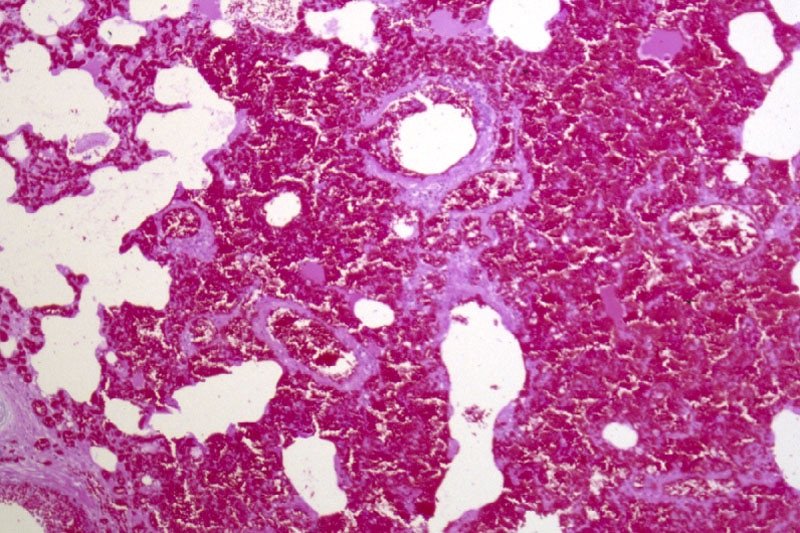
The glandular region of the horse’s stomach contains glands that secrete hydrochloric acid, pepsin, bicarbonate and mucus. The stomach secretes acid continuously; as the horse is a trickle feeder it has evolved to do so continuously and it is important to note that this process continues even when the horse isn’t eating. This is why periods of more than 6 hours without access to forage are a risk factor for ulcers. The volume of secretion has been shown to be around 1.5l of gastric juice per hour although this does vary at different times during the day. Consuming too little fiber and eating materials that are high in starch, means acidity levels increase in the stomach. This not only increases the risk of ulcers but also changes the environment in the stomach sufficiently to impact the microbes that live there. Microbial dysbiosis in the stomach is increasingly being linked to an increased risk of gastric disease, particularly in the glandular region which is now recognized as an inflammatory disease rather than an ulcerative one.   

There are other potential health issues to consider too. It has been shown in trials in mice for example, that a low fiber diet increases the permeability of the gut – a phenomenon known as leaky-gut syndrome. When fibre is fermented in the hind gut, one of the volatile fatty acids produced is butyrate and this is the energy used by the colonocytes (gut cells) themselves. Insufficient fiber and therefore butyrate, can compromise the health of the cells creating bigger gaps between them which allows contents of the gut that shouldn’t pass through, to do so. The racehorse is repeatedly exposed to new and different environments when travelling to different racecourses and encounters pathogens they might have no previous immunity to. Their reduced defensive barriers in the gut mean they are more vulnerable to these pathogens which can result in digestive upsets.  

So can more fiber be fed without compromising performance? 

Researchers at the Lab to Field research center in Dijon, France believe so.  In work funded by the French government and published in Frontiers in Physiology, they found that Standardbred horses in training fed a third of their total ration as alfalfa with just 7% oats, performed comparably with those fed 33% oats (the remainder of the diet was hay). The horses were monitored over an 8 week period rather than just in a one-off standardized exercise test (SET). The replacement of a significant proportion of oats with alfalfa had no detrimental effects on performance or muscle tone and in fact, altered energy metabolism in such a way as to potentially improve performance and recovery the authors suggest (Martin et al., 2023). Studies back in the early 2000s (Nadeua et al, 2000; Lybbert et al, 2007) showed that alfalfa was more beneficial for horses with ulcers compared to grass forages because it helps counter the increased acidity that occurs when feeding cereals. This latest study suggests that alfalfa can actually replace a significant proportion of the cereals as an energy source too. 

The prevalence of gastric ulcers means it is an issue that needs to be addressed especially when viewed in the context of equine welfare in sport. Two recent studies have again shown how alfalfa has a key role to play in this regard too. The Lab to Field research group demonstrated that clinical success with horses with EGGD was 47.7 times more likely in horses fed alfalfa pellets as part of their ration compared to those on concentrate only rations (Julliand et al., 2023). 

In addition, a study published in 2024 showed that a combination of alfalfa, sugar beet and cereal fiber fed alongside the existing ration, aided the reduction in recurrence of gastric ulcers when fed during the healing and post-medication periods. This is key for when ulcer medication is stopped and the recognized rebound increase in acid production can occur (Menzies-Gow and Shurlock, 2024). 

A key point from these studies is that the quality of fiber matters. Alfalfa and sugar beet both contain higher proportions of digestible fiber such as pectin and hemi-cellulose, rather than indigestible fibrous elements such as lignin. This means they aren’t sitting in the gut for so long but they are being digested and utilized as an energy source. If fed in chopped forms they help to increase the amount of chewing the horse does and more chew time might actually be a relatively simple step in the right direction from a welfare perspective. The pros and cons of turning out racehorses have been widely debated but for those where it isn’t (currently) practical, it is surely a positive action to at least provide the horse with high fiber materials to eat when stabled, especially when it isn’t having a negative impact on their performance.

Dispelling Myths - Facts about Fibre

How much sugar does sugar beet contain?

The pulp fed to horses is actually really low in sugar – less than 5% assuming no molasses has been added back in. This is because the sugar has been extracted for use in the human food industry and the fibrous pulp is used for animal feed. 

Why does alfalfa contain more calcium than grass forages? 

Alfalfa has really deep roots – about 3 to 4 meters – and the calcium at this depth in the soil is more available for absorption. This means that alfalfa plants can take up more calcium than grass – chopped alfalfa contains between 30 - 50% more calcium than grass forages. Early studies suggest that omeprazole is reducing calcium absorption in the horse as is seen in humans and in Swanhall et al’s (2018) study, they recommend using bio-available calcium sources in the diet to help counteract this effect. Plant based sources of calcium such as alfalfa are much easier for the horse to absorb than inorganic sources such as limestone flour. 

Why is alfalfa so low in starch?

Like other plants alfalfa makes sugar when photosynthesizing but it stores any surplus sugar as starch in its roots – the part that horses don’t eat! Grass plants tend to store sugar as fructan in leaves and the stem which is why they supply the horse with more sugar. 

What contribution can forage make to a racehorse’s requirements? 

Remember that grass-based forages contain sugar, both simple sugar (glucose, fructose etc) and as water soluble carbohydrates or storage sugars (fructan). 10kgs of hay can provide around 1kg of simple sugar and in the region of 2-2.5kgs of storage sugar. This supplies around 20% of the energy required by a 500kgs horse in intense training. Additionally, forage provides energy from the fiber it contains and so overall, including the contribution from sugar, 10kgs of hay would supply around 60-70% of the horse’s energy needs depending on the quality of the forage. 

Earlier cut forages tend to be more digestible and therefore supply more energy. These tend to be the wrapped forages in the UK and other wetter and colder European countries as there just aren’t long enough periods of dry weather to make good hay very often. Why is this significant? The way forages are conserved has changed over the years so now, a more accurate description of many forages previously defined as haylages, would be ‘wrapped hay’ as they are often very dry which has meant that little or no fermentation has occurred. This means the levels of acidity are no different to a normal hay which can be seen from the analysis results in table 1. Using lactic acid levels as a marker of acidity levels shows that most of the wrapped forages analyzed in the UK are too dry for fermentation to occur and so the level of acidity is no different to hay. 

Table 1 A comparison of different forages 

Concern about using a true haylage for horses with ulcers relates to the increased acidity from the fermentation that occurs. Clearly this doesn’t apply if the forage hasn’t fermented and so a wrapped hay may well be a really useful option for a horse with ulcers. They tend to be more palatable and softer than hay. It is important to know the level of acidity before making the decision to use a wrapped forage and having it analyzed is therefore advisable. 

So if the paradigm shift happens, what will a racehorse’s diet look like in years to come? 

The basis would be a good, early cut wrapped hay. The daily bucket feed would consist of 1-2kgs of oats with 1.5kgs of alfalfa pellets, 1.5kgs of chopped alfalfa and 0.5kgs of soaked sugar beet. The chopped alfalfa  contributes to the horse’s overall forage requirement so if the dry matter of the wrapped hay is around 75%, a 500kgs horse would need a minimum of 8kgs per day to supply 6kgs of additional fiber on a dry matter basis. 

Key takeaways 

  • ESGD risk factors are well established and include too little fiber and too much starch 

  • Feeding at least 1.5% of bodyweight on a dry matter basis is the minimum amount of forage required for long term gastric and digestive health

  • Wrapped hays that have not fermented and so are no more acidic than hay are also appropriate to use for horses with ulcers

  • EGGD is still not fully understood but increasingly it is acknowledged by researchers that stress is a key contributing factor

  • Studies have shown alfalfa to be beneficial as an alternative energy source compared to cereals for horses in training


References 

Julliand et al (2023) Effect of diet composition on glandular gastric disease in horses. Journal of Veterinary Internal Medicine

Lybbert et al (2007), Proceedings of Annual Convention of the AAEP, Orlando, Florida, 2007. 

Martin et al (2023) Effect of high-starch or high-fibre diets on the energy metabolism and physical performance of horses during an 8-week training period. Front. Physiol. 14:1213032. doi: 10.3389/fphys.2023.1213032

Menzies-Gow and Shurlock (2024) The effect of feeding a commercial feedstuff on equine gastric squamous disease. Journal of Equine Veterinary Science. 133. 

Muller and Uden (2007) Preference of horses for grass conserved as hay, haylage or silage. Animal Feed Science and Technology, 132, (1-2) 66-78

Nadeau et al (2000) Evaluation of diet as a cause of gastric ulcers in horses. American Journal of Veterinary Research. Jul;61(7):784-90.

Pratt et al, (2022) Assessment of agreement using the equine glandular gastric disease grading system in 84 cases. Veterinary Medicine Science, 8 (4) 1472-1477doi: 10.1002/vms3.807

Swanhall et al (2018) Mineral and Vitamin Supplementation Including Marine Derived Calcium Increases Bone Density in Thoroughbreds. Proceedings of the Australasian Equine Science Symposium

EIPH - could there be links to sudden death and pulmonary haemorrhage?

Dr Peter W. Physick-Sheard, BVSc, FRCVS, explores preliminary research and hypotheses, being conducted by the University of Guelph, to see if there is a possibility that these conditions are linked and what this could mean for future management and training of thoroughbreds. 

Thoroughbred EIPH/bleeder.jpg

"World's Your Oyster,” a three-year-old thoroughbred mare, presented at the veterinary hospital for clinical examination. She won her maiden start as a two-year-old and placed once in two subsequent starts. After training well as a three-year-old, she failed to finish her first start, easing at the top of the stretch, and was observed to fade abruptly during training. Some irregularity was suspected in heart rhythm after exercise. Thorough clinical examination, blood work, ultrasound of the heart and an ECG during rest and workout revealed nothing unusual. 

Returning to training, Oyster placed in six of her subsequent eight starts, winning the last two. She subsequently died suddenly during early training as a four-year-old. At post-mortem, diagnoses of pulmonary haemorrhage and exercise-induced pulmonary haemorrhage were established—a very frustrating and unfortunate outcome.

Across the racing world, a case like this probably occurs daily. Anything that can limit a horse's ability to express its genetic potential is a major source of anxiety when training. The possibility of injury and lameness is the greatest concern, but a close second is respiratory disease, with bleeding  from the lungs (most often referred to as exercise induced pulmonary [lung] haemorrhage or EIPH) being high on the list. 

EIPH is thought to occur in as many as 85 percent of racehorses, and may initially be very mild without obvious clinical consequences. In some cases it can be associated with haemorrhage of sufficient severity for blood to appear at the nostrils, even at first occurrence. In many racing jurisdictions this is a potentially career-ending problem. In these horses, an impact on performance is unquestionable. Bleeding from the lungs is the reason for the existence of ‘Lasix programs,’ involving pre-race administration of a medication considered to reduce haemorrhage. Such programs are controversial—the justifications for their existence ranging from addressing welfare concerns for the horse to dealing with the performance impacts. 

Much less frequently encountered is heavy exercise-associated bleeding from the nostrils (referred to as epistaxis), which can sometimes be accompanied by sudden death, during or shortly after exercise. Some horses bleed heavily internally and die without blood appearing at the nostrils. Haemorrhage may only become obvious when the horse is lying on its side, or not until post-mortem. Affected animals do not necessarily have any history of EIPH, either clinically or sub-clinically. There is an additional group of rare cases in which a horse simply dies suddenly, most often very soon after work and even after a winning performance, and in which little to nothing clearly explains the cause on post-mortem. This is despite the fact most racing jurisdictions study sudden death cases very closely.

EIPH is diagnosed most often by bronchoscopy—passing an endoscope into the lung after work and taking a look. In suspected but mild cases, there may not be sufficient haemorrhage to be visible, and a procedure called a bronchoalveolar lavage is performed. The airways are rinsed and fluid is collected and examined microscopically to identify signs of bleeding. Scoping to confirm diagnosis is usually a minimum requirement before a horse can be placed on a Lasix program. 


Are EIPH, severe pulmonary haemorrhage and sudden death related? Are they the same or different conditions? 

At the University of Guelph, we are working on the hypothesis that most often they are not different—that it’s degrees of the same condition, or closely related conditions perhaps with a common underlying cause. We see varying clinical signs as being essentially a reflection of severity and speed of onset of underlying problems. 

Thoroughbred lung size.jpg

Causes in individual cases may reflect multiple factors, so coming at the issues from several different directions, as is the case with the range of ongoing studies, is a good way to go so long as study subjects and cases are comparable and thoroughly documented. However, starting from the hypothesis that these may all represent basically the same clinical condition, we are approaching the problem from a clinical perspective, which is that cardiac dysfunction is the common cause. 

Numerous cardiac disorders and cellular mechanisms have the potential to contribute to transient or complete pump (heart) failure. However, identifying them as potential disease candidates does not specifically identify the role they may have played, if any, in a case of heart failure and in lung haemorrhage; it only means that they are potential primary underlying triggers. It isn't possible for us to be right there when a haemorrhage event occurs, so almost invariably we are left looking at the outcome—the event of interest has passed. These concerns influence the approach we are taking.


Background

The superlative performance ability of a horse depends on many physical factors:

  • Huge ventilatory (ability to move air) and gas exchange capacity

  • Body structure including limb length and design - allows it to cover ground rapidly with a long stride

  • Metabolic adaptations - supports a high rate of energy production by burning oxygen, tolerance of severe metabolic disruptions toward the end of race-intensity effort

  • High cardiovascular capacity - allows the average horse to pump roughly a brimming bathtub of blood every minute

At race intensity effort, these mechanisms, and more, have to work in coordination to support performance. There is likely not much reserve left—two furlongs (400m) from the winning post—even in the best of horses. There are many wild cards, from how the horse is feeling on race day to how the race plays out; and in all horses there will be a ceiling to performance. That ceiling—the factor limiting performance—may differ from horse to horse and even from day to day. There’s no guarantee that in any particular competition circumstances will allow the horse to perform within its own limitations. One of these factors involves the left side of the heart, from which blood is driven around the body to the muscles.


A weak link - filling the left ventricle

The cardiovascular system of the horse exhibits features that help sustain a high cardiac output at peak effort. The feature of concern here is the high exercise pressure in the circulation from the right  ventricle, through the lungs to the left ventricle. At intense effort and high heart rates, there is very little time available to fill the left ventricle—sometimes as little as 1/10 of a second; and if the chamber cannot fill properly, it cannot empty properly and cardiac output will fall. The circumstances required to achieve adequate filling include the readiness of the chamber to relax to accept blood—its ‘stiffness.’ Chamber stiffness increases greatly at exercise, and this stiffened chamber must relax rapidly in order to fill. That relaxation seems not to be sufficient on its own in the horse at high heart rates. Increased filling pressure from the circulation draining the lungs is also required. But there is a weak point: the pulmonary capillaries.

Thoroughbred inflated lungs.jpg

These are tiny vessels conducting blood across the lungs from the pulmonary artery to the pulmonary veins. During this transit, all the gas exchange needed to support exercise takes place. The physiology of other species tells us that the trained lung circulation achieves maximum flow (equivalent to cardiac output) by reducing resistance in those small vessels. This process effectively increases lung blood flow reserve by, among other things, dilating small vessels. Effectively, resistance to the flow of blood through the lungs is minimised. We know this occurs in horses as it does in other species; yet in the horse, blood pressure in the lungs still increases dramatically at exercise. 

If this increase is not the result of resistance in the small vessels, it must reflect something else, and that appears to be resistance to flow into the left chamber. This means the entire lung circulation is exposed to the same pressures, including the thin-walled capillaries. Capillaries normally work at quite low pressure, but in the exercising horse, they must tolerate very high pressures. They have thin walls and little between them, and the air exchange sacs in the lung. This makes them vulnerable. It's not surprising they sometimes rupture, resulting in lung haemorrhage.

Thoroughbred lungs effected by EIPH.jpg

Recent studies identified changes in the structure of small veins through which the blood flows from the capillaries and on toward the left chamber. This was suspected to be a pathology and part of the long-term consequences of EIPH, or perhaps even part of the cause as the changes were first identified in EIPH cases. It could be, however, that remodelling is a normal response to the very high blood flow through the lungs—a way of increasing lung flow reserve, which is an important determinant of maximum rate of aerobic working. 

The more lung flow reserve, the more cardiac output and the more aerobic work an animal can perform. The same vein changes have been observed in non-racing horses and horses without any history or signs of bleeding. They may even be an indication that everything is proceeding as required and a predictable consequence of intense aerobic training. On the other hand, they may be an indication in some horses that the rate of exercise blood flow through their lungs is a little more than they can tolerate, necessitating some restructuring. We have lots to learn on this point.

If the capacity to accommodate blood flow through the lungs is critical, and limiting, then anything that further compromises this process is likely to be of major importance. It starts to sound very much as though the horse has a design problem, but we shouldn't rush to judgement. Horses were probably not designed for the very intense and sustained effort we ask of them in a race. Real-world situations that would have driven their evolution would have required a sprint performance (to avoid ambush predators such as lions) or a prolonged slower-paced performance to evade predators such as wolves, with only the unlucky victim being pushed to the limit and not the entire herd. 

Lung blood flow and pulmonary oedema

There is another important element to this story. High pressures in the capillaries in the lung will be associated with significant movement of fluid from the capillaries into lung tissue spaces. This movement in fact happens continuously at all levels of effort and throughout the body—it's a normal process. It's the reason the skin on your ankles ‘sticks’ to the underlying structures when you are standing for a long time. So long as you keep moving a little, the lymphatic system will draw away the fluid. 

Lung blood flow and pulmonary odema.jpg

In a diseased lung, tissue fluid accumulation is referred to as pulmonary oedema, and its presence or absence has often been used to help characterise lung pathologies. The lung lymphatic system can be overwhelmed when tissue fluid is produced very rapidly. When a horse experiences sudden heart failure, such as when the supporting structures of a critical valve fail, one result is massive overproduction of lung tissue fluid and appearance of copious amounts of bloody fluid from the nostrils. 

The increase in capillary pressure under these conditions is as great as at exercise, but the horse is at rest. So why is there no bloody fluid in the average, normal horse after a race? It’s because this system operates very efficiently at the high respiratory rates found during work: tissue fluid is pumped back into the circulation, and fluid does not accumulate. The fluid is pumped out as quickly as it is formed. An animal’s level of physical activity at the time problems develop can therefore make a profound difference to the clinical signs seen and to the pathology.

Usual events with unusual consequences 

If filling the left ventricle and the ability of the lungs to accommodate high flow at exercise are limiting factors, surely this affects all horses. So why do we see such a wide range of clinical pictures, from normal to subclinical haemorrhage to sudden death? 

Variation in contributing factors such as type of horse, type and intensity of work, sudden and unanticipated changes in work intensity, level of training in relation to work and the presence of disease states are all variables that could influence when and how clinical signs are seen, but there are other considerations.

Although we talk about heart rate as a fairly stable event, there is in fact quite a lot of variation from beat to beat. This is often referred to as heart rate variability. There has been a lot of work performed on the magnitude of this variability at rest and in response to various short-term disturbances and at light exercise in the horse, but not a lot at maximal exercise. Sustained heart rate can be very high in a strenuously working horse, with beats seeming to follow each other in a very consistent manner, but there is in fact still variation. 

Some of this variation is normal and reflects the influence of factors such as respiration. However, other variations in rate can reflect changes in heart rhythm. Still other variations may not seem to change rhythm at all but may instead reflect the way electrical signals are being conducted through the heart. 

These may be evident from the ECG but would not appear abnormal on a heart rate monitor or when listening. These variations, whether physiologic (normal) or a reflection of abnormal function, will have a presently, poorly understood influence on blood flow through the lungs and heart—and on cardiac filling. Influences may be minimal at low rates, but what happens at a heart rate over 200 and in an animal working at the limits of its capacity?

Normal electrical activation of the heart follows a pattern that results in an orderly sequence of heart muscle contraction, and that provides optimal emptying of the ventricles. Chamber relaxation complements this process. 

An abnormal beat or abnormal interval can compromise filling and/or emptying of the left ventricle, leaving more blood to be discharged in the next cycle and back up through the lungs, raising pulmonary venous pressure. A sequence of abnormal beats can lead to a progressive backup of blood, and there may not be the capacity to hold it—even for one quarter of a second, a whole cardiac cycle at 240 beats per minute. 

For a horse that has a history of bleeding and happens to be already functioning at a very marginal level, even minor disturbances in heart rhythm might therefore have an impact. Horses with airway disease or upper airway obstructions, such as roarers, might find themselves in a similar position. An animal that has not bled previously might bleed a little, one that has a history of bleeding may start again, or a chronic bleeder may worsen. 

Relatively minor disturbances in cardiac function, therefore, might contribute to or even cause EIPH. If a horse is in relatively tough company or runs a hard race, this may also contribute to the onset or worsening of problems. Simply put, it's never a level playing field if you are running on the edge.


Severe bleeding

It has been suspected for many years that cases of horses dying suddenly at exercise represent sudden-onset cardiac dysfunction—most likely a rhythm disturbance. If the rhythm is disturbed, the closely linked and carefully orchestrated sequence of events that leads to filling of the left ventricle is also disturbed. A disturbance in cardiac electrical conduction would have a similar effect, such as one causing the two sides of the heart to fall out of step, even though the rhythm of the heart may seem normal. 

The cases of horses that bleed profusely at exercise and even those that die suddenly without any post-mortem findings can be seen to follow naturally from this chain of events. If the changes in heart rhythm or conduction are sufficient, in some cases to cause massive pulmonary haemorrhage, they may be sufficient in other cases to cause collapse and death even before the horse has time to exhibit epistaxis or even clear evidence of bleeding into the lungs. 

ECG.jpg

EIPH and dying suddenly

If these events are (sometimes) related, why is it that some horses that die of pulmonary haemorrhage with epistaxis do not show evidence of chronic EIPH? This is one of those $40,000 questions. It could be that young horses have had limited opportunity to develop chronic EIPH; it may be that we are wrong and the conditions are entirely unrelated. But it seems more likely that in these cases, the rhythm or conduction disturbance was sufficiently severe and/or rapid in onset to cause a precipitous fall in blood pressure with the animal passing out and dying rapidly. 

In this interpretation of events, the missing link is the heart. There is no finite cutoff at which a case ceases to be EIPH and becomes pulmonary haemorrhage. Similarly, there is no distinct point at which any case ceases to be severe EIPH and becomes EAFPH (exercise-associated fatal pulmonary haemorrhage). In truth, there may simply be gradation obscured somewhat by variable definitions and examination protocols and interpretations.


The timing of death

It seems from the above that death should most likely take place during work, and it often does, but not always. It may occur at rest, after exercise. Death ought to occur more often in racing, but it doesn't. 

The intensity of effort is only one factor in this hypothesis of acute cardiac or pump failure. We also have to consider factors such as when rhythm disturbances are most likely to occur (during recovery is a favourite time) and death during training is more often a problem than during a race. 

A somewhat hidden ingredient in this equation is possibly the animal's level of emotional arousal, which is known to be a risk factor in humans for similar disturbances. There is evidence that emotions/psychological factors might be much more important in horses than previously considered. Going out for a workout might be more stimulating for a racehorse than a race because before a race, there is much more buildup and the horse has more time to adequately warm up psychologically. And then, of course, temperament also needs to be considered. These are yet further reasons that we have a great deal to learn.

Scoping Thoroughbred.jpg

Our strategy at the University of Guelph

These problems are something we cannot afford to tolerate, for numerous reasons—from perspectives of welfare and public perception to rider safety and economics. Our aim is to increase our understanding of cardiac contributions by identifying sensitive markers that will enable us to say with confidence whether cardiac dysfunction—basically transient or complete heart failure—has played a role in acute events. 

We are also looking for evidence of compromised cardiac function in all horses, from those that appear normal and perform well, through those that experience haemorrhage, to those that die suddenly without apparent cause. Our hope is that we can not only identify horses at risk, but also focus further work on the role of the heart as well as the significance of specific mechanisms. And we hope to better understand possible cardiac contributions to EIPH in the process. This will involve digging deeply into some aspects of cellular function in the heart muscle, the myocardium of the horse, as well as studying ECG features that may provide insight and direction. 

Fundraising is underway to generate seed money for matching fund proposals, and grant applications are in preparation for specific, targeted investigations. Our studies complement those being carried out in numerous, different centres around the world and hopefully will fill in further pieces of the puzzle. This is, indeed, a huge jigsaw, but we are proceeding on the basis that you can eat an elephant if you're prepared to process one bite at a time.

How can you help? Funding is an eternal issue. For all the money that is invested in horses there is a surprisingly limited contribution made to research and development—something that is a mainstay of virtually every other industry; and this is an industry. 

Look carefully at the opportunities for you to make a contribution to research in your area. Consider supporting studies by making your experience, expertise and horses available for data collection and minimally invasive procedures such as blood sampling. 

Connect with the researchers in your area and find out how you can help. Watch your horses closely and contemplate what they might be telling you—it's easy to start believing in ourselves and to stop asking questions. Keep meticulous records of events involving horses in your care— you never know when you may come across something highly significant. And work with researchers (which often includes track practitioners) to make your data available for study. 

Remember that veterinarians and university faculty are bound by rules of confidentiality, which means what you tell them should never be ascribed to you or your horses and will only be used without any attribution, anonymously. And when researchers reach out to you to tell you what they have found and to get your reactions, consider actually attending the sessions and participating in the discussion; we can all benefit—especially the ultimate beneficiary which should be the horse. We all have lots to learn from each other, and finding answers to our many challenges is going to have to be a joint venture.  

Finally, this article has been written for anybody involved in racing to understand, but covering material such as this for a broad audience is challenging. So, if there are still pieces that you find obscure, reach out for help in interpretation. The answers may be closer than you think!


Oyster

And what about Oyster? Her career was short. Perhaps, had we known precisely what was going on, we might have been able to treat her, or at least withdraw her from racing and avoid a death during work with all the associated dangers—especially to the rider and the associated welfare concerns. 

Had we had the tools, we might have been able to confirm that whatever the underlying cause, she had cardiac problems and was perhaps predisposed to an early death during work. With all the other studies going on, and knowing the issue was cardiac, we might have been able to target her assessment to identify specific issues known to predispose. 

In the future, greater insight and understanding might allow us to breed away from these issues and to better understand how we might accommodate individual variation among horses in our approaches to selection, preparation and competition. There might be a lot of Oysters out there!

For further information about the work being undertaken by the University of Guelph

Contact - Peter W. Physick-Sheard, BVSc, FRCVS.

Professor Emeritus, Ontario Veterinary College, University of Guelph - pphysick@uoguelph.ca

Research collaborators - Dr Glen Pyle, Professor, Department of Biomedical Sciences, University of Guelph - gpyle@uoguelph.ca

Dr Amanda Avison, PhD Candidate, Department of Biomedical Sciences, University of Guelph. ajowett@uoguelph.ca

References

Caswell, J.I. and Williams K.J. (2015), Respiratory System, In ed. Maxie, M. Grant, 3 vols., 6th edn., Jubb, Kennedy and Palmer’s Pathology of Domestic Animals, 2; London: Elsevier Health Sciences, 490-91.

Hinchcliff, KW, et al. (2015), Exercise induced pulmonary hemorrhage in horses: American College of Veterinary Internal Medicine consensus statement, J Vet Intern Med, 29 (3), 743-58.

Rocchigiani, G, et al. (2022), Pulmonary bleeding in racehorses: A gross, histologic, and ultrastructural comparison of exercise-induced pulmonary hemorrhage and exercise-associated fatal pulmonary hemorrhage, Vet Pathol, 16:3009858221117859. doi: 10.1177/03009858221117859. Online ahead of print.

Manohar, M. and T. E. Goetz (1999), Pulmonary vascular resistance of horses decreases with moderate exercise and remains unchanged as workload is increased to maximal exercise, Equine Vet. J., (Suppl.30), 117-21.

Vitalie, Faoro (2019), Pulmonary Vascular Reserve and Aerobic Exercise Capacity, in Interventional Pulmonology and Pulmonary Hypertension, Kevin, Forton (ed.), (Rijeka: IntechOpen), Ch. 5, 59-69.

Manohar, M. and T. E. Goetz (1999), Pulmonary vascular resistance of horses decreases with moderate exercise and remains unchanged as workload is increased to maximal exercise, Equine Vet. J., (Suppl.30), 117-21.