Planning a diagnostic-led worm control programme

Planning a diagnostic-led worm control programmeBy Dr Corrine Austin (Austin Davis Biologics) and Prof Jacqui Matthews (Roslin Technologies)All horses are exposed to worms while grazing, but how we control these parasites is essential to horse healt…

By Dr Corrine Austin (Austin Davis Biologics) and Prof Jacqui Matthews (Roslin Technologies)

Planning a diagnostic-led worm control programme

All horses are exposed to worms while grazing, but how we control these parasites is essential to horse health and performance. Most horse owners are aware of testing to determine whether their horse needs deworming. The tests comprise faecal worm egg counts (FEC) for redworm/roundworm detection and saliva testing to detect tapeworm infections (standard FEC methods are unreliable for tapeworm). Until now, encysted small redworm larvae have remained undetectable as FEC only determines the presence of egg laying adult worms. This has meant that routine winter moxidectin treatment has become recommended practice to target potentially life-threatening burdens of small redworm encysted larvae. Excitingly, a new small redworm blood test is being commercialised* which detects all stages of the small redworm life cycle, including the all-important encysted larval phase. Together, these tests offer a complete worm control programme for common horse worms using diagnostic information. This is known as ‘diagnostic-led worm control’ (see Figure 1 for common worms in horses). Essentially, testing is used to tell you whether your horse needs deworming or not.

Common horse worms

Common horse worms

Why should you use testing to determine whether you should use dewormers or not?

Gone are the days of routinely administering dewormers to every horse and hoping for the best. That strategy is outdated as it has caused widespread drug resistance in worms (i.e., worms are able to survive the killing effects of dewormers and remain in place after treatment, which can lead to disease and in worst cases, death). To reduce the risk of further resistance occurring, we need to ensure that dewormers are only used when they are genuinely needed—when testing detects that horses have a worm burden requiring treatment. Regular testing also helps identify horses likely to be more susceptible to infection and thus at risk of disease in the future.

How to plan your horse’s worm control programme ….

TO READ MORE —

BUY THIS ISSUE IN PRINT OR DOWNLOAD

October - December 2019, issue 67 (PRINT)
£8.95
Quantity:
Add to Cart

WHY NOT SUBSCRIBE?

DON'T MISS OUT AND SUBSCRIBE TO RECEIVE THE NEXT FOUR ISSUES!

Bleeders - the facts, fiction and future direction

By Dr. David Marlin

Bleeders - the facts, fiction and future directionWe are now approaching half a century since Bob Cook pioneered the use of the flexible fibreoptic endoscope, which allowed examination of the respiratory tract in the conscious horse. One of the impo…

We are now approaching half a century since Bob Cook pioneered the use of the flexible fibreoptic endoscope, which allowed examination of the respiratory tract in the conscious horse. One of the important outcomes of this technique was that it opened the door to the study of ‘bleeding’ or exercise-induced pulmonary haemorrhage (EIPH). But nearly 50 years on the irony is perhaps that whilst we have become good at describing the prevalence of EIPH and some of the factors that appear to increase the severity of EIPH within individual horses, we still lack a clear understanding of the condition and how to manage it. I use the term manage rather than treat or prevent as our knowledge of EIPH must show us that EIPH cannot be stopped entirely; it is a consequence of intense exercise. The other irony is that in the past 50 years, by far the majority of research into the management of EIPH has focussed on the use of the diuretic furosemide. Whilst we have good evidence from controlled studies that furosemide reduces the severity of EIPH on a single occasion, we still lack good evidence to suggest that furosemide is effective when used repeatedly during training and or racing; and there is also evidence to the contrary.

Let’s review some basic facts about EIPH, which should not be contentious.

  • EIPH is the appearance of blood in the airways associated with exercise.

  • EIPH occurs as a result of moderate to intense exercise. In fact, EIPH has been found after trotting when deep lung wash (bronchoalveolar lavage or BAL) is done after exercise. 

  • EIPH most often involves the smallest blood vessels (capillaries) but can sometimes and less commonly be due to the rupture of larger blood vessels.

  • The smallest blood vessels are extremely thin. Around 1/100th the thickness of a human hair. But this extremely thin membrane is also what allows racehorses such as thoroughbreds, standardbreds and Arabs to use oxygen at such a high rate and is a major reason for their athleticism. 

  • EIPH is a progressive condition. The chance of seeing blood in the trachea after exercise increases with time in racing.

  • EIPH is variable over time, even when horses are scoped after the same type of work.

  • If you ‘scope a horse after three gallops in a row, you can expect to see blood in the trachea on at least one occasion.

  • EIPH damage to the lungs starts at the back and top, and over time moves forward and down and is approximately symmetrical.

  • Following EIPH the lung becomes fibrotic (as scar tissue), stiffer and does not work as well. The iron from the blood is combined with protein and stored permanently in the lung tissue where it can cause inflammation.

  • High blood pressure within the lung is a contributing factor in EIPH. Horses with higher blood pressure appear to suffer worse EIPH.

  • There is also evidence that upper airway resistance and breathing pattern can play a role in EIPH.

  • Airway inflammation and poor air quality may increase the severity of EIPH within individual horses.

  • Increasing severity of EIPH appears to have an increasing negative effect on performance.

  • Visible bleeding (epistaxis) has a very clear and marked negative effect on performance.

In order to make progress in the management of EIPH (i.e., to minimise the severity of EIPH in each individual), there are certain steps that trainers can take based on the information we have to date. 

These include:

Screenshot 2019-09-19 at 17.04.30.png
  • Ensuring good air quality in stables

  • Regular respiratory examination and treatment of airway inflammation

  • Reduced intensity of training during periods of treatment for moderate to severe airway inflammation 

  • Extended periods of rest and light work with a slower return to work for horses following viral infection

  • Addressing anything that increases upper airway resistance (e.g., roaring, gurgling)

  • Avoiding intense work in cold weather

  • Avoiding extremes of going

  • Limiting number of training days in race preparation and increasing interval between races

Endoscopy

Endoscopy

FUTURE OPPORTUNITIES IN UNDERSTANDING AND MANAGING EIPH

We have to accept EIPH as a normal consequence of intense exercise in horses. Our aim should be to reduce the severity to a minimum in each individual horse. However, there are areas in which we still need a much greater scientific understanding.   

What actually causes the capillaries to leak or rupture?…

TO READ MORE —

BUY THIS ISSUE IN PRINT OR DOWNLOAD

October - December 2019, issue 67 (PRINT)
£8.95
Quantity:
Add to Cart

WHY NOT SUBSCRIBE?

DON'T MISS OUT AND SUBSCRIBE TO RECEIVE THE NEXT FOUR ISSUES!

IF YOU LIKE THIS ARTICLE

WHY NOT SUBSCRIBE - OR ORDER THE CONTENT FROM THIS ISSUE IN PRINT?

An Introduction to the Functional Aspects of Conformation

An Introduction to the Functional Aspects of ConformationJudy Wardrope Why is one horse a sprinter and another a stayer? Why is one sibling a star and another a disappointment? Why does one horse stay sound and another does not? Over the course of t…

By Judy Wardrope

Why is one horse a sprinter and another a stayer? Why is one sibling a star and another a disappointment? Why does one horse stay sound and another does not? Over the course of the next few issues, we will delve into the mechanics of the racehorse to discern the answer to these questions and others. We will be learning by example, and we will be using objective terminology as well as repeatable measures. This knowledge can be applied to the selection of racing prospects, to the consideration of distance or surface preferences and, of course, to mating choices.

Introducing a different way of looking at things requires some forethought. Questions need to be addressed in order to provide educational value for the audience. How does one organise the information, and how does one back up the information? In the case of equine functionality in racing, which horses will provide the best corroborative visuals?

After considerable thought, these three horses were selected: Tiznow (Horse #1) twice won the Breeders’ Cup Classic (1¼ miles) ; Lady Eli (Horse #2) won the Juvenile Fillies Turf and was twice second in the Filly and Mare Turf (13/8 miles); while our third example (Horse #3) did not earn enough to pay his way on the track. Let’s see if we can explain the commonalities and the differences so that we can apply that knowledge in the future.

Factors for Athleticism

If we consider the horse’s hindquarters to be the motor, then we should consider the connection between hindquarters and body to be the horse’s transmission. Like in a vehicle, if the motor is strong, but the transmission is weak, the horse will either have to protect the transmission or damage it.

According to Dr. Hilary M. Clayton (BVMS, PhD, MRCVS), the hind limb rotates around the hip joint in the walk and trot and around the lumbosacral joint in the canter and gallop. “The lumbosacral joint is the only part of the vertebral column between the base of the neck and the tail that allows a significant amount of flexion [rounding] and extension [hollowing] of the back. At all the other vertebral joints, the amount of motion is much smaller. Moving the point of rotation from the hip joint to the lumbosacral joint increases the effective length of the hind limbs and, therefore, increases stride length.” From a functional perspective, that explains why a canter or gallop is loftier in the forehand than the walk or the trot.

In order to establish an objective measure, I use the lumbosacral (LS) gap, which is located just in front of the high point of the croup. This is where the articulation of the spine changes just in front of the sacrum, and it is where the majority of the up and down motion along the spine occurs. The closer a line drawn from the top point of one hip to the top point of the other hip comes to bisecting this palpable gap, the stronger the horse’s transmission. In other words, the stronger the horse’s coupling.

We can see that the first two horses have an LS gap (just in front of the high point of the croup as indicated) that is essentially in line with a line drawn from the top of one hip to the top of the opposing hip. This gives them the ability to transfer their power both upward (lifting of the forehand) and forward (allowing for full extension of the forehand and the hindquarters). Horse #3 shows an LS gap considerably rearward of the top of his hip, making him less able to transfer his power and setting him up for a sore back.

You may also notice that all three of these sample horses display an ilium side (point of hip to point of buttock), which is the same length as the femur side (point of buttock to stifle protrusion)—meaning that they produce similar types of power from the rear spring as it coils and releases when in stride. We can examine the variances in these measures in more detail in future articles, when we start to delve into various ranges of motion as well as other factors for soundness or injury.

Factors for Distance Preferences…

TO READ MORE —

BUY THIS ISSUE IN PRINT OR DOWNLOAD

July - September 2019, issue 66 (PRINT)
£8.95
Quantity:
Add to Cart

WHY NOT SUBSCRIBE?

DON'T MISS OUT AND SUBSCRIBE TO RECEIVE THE NEXT FOUR ISSUES!

Equine Pain: how can we recognise it and which painkiller should we use?

Screen Shot 2019-06-19 at 17.11.25.png

By Professor Celia Marr

We can all agree that alleviating pain in our patients is an important goal, but we may not be as good as we might hope at recognising pain in horses. Studies have shown that there is considerable variation in the scores vets assign when asked to predict how much pain they expect to see with specific clinical conditions.


Acute severe pain is perhaps most easily recognised by horsemen and vets; signs of severe colic, such as rolling, are usually very obvious. Low-grade pain, and pain not associated with abdominal disease can be more difficult to detect and go unrecognised. In particular, intra-thoracic pain and pain associated with injuries to the thoracic cage, withers and spine can be difficult to pinpoint.

This horse is clearly showing signs of abdominal pain—colic. It is lying down, has been rolling and is looking at its flank.

This horse is clearly showing signs of abdominal pain—colic. It is lying down, has been rolling and is looking at its flank.

Comfortable horses interact with their environment, look out over their stable door and eat willingly. Reluctance to move and restlessness indicate pain while looking at the flank, and kicking at the abdomen all suggest localised pain. Behaviours such as lifting hindlimbs, extending head, lateral and/or vertical head movements and pawing are also observed in uncomfortable horses.

Facial expression and pain

In humans, facial expressions are an important part of nonverbal communication. The Horse Grimace Scale has been developed to help identify subtle pain in horses. The grimace scale is easy to learn, can be applied quickly and takes into account our natural human tendency to focus on the face when evaluating both human and non-humans around us. This scale looks at ear position, tension around the eyes, tension in the chewing muscles and shape of the nostrils which tend to be held in a strained position if in pain. More complex pain scales incorporate facial expression with head position, flehmen, yawning, teeth grinding and interaction with people.

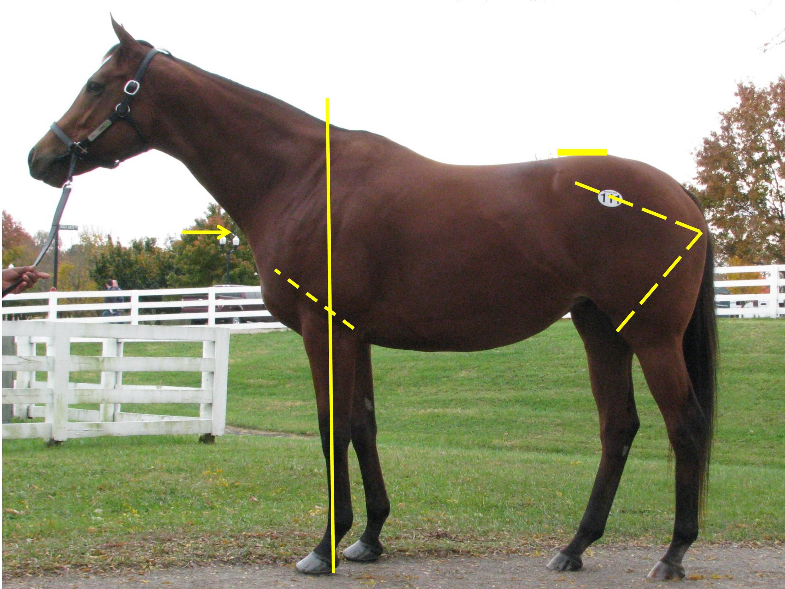
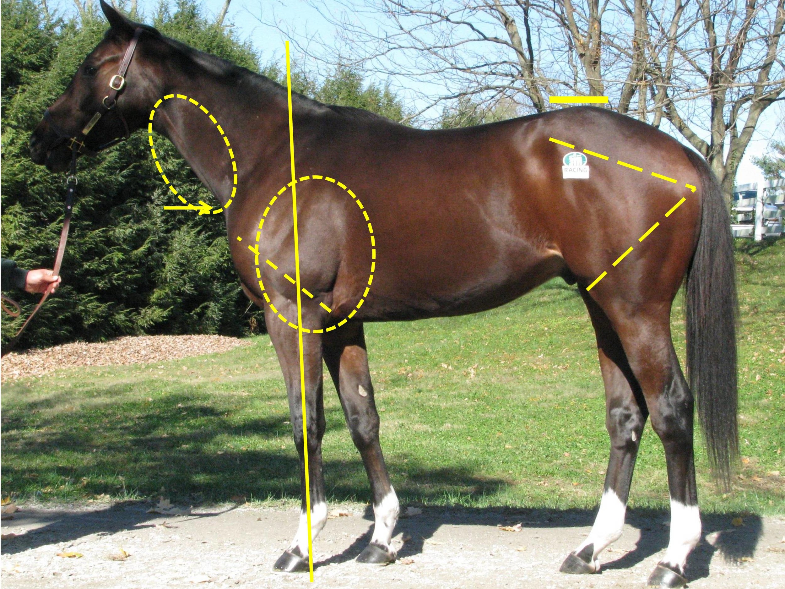
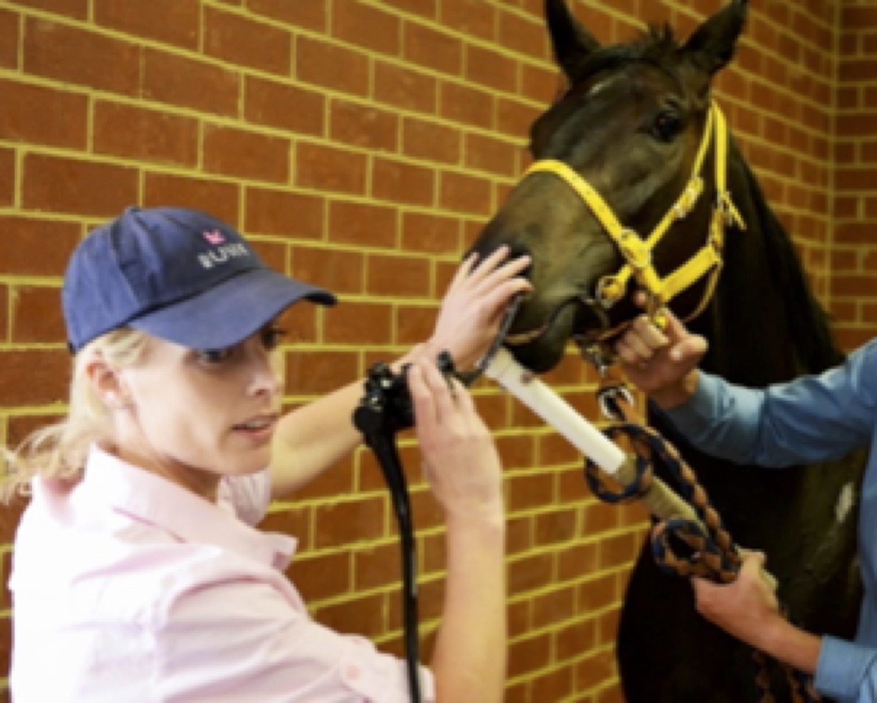
These scales were used in a recent Equine Veterinary Journal article looking at optimal methods to provide anaesthesia for castration. But, the focus on a strained facial expression, ears held back and lack of interaction with people can easily be misinterpreted as poor temperament. It is well worth trainers taking time to make sure their staff are educated on how to recognise signs of pain, as these sorts of clinical signs might indicate important conditions such as gastric ulcers, pneumonia or even musculoskeletal conditions such as fractured ribs. Yard staff should be encouraged to give horses the benefit of the doubt and report any apparent poor temperament so that veterinary investigations can be undertaken to get to the bottom of the problem. Similarly, these signs can be used to monitor horses after potentially painful procedures such as following surgery or castration.

What do we know about analgesic use in equine practice?

There is an increasingly large number of painkillers, also known as analgesics, which are either licensed for use in the horse or supported by research evidence. But it is likely that most equine vets use a relatively small range. British Equine Veterinary Association (BEVA) has recently tasked a team of its members to look at the evidence with underpin best practice for selections of analgesics in common clinical scenarios. This group is chaired by Professor Mark Bowen of the University of Nottingham and has been working for two years now and has collected evidence from the veterinary literature; and in parallel the group has consulted BEVA members to develop robust recommendations. The BEVA Clinical Practice Guidelines report on analgesia will be published soon and looks at the most effective analgesia in horses undergoing routine castration, horses with acute colic, orthopaedic pain and in horses with chronic pain that does not respond to standard non-steroidal anti-inflammatory drugs (NSAIDs) such as phenylbutazone (aka “Bute”). In making their recommendations around use of analgesics in horses, the BEVA team considered both the effectiveness of each analgesic drug, its safety and potential for side-effects.

What are the desirable characteristics of analgesic drugs?

The ideal analgesic has predictable effect and duration, minimal side effects and is easy to prescribe, purchase and administer, lacking any impact on the horse’s future status for human consumption. Of course, the ideal analgesic does not exist. To a large extent, the most appropriate analgesic will be dictated by the specific clinical indication.

Analgesia in colic

With colic, predictable level of analgesia and duration of action are key characteristics. The BEVA team found moderate evidence that flunixin provides superior analgesia to meloxicam and phenylbutazone in horses with colic. However, effective analgesia is desirable but very potent drugs are usually avoided for fear of masking declining clinical status in a horse which would be best served by surgical exploration rather than controlled with extremely potent analgesics. Potential damage to the gastrointestinal tract and effects on gastrointestinal motility are critical and the impact of concurrent shock and volume depletion must be considered. Similar considerations come into play with peri-operative pain but here, the level of analgesia required may be modified by the exact surgical indication and specific procedure and with some procedures, it will be appropriate to provide very potent analgesia, for example with surgical repair of fractures or other painful orthopaedic surgeries. In these cases, multimodal analgesia may well be indicated.

Analgesia following castration

The BEVA team found robust evidence to support a recommendation that pre-operative NSAIDs should be administered prior to surgery. They also recommended that analgesia should be given for at least three days after surgery and that local anaesthetic should be infused into the testicle even when a general anaesthetic is administered. Finally, they counselled that pre-operative use of butorphanol (a commonly used component of sedative protocols) alone should not be considered adequate analgesia for horses undergoing castration.

Selection of NSAIDs for musculoskeletal pain

There is moderate evidence to indicate that phenylbutazone provides superior analgesia for hoof pain / laminitis, compared to firocoxib and meloxicam but strong evidence to show that Suxibuzone can be used as a direct replacement for phenylbutazone in chronic orthopaedic pain. The evidence supporting the use of other NSAIDs is less definitive. Meloxicam and firocoxib may be equivalent to phenylbutazone for pain associated with inflammation of the joint lining and, although studies are not conclusive, the group came to the conclusion that ketoprofen is not as effective for addressing musculoskeletal pain.

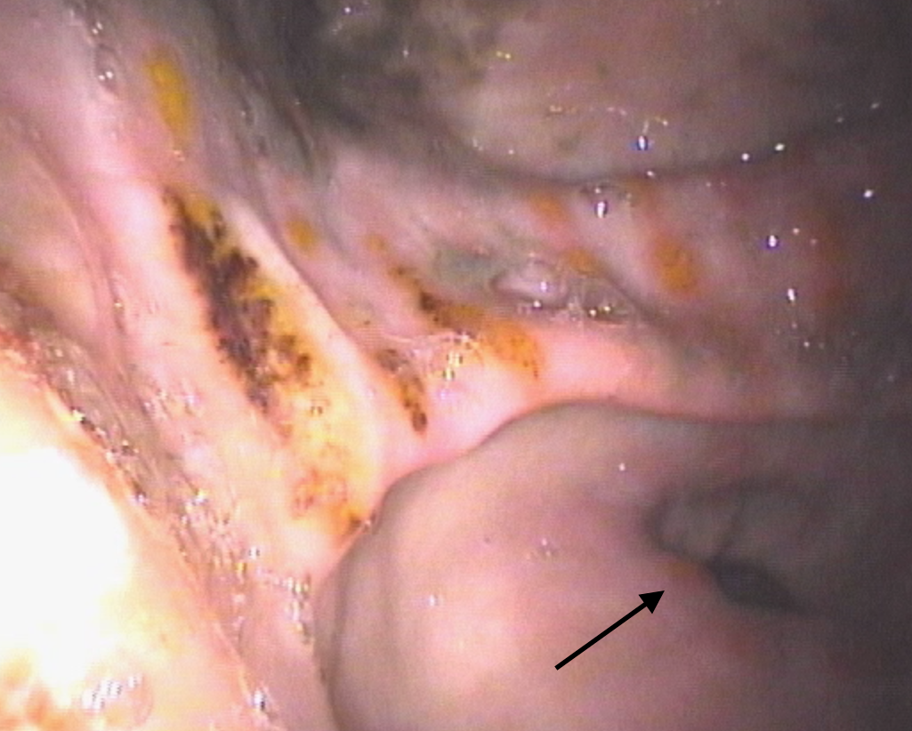
One of the key safety recommendations relating to the use of phenylbutazone was that it is the NSAID that is most likely to induce gastrointestinal adverse events (right dorsal colitis or gastric glandular ulceration). However, although other NSAIDs have less adverse intestinal effects but can all be considered as potentially ulcerogenic. Horses on long-term analgesic therapy should be monitored carefully and further investigations undertaken if they show weight loss, poor appetite or develop the more general signs of pain described above, as this might indicate that the NSAID is having adverse effects on the intestine.

The BEVA team also concluded that giving NSAIDs at doses above those generally recommended in veterinary texts and stacking (i.e., combining maximal doses of different NSAIDs) should be avoided. These practices simply increase risk with no analgesic benefit.

Alternative analgesics

Unfortunately, there are not many practical alternatives to NSAIDs. There is research ongoing looking at topical NSAIDs and alternative drugs such as paracetamol, tramadol and fentanyl. These drugs are unlikely to be in common use in horses in training but do have a place in management of horses with more severe clinical problems.

Similarly, morphine and methadone are used commonly in equine hospitals, but these Schedule 2 controlled drugs are generally not used widely in practice. Buprenorphine has been extensively researched recently and evidence is accumulating supporting its use particularly in the peri-operative patient.

Final warnings

The BEVA group’s report contained a clear warning that highly potent analgesia should only be utilised under the direct control of a veterinary surgeon who has fully evaluated a horse and having developed a therapeutic, analgesic plan that includes ongoing monitoring. It is also important to bear in mind that the best way to alleviate pain associated with a specific clinical condition is to cure the underlying cause. Painkillers should always be used with respect and not be seen as a way to patch up a horse that has an undiagnosed musculoskeletal problem or internal condition.

BUY THIS ISSUE IN PRINT OR DOWNLOAD

July - September 2019, issue 66 (PRINT)
£8.95
Quantity:
Add to Cart

WHY NOT SUBSCRIBE?

DON'T MISS OUT AND SUBSCRIBE TO RECEIVE THE NEXT FOUR ISSUES!

Post-Race Collapse - Prevention & Management

POST-RACE COLLAPSE: PREVENTION & MANAGEMENTDr David MarlinFortunately, incidents of post-race collapse are relatively rare following racing, however if they do occur, it’s important to know what steps can be taken. Common causes of post-race col…

By Dr. David Marlin

Fortunately, incidents of post-race collapse are relatively rare following racing, however if they do occur, it’s important to know what steps can be taken. Common causes of post-race collapse include cardiac arrhythmias, neurologic events, internal bleeding due to large blood vessel rupture, airway obstruction and overheating. All of these are a serious cause for concern and likely to require veterinary support. However, overheating is likely to be one of the most common reasons for post-race collapse, but it is often not recognised as such and can lead to horses not receiving prompt treatment that may ensure a swift and uneventful recovery with no long-lasting injury.

During races, horses get hot because for every unit of energy they use which makes the muscles contract, four times as much energy is produced as heat. The harder and longer the horse works, the more heat it produces. Although horses lose heat by sweating (around 85%) and through breathing (around 15%) during a race, around 90% or more of the heat produced is stored in the muscles. Even so, on a hot day, horses may come in at the end of a race with body temperatures 1-2°C higher than they would for the same race in cool conditions. 

It would not be unusual for horses to finish races with rectal temperatures of 40-41°C. But taking rectal temperature can also mislead us as the temperature inside the working muscles may be much higher; and it can take five minutes for the rectal temperature to reach a peak after a horse pulls up, increasing by another 1-2°C. It’s as we get to rectal temperatures of 42°C that the risk of collapse due to hyperthermia (high body temperature) becomes significant. Let’s look at why high body temperature can lead to collapse.

Firstly, very high body temperature leads to direct and damaging effects on the brain, the nervous system as a whole and the heart, which may lead to collapse. These effects are related to how high the temperature is and how long the horse stays at that elevated body temperature. For example, if a horse was not cooled off following a race, then it may take 5-10 minutes for the onset of collapse. However, post-race collapse on pulling-up and/or returning to the winners enclosure or stables is not uncommon, and this has a different underlying cause.  

During the race, the horse actually reduces blood flow to the skin and chooses instead to send as much as possible to the muscles. This is very different to the situation in people where a significant amount of blood is always sent to the skin to help cooling (thermoregulation). The consequence of blood being directed to the muscles is that the muscle temperatures increase rapidly even over a few minutes of a race. When the horse starts to pull-up, this is reversed and blood is suddenly redirected to the skin. This is most pronounced when the horse comes to a stop. The effect is similar to fainting in people; the flow of blood to the surface causes a fall in blood pressure and effectively the horse faints. 

Clearly, collapse of horses is undesirable and has the potential to cause further injury, so it is important to recognise the risks for post-race collapse with respect to overheating and what to do if the situation arises. One of the common misconceptions of post-race collapse is that this is due to “lack of oxygen”. Whilst this could be true in some cases, this is likely to occur in a very small number of horses and only in those with airway obstruction. From studies on treadmills, for example, we know that within a few seconds of starting to slow down, the low oxygen levels in the blood are immediately reversed and even become higher than they were before exercise. People will often cite the ‘blowing’ of horses after a race as an attempt by the horse ‘to get more oxygen in’, however, it’s clear from a number of studies that blowing/breathing after exercise is directly related to body temperature and not oxygen levels. Rapidly reducing body temperature by aggressive cooling results in a more rapid cessation of blowing.   

HRJTC3.jpg

When should heat stress and overheating be suspected? A horse that is hot to touch, blowing very hard and also ataxic (wobbly) when pulled up should be suspected as suffering from overheating, and cooling should be started immediately. If possible avoid turning in tight circles but keep walking as this helps increase blood pressure. Even in cases where overheating is not the main problem, cooling is extremely unlikely to have any negative effects. As mentioned previously, overheating is frequently not considered as a possible cause for post-race ataxia/collapse and may therefore not be recorded as such. 

Some time ago Professor Tim Parkin and I examined data from the British Horseracing Authority over three seasons of diagnosed cases of post-race heat stress. Over a three-year period, 108 cases had been recorded by on-course veterinary surgeons post-race. A number of factors significantly increased the risk of a horse suffering overheating. Perhaps not surprisingly, races run in the spring or summer were eight times more likely to include at least one horse with heat stress as races run in the autumn or winter. National Hunt races were almost three times more likely to have a horse with heat stress due to the longer duration of the races compared with the flat. Also, for any type of race, there was an increasing risk for every five furlongs (1,000m). Races run in the afternoon were also three times more likely to have a heat stress case than a race run in the evening. Finally, faster races also increased the risk of horses suffering overheating. 

This should all be expected: long races and/or faster races in the afternoon on warm days in spring and summer carry an increased risk of overheating/heat stress and collapse. As not racing in such conditions is not likely to be an option, it’s essential that racecourses and trainers are aware of the signs and risks of overheating and the risk of post-race collapse and take appropriate and prompt action if necessary. 

CNYK67.jpg

Aggressive cooling is now used extensively in professional endurance racing and eventing, as well as in all equestrian disciplines at major events such as FEI World Championships and Olympic Games. The principle is simple. Applying cold water (0-5°C), either from a hose or from a large container of ice in water, rapidly cools the blood in the skin which in turn more slowly cools the muscles. In horses that are very hot and at risk of heat stroke/collapse, there is no requirement or benefit to scraping water. The key to minimising risk is continuous application of water over as much of the body surface as possible until the breathing starts to recover (i.e., until the blowing reduces). This is the best and most practical indicator of the effectiveness of the cooling. It’s also essential to continue aggressive cooling for 5-10 minutes to bring a horse’s temperature down 1-2°C. As mentioned previously, the cessation of blowing is the best indicator of effective cooling.   

Applying cold towels, fans, shade, ice packs on large blood vessels, ice in the rectum, spraying with alcohol are all minimally effective in comparison with continuous application of large volumes of cold water all over the body. In contrast to widely held misconceptions, this approach to cooling does not cause the horse to heat up due to constriction of blood vessels in the skin nor does it cause muscle or kidney damage. The key to preventing collapse and or permanent injury due to heat stroke is rapid instigation of cooling. Literally, seconds count. Delaying cooling by thirty seconds may result in a collapsed horse.

Even in cases where the cause of ataxia/collapse is not primarily due to overheating, starting cooling until veterinary help arrives will not make the situation worse. 

Compared with their jockeys, horses are actually able to tolerate much higher body temperatures. A jockey with a temperature of 41°C would be comatose and at risk of serious injury or even death, whilst a horse at 41°C would still be running. However, it is possible for both jockeys and horses to acclimatise to heat. Acclimatisation is the process whereby the body becomes more tolerant of heat as a result of regular daily exercise in the heat. Of course racehorses are most commonly trained early in the morning in the cooler part of the day, yet the majority of races are held in the warmer times of the day, so it’s conceivable that most racehorses are not heat acclimatised. It may also be of interest that heat acclimatisation also improves performance.

In summary, overheating of horses during races is more likely in longer, faster races at warmer times of the year. Horses that are hotter than normal are at an increased risk of heat-related collapse, often when returning to the paddock and standing. Horses that are very hot to touch, blowing hard, wobbly and possibly ‘excited’ are likely to be at risk for collapse. Starting cooling aggressively immediately can lead to rapid recovery and prevent collapse and the risk of more serious injury. 


BUY THIS ISSUE IN PRINT OR DOWNLOAD

July - September 2019, issue 66 (PRINT)
£8.95
Quantity:
Add to Cart

WHY NOT SUBSCRIBE?

DON'T MISS OUT AND SUBSCRIBE TO RECEIVE THE NEXT FOUR ISSUES!

Advances in Imaging of the Equine Athletic Heart

Advances in imaging of the equine athletic heartBy Francesca Worsman BVM&S MRCVSHorserace Betting and Levy Board Senior Clinical Training Scholar in Equine Internal Medicine, Royal (Dick) School of Veterinary Studies, University of Edinburgh Hor…

By Francesca Worsman BVM&S MRCVS

Horserace Betting and Levy Board Senior Clinical Training Scholar in Equine Internal Medicine, Royal (Dick) School of Veterinary Studies, University of Edinburgh

Horses, through selective breeding for athletic excellence, have well-developed hearts that rarely cause problems compared to those encountered by humans. On occasions however, things go wrong. Due to their well-developed physiology, horses are at risk of a fibrillating heart (atrial fibrillation), while circumstantial evidence suggests that more severe heart rhythm abnormalities, somewhat akin to those experienced by human athletes, may cause sudden death. Ultrasound examination of the heart, known as echocardiography is a readily available tool for examining the heart and significant advances in ultrasound technology are likely to provide exciting information about the detailed function of the equine heart. A huge benefit of ultrasound is that it is non-invasive and can be carried out on a standing unsedated horse, so normal heart function is maintained during the examination. 

The equine heart, like all mammalian hearts, has four chambers. The right atrium, which receives oxygen-depleted blood from the organs, passes it into the right ventricle which then pumps it to the lungs. Blood picks up oxygen in the lungs and then returns it to the left atrium, which then passes it onto the left ventricle for pumping to the organs of the body, including the muscles. Oxygen is thus delivered to the tissues and then the cycle repeats, more than 50,000 times per day! The left and right atria work in unison during heart filling (diastole), and the left and right ventricles work in unison during evacuation of blood from the heart (systole). Murmurs, often detected by veterinary surgeons when listening to hearts, are either caused by normal forward blood flow through the heart or by backflow leakage across the valves within the heart (regurgitation). Many of these murmurs are not a cause for concern, although some regurgitant murmurs are more severe and can cause problems with heart function. Similarly most heart rhythm abnormalities are innocuous and do not affect performance while others are more serious. In some horses, due to suspicion by a veterinary surgeon of a more significant problem, extensive evaluation of the heart is required; echocardiography is one of the key tools for diagnostic evaluation of the heart to assess the impact of any problems on athletic performance.


Image taken from the right side of the horse. Biplane views are on the left (4 chamber standard long axis view at top, short axis view at the bottom of the picture) and 3DE image on the right of the picture. In real-time the image can be manipulated…

Image taken from the right side of the horse. Biplane views are on the left (4 chamber standard long axis view at top, short axis view at the bottom of the picture) and 3DE image on the right of the picture. In real-time the image can be manipulated as the heart is beating to visualise the cardiac structures from different angles. RV = right ventricle, TV = tricuspid valve, RA = right atrium, LV = left ventricle, MV = mitral valve, LA = left atrium.

Real-time three-dimensional echocardiography (3DE) is an exciting new tool that has recently become available in equine medicine and may shed light on important heart problems in horses, including those that cause poor performance. Compared to standard two dimensional echocardiography (2DE), which evaluates a single scan plane, 3DE involves the simultaneous collection of multiple scan planes from the heart to create a pyramid of scan data. 3DE is preferable to 2DE because in theory it does not rely on geometric assumptions on chamber shape to calculate chamber volumes. Also assessment of heart architecture and function (including that of valves) is likely to be more accurate as the technique provides unlimited viewing planes. Finally, for assessment of regurgitant flow, this technique should also be better because the image can be manipulated to better assess the leaky flow from those valves. In humans, for certain heart volume and muscle mass measurements, 3DE is more comparable than 2DE to cardiac magnetic resonance imaging (MRI), which is the acknowledged gold standard. Unfortunately, owing to the significantly larger size of horses, there is currently no equipment available for equine cardiac MRI. Therefore, 3DE could provide the next best option for more detailed equine cardiac evaluation. 

Mitral valve regurgitation for example is commonly encountered in the equine athlete and, while often of no consequence, in more severe cases, it may lead to poor performance. Pathological consequences are due to backflow leakage causing secondary left atrial volume overload, and this will lead to an increased likelihood of atrial fibrillation. Potential advantages of 3DE in this context are more accurate assessment of the degree of volume overload, the regurgitant orifice (i.e., size of the hole!), and the valve structure and motion. Mitral valve regurgitation can be easily confirmed by 2DE, however it can be more difficult to ascertain the cause and severity. With 3DE, the software allows manipulation and therefore anatomic evaluation of the valves from many angles including ‘face-on‘ views. This results in more detailed evaluation of subtle abnormal valve motion and confirmation of suspected findings, which can’t be reliably detected by 2DE -  eg. mitral valve thickening, mitral valve prolapse or ruptured chordae tendineae as the cause of mitral valve disease. Geometric assumptions on volume are avoided as much more structural data is obtained.

A few specialist centres worldwide currently offer 3DE imaging for horses including the Equine Hospital at the Royal (Dick) School of Veterinary Studies, University of Edinburgh. In equine medicine we are still at an early stage of using 3DE as it is not validated in horses, therefore it is mostly used in research at the moment as opposed to routine diagnosis. As part of my research at Edinburgh, sponsored by the Horserace Betting and Levy Board, I have been assessing the left atrial volume using 3DE from thoroughbreds in training. One of my aims was to determine the variability of equine left atrial volume measurement using a special 3DE software analysis package to see how much variation there was between successive 3DE measurements by the same person. Forty-four National Hunt thoroughbreds in training were scanned to obtain the 3DE views of this chamber. We then graded them to exclude images of reduced image quality so that we were only assessing good quality images of the left atrium. In total 24 horses were included—aged 4-9 yrs, weighing 411-534kg. I analysed the images retrospectively, after the horses were scanned. I didn’t include any horses with grade >3/6 heart murmurs. This was because we first need to validate 3DE with normal, healthy hearts. Random generated order measurements were obtained by a single person on four occasions. Real-time three-dimensional end-systolic (ESV) and end-diastolic (EDV) left atrial volumes were measured using 3DE software, and the results were then statistically analysed.

TO READ MORE —

BUY THIS ISSUE IN PRINT OR DOWNLOAD

July - September 2019, issue 66 (PRINT)
£8.95
Quantity:
Add to Cart

WHY NOT SUBSCRIBE?

DON'T MISS OUT AND SUBSCRIBE TO RECEIVE THE NEXT FOUR ISSUES!

EIPH: An Australian Perspective with Worldwide Implications

Exercise-induced pulmonary haemorrhage (EIPH) is a common disease of racehorses. The precise cause of EIPH is yet to be fully determined, but a well-accepted theory is that lung blood vessels rupture in response to the extremely high blood pressure …

By Ellie Crispe and Guy Lester

Exercise-induced pulmonary haemorrhage (EIPH) is a common disease of racehorses. The precise cause of EIPH is yet to be fully determined, but a well-accepted theory is that lung blood vessels rupture in response to the extremely high blood pressure and low airway pressure experienced during strenuous exercise. The barrier that separates the airway from the blood vessels is ultra-thin to facilitate the efficient exchange of gases, but this predisposes to breakage. The condition is most frequently described in Thoroughbred and Standardbred racehorses, but it has also been identified in racing Appaloosas and Quarter Horses, as well as horses involved in other high intensity athletic activities, including showjumpers, 3-day eventers, barrel racers, steeplechasers and polo horses.

EIPH is not unique to horses and has been reported in human athletes, as well as racing greyhounds and camels. Our group at Murdoch University in Perth Australia has had an interest in EIPH, which has led to three recent publications in the Equine Veterinary Journal.1-3

How common is EIPH?

Fig 1.jpg

Blood from both nostrils—also known as epistaxis—is the most obvious manifestation of EIPH and occurs between 1.5 and 8.4/1000 race starts, varying with racing jurisdiction. Epistaxis represents a severe manifestation of EIPH, and basing surveys on its presence vastly underestimates the true prevalence of lung haemorrhage. There are several techniques used to diagnose EIPH, but endoscopy of the trachea 30-120 minutes after racing or galloping is a common and reliable method. Occurrence and severity of pulmonary haemorrhage is typically graded using a 0-4 scale. Using endoscopy, we reported a prevalence of EIPH post-race in Australian thoroughbreds racing on turf tracks of around 55%, with most positive horses having low to moderate volumes of blood in the trachea. EIPH is less common if horses are examined after trialling, and reduced further if examined after track gallops. The prevalence of EIPH increases when horses are examined on multiple occasions after racing, and in fact all horses in our research population that had seven monitored race-starts experienced EIPH on at least one occasion.

What is the effect of EIPH on race-day performance?

It is generally considered that EIPH has a negative impact on racing performance, but evidence for this assertion is surprisingly lacking. We performed 3,794 post-race endoscopy exams on over 1,500 Australian horses and reported that inferior race-day performance was limited to horses with severe EIPH (grades 3 and 4); this reflected only 6.3% of all examinations. Horses with the highest grades of EIPH (grade 4) were less likely to finish in the first three, finished further from the winner, were less likely to collect race earnings, were slower over the final stages of the race, and were more likely to be overtaken by other competitors in the home straight than horses without EIPH. Interestingly, horses with EIPH grade 1 or 2 were more likely to overtake others in the home straight, compared to horses without EIPH (grade 0).

Screen Shot 2019-03-21 at 11.31.49.png

It is highly unlikely that low-grade EIPH (grade 1 or 2) confers an athletic advantage; a plausible explanation is that horses that are ridden competitively to the finish are functioning at their maximal physiological limit, compared to horses that are eased up, and overtaken, during the finishing stages of the race because they are not in prize contention or are affected by interference in the home straight.  Another interesting finding was that horses with moderate to severe EIPH (grades 3 or 4) raced the early and mid-sections of the race faster than horses without EIPH. It is possible that these horses reach the breaking threshold of the small lung blood vessels at an earlier stage in the race compared to horses that start the race slower, compounding the severity thereafter. A study of barrel racing horses reported that horses with the most severe grade of EIPH were faster than horses without EIPH, a finding which may also reflect this rapid acceleration increasing the risk of EIPH. It may be wise for trainers to instruct jockeys riding horses with a history of moderate to severe EIPH to refrain from racing in this manner.

What is the effect of a one-off diagnosis of EIPH over a horse’s career?…

TO READ MORE —

BUY THIS ISSUE IN PRINT OR DOWNLOAD

April - June 2019, issue 65 (PRINT)
£8.95
Quantity:
Add to Cart

WHY NOT SUBSCRIBE?

DON'T MISS OUT AND SUBSCRIBE TO RECEIVE THE NEXT FOUR ISSUES!

IF YOU LIKE THIS ARTICLE

WHY NOT SUBSCRIBE - OR ORDER THE CONTENT FROM THIS ISSUE IN PRINT?

Skin-deep: overcoming barriers for effective transdermal drug delivery

Skin-deep: overcoming barriers for effective transdermal drug delivery Ancient art, modern scienceOne shared medicinal practice amongst disparate ancient societies was the application of primitive ointments to the skin to treat almost all and any ai…

By Roger Smith

Ancient art, modern science

One shared medicinal practice amongst disparate ancient societies was the application of primitive ointments to the skin to treat almost all and any ailments. A vast plethora of poultices and plasters have been described, including in Babylonian and Greek medicine texts1 amongst others, suggesting that the magical health-restoring powers of ointments were well-recognised to traverse the skin. Thus, it was no coincidence that the skin was the preferred therapeutic route over surgical (and oral) intervention since the former method was likely to result in reduced mortality rates compared to the latter; undoubtedly an important consideration, given that the top ancient physicians were likely charged with the health of the royal courts.

Although the art of transdermal delivery of medicines dates back millennia, it is only in more recent times that the science of transdermal drug delivery in man has advanced significantly2.  The choice of modern drugs for topical applications is, however, relatively limited compared to the seemingly infinite choice available for oral delivery.  This is perhaps not surprising since the gut is an organ that has evolved with the main purpose of absorbing food (chemicals when it comes to it) whereas the skin, despite being the largest organ, has evolved primarily as a protective layer to prevent desiccation of underlying tissues and to keep out harmful environmental chemicals. As this includes medicinal drugs, the pursuit of transdermal administration would appear, at first sight, to be an illogical choice. However, there are several compelling reasons why transdermal delivery routes are an important alternative to pills, injections or inhalation routes:

  • It avoids poor absorption after oral ingestion—especially in animals, the absorption of a drug can vary between the omnivore (e.g., human) and herbivore (e.g., horse) stomach.  

  • It avoids first-pass effect where the blood circulation from the gut passes through the liver to remove absorbed drugs.

  • It can reduce systemic drug levels to minimise adverse effects.

  • The design of sustained release formulations overcomes the frequent dosing necessitated by oral and injectables to achieve constant drug levels.

  • It enables ease and efficacy of drug withdrawal.

  • Transdermal drug delivery is painless and non-invasive, thereby potentially allowing longer treatment when daily injection is unacceptable or impractical.

  • It has the potential to target local administration such as for the treatment of flexor tendon disease because the tendons are subcutaneous.

Challenges for transdermal drug applications

The skin is made up of three key layers: the epidermis, dermis and hypodermis (figure 1) and the water-attracting (hydrophilic) or water-repelling (hydrophobic) properties within each raise unique challenges for topical or transdermal drug applications.  

Figure 1 – Anatomy of the skin with expanded illustration showing the cells of the stratum corneum (‘bricks’) embedded in lipid matrix (‘mortar’).

Figure 1 – Anatomy of the skin with expanded illustration showing the cells of the stratum corneum (‘bricks’) embedded in lipid matrix (‘mortar’).

Topical applications, such as insect repellents and sunscreen creams, target the surface of the skin or deliver a drug locally such as for the control of inflammation (insect bite or reaction to an allergen). In contrast the aim of transdermal, or subcutaneous, applications are to deliver the drug deeper to either an adjacent organ, or, more commonly, to the blood circulation as an alternative to oral or needle routes to reach distant organs. The main barrier to local or transdermal delivery is the outermost layer of the skin, called the stratum corneum in the epidermis (figure 1). This consists of dead skin cells, the corneocytes, that combine with lipid bilayers into a tightly packed “bricks-and-mortar” layer that form alternating hydrophilic (the water rich corneocytes) and hydrophobic (lipid bilayer) regions (figure 1). The stratum corneum therefore not only forms a mechanically robust layer but also presents a challenge in designing drugs with chemical properties that can negotiate their way into and through these contrasting hydrophobic and hydrophilic environments to reach the lower region of the epidermis. The epidermis consists of living skin cells but has no blood vessels for the drug to diffuse into, so instead the drug must penetrate further to the dermis where it can finally enter the bloodstream or the subcutaneous layers.

Routes for drugs through the skin

shutterstock_625969397.jpg

Most transdermal drugs are designed so that they diffuse through the skin in a passive fashion. The routes for drug can be through the skin cells (transcellular), around them (intercellular) or using the skin components hair follicles, sweat glands and sebaceous glands (produce lipids) to bypass the stratum corneum (so-called ‘appendageal’ routes).

Transcellular route: Drugs pass through the corneocytes of the stratum corneum rather than the lipid ‘mortar’ that surrounds them (figure 2). However, the drug has to exit the cell to enter the next corneocyte and therefore through the skin. It means that it has to encounter the external hydrophobic environment between the cells multiple times as it moves through the alternating cell and lipid layers of the epidermis. Drugs therefore have to have balanced hydrophilic and hydrophobic properties to enable this to happen.

Figure 2 – Path of molecules through (A) the stratum corneum for the transcellular route (Note: the drug has to enter and exit the aqueous environment of the cells into the surrounding lipid matrix requiring an ability to be soluble in both); (B) In…

Figure 2 – Path of molecules through (A) the stratum corneum for the transcellular route (Note: the drug has to enter and exit the aqueous environment of the cells into the surrounding lipid matrix requiring an ability to be soluble in both); (B) Intercellular route (Note: the tortuous path for molecules passing through the stratum corneum via this route which delays diffusion.

Intercellular route: The drug predominantly diffuses through the lipid rich ‘mortar’ around the corneocytes of the epidermis. This lipid matrix can form a continuous route through the epidermis (avoiding entering the cells), but this route has been suggested to be less efficient because it increases the distance 50-fold3 compared to the direct route through the stratum corneum due to the interdigitating brick and mortar arrangement (figure 2). Again, the chemical formulation used to carry the drug is important and drugs that more readily dissolve in lipids benefit from this route.

Appendageal route: The hair, sweat glands and sebaceous glands provide a direct channel to the deep layers of the skin circumventing the hazardous barriers of the epidermis and dermis. The main challenge for this relatively easy route is that the amount of drug that can be taken up is limited by the density of hair follicles and sweat glands, although in haired animals, such as the horse, the density can be as high as 1-5% of the skin surface area. Furthermore, sweat from an active sweat gland would be travelling against the direction of drug flow, washing out the drug and its carrier and severely limit drug uptake. It is likely that all skin applications use this appendageal route as it’s unavoidable but probably more efficient for drugs that are large molecules.

TO READ MORE —

BUY THIS ISSUE IN PRINT OR DOWNLOAD

April - June 2019, issue 65 (PRINT)
£8.95
Quantity:
Add to Cart

WHY NOT SUBSCRIBE?

DON'T MISS OUT AND SUBSCRIBE TO RECEIVE THE NEXT FOUR ISSUES!

IF YOU LIKE THIS ARTICLE

WHY NOT SUBSCRIBE - OR ORDER THE CONTENT FROM THIS ISSUE IN PRINT?

Fungi - the invisible health risk

Fungi - the invisible health risk Dr Emmanuelle van Erck, DVM, PhD, ECEIM explains her work looking at the link between the presence of fungi and lower airway inflammation Horses are incredible athletes. Their physiology—the way their body functions…

Dr. Emmanuelle van Erck, DVM, PhD, ECEIM explains her work looking at the link between the presence of fungi and lower airway inflammation

Horses are incredible athletes. Their physiology—the way their body functions—is truly fascinating. They can adapt to training at a phenomenal rate, they have massive hearts that fuel their powerful muscles and pushes them to peak speeds. So what could stop them? Oxygen, or rather the lack of it. Horses experience hypoxemia during racing, which means they enter a state of deficiency in oxygen. The reason for this deficiency is a failure of the respiratory system to effectively ventilate and adequately fuel oxygen to the muscles. Horses are obligate nasal breathers and were endowed with particularly long and narrow upper airways in relation to their body size. These factors increase the resistance to breathing. They are also constrained by the fact that they ventilate at very high rates, which does not allow for effective and rapid renewal of oxygen in the lungs. Even the fittest, best thoroughbreds crave oxygen from mid-race onwards. So maintaining horses in optimal respiratory health is absolutely essential for them to achieve an efficient sprint and optimal performance.

Respiratory diseases are highly prevalent in horses. It is inherent to their living and working conditions. The mere fact that a horse is housed in a box increases his risk of developing airway inflammation. The content in fine dust is naturally high in a horse’s box. Closed or poorly ventilated barns further deteriorate air quality in the horse’s immediate environment. Several studies have shown that horses housed indoors are exposed not only to high amounts of organic dust and ammonia but also germs and endotoxin they produce that trigger a detrimental reaction from the immune system. The problem is that even low-grade respiratory diseases will directly affect the horse’s capacity to perform and recover from strenuous exercise.

Microscope picture of a germinating fungal spore in a respiratory sample - an indication of inhalation of fungal spores in the airways and subsequent infection from the mould.

Microscope picture of a germinating fungal spore in a respiratory sample - an indication of inhalation of fungal spores in the airways and subsequent infection from the mould.

With my colleagues, Dr Dauviller and Dr ter Woort, specialists in equine internal medicine, we have investigated the link between the presence of fungi and lower airway inflammation. In our ambulatory referral practice, we go out to the stables and have the opportunity not only to examine the horse but also attentively assess his environment. As we collected respiratory samples and analysed them ourselves, we became aware that the presence of microscopic moulds or fungal elements was frequently associated to lung issues. To investigate this further, we decided to systematically record clinical and environmental data and link it to our findings in the respiratory samples of the horses referred for investigation.

We collected more than 700 cases; the horses included in the study were either referred routine examinations, unexplained poor performance or respiratory symptoms such as coughing or breathing heavily during exercise. All horses had a tracheal and a bronchoalveolar lavage done which allowed us to evaluate their level of respiratory inflammation, as well as estimate the presence of fungi within the airways. We also looked at the state of activation of fungi: if they were inert particles or if they showed signs of active proliferation. Our results were without appeal; the presence of inhaled fungi significantly and negatively affected respiratory health in horses, causing inflammation and in some cases, infection.

In this population, inflammatory airway disease (IAD) was diagnosed in 88% of cases, confirming that respiratory inflammation is very common and often under-diagnosed. Of these positive cases, 81% had evidence of fungi in their airways. The presence of fungi more than doubled the odds of having lung inflammation.

TO READ MORE —

BUY THIS ISSUE IN PRINT OR DOWNLOAD

April - June 2019, issue 65 (PRINT)
£8.95
Quantity:
Add to Cart

WHY NOT SUBSCRIBE?

DON'T MISS OUT AND SUBSCRIBE TO RECEIVE THE NEXT FOUR ISSUES!

IF YOU LIKE THIS ARTICLE

WHY NOT SUBSCRIBE - OR ORDER THE CONTENT FROM THIS ISSUE IN PRINT?

Ulcer medication: are the products to treat that different?

Stomach ulcers are not all the sameRacehorse trainers and their vets first began to be aware of stomach ulcers over 20 years ago. The reasons why we became aware of ulcers are related to technological advances, which produced endoscopes long enough …
EVJ banner 1 (1).png

By Celia Marr

Stomach ulcers are not all the same

Racehorse trainers and their vets first began to be aware of stomach ulcers over 20 years ago. The reasons why we became aware of ulcers are related to technological advances, which produced endoscopes long enough to get into the equine stomach. At that time, scopes were typically about 2.5m long and were most effective in examining the upper area of the stomach, which is called the squamous portion. Once this technology became available, it was quickly appreciated that it is very common for racehorses to have ulcers in the squamous portion of the stomach.

Fig 1. The equine stomach has two regions: the upper region is the squamous portion and the lower region is the glandular portion. The squamous portion is lined by pale pink tissue which is susceptible to acid damage. The glandular portion is lined …

Fig 1. The equine stomach has two regions: the upper region is the squamous portion and the lower region is the glandular portion. The squamous portion is lined by pale pink tissue which is susceptible to acid damage. The glandular portion is lined by darker purple tissue. Acid is produced in this region. In this horse, the stomach lining is healthy and unblemished. The froth is due to saliva which is continuously swallowed.

The equine stomach has two main areas: the squamous portion and the glandular portion. The stomach sits more or less in the middle of the horse, immediately behind the diaphragm and in front of and above the large colon. Imagine the stomach as a large balloon with the oesophagus—the gullet—entering halfway up the front side and slightly to the left of the balloon-shaped stomach and the exit point also coming out the front side but slightly lower and to the right side. The tissue around the exit—the pylorus—and the lower one-third, the glandular portion, has a completely different lining to the top two-thirds, the squamous portion.

The stomach produces acid to start the digestive process. Ulceration of the squamous portion is caused by this acid. Like the human oesophagus, the lining of the squamous portion has very limited defences against acid.  But, the acid is actually produced in the lower, glandular portion. The position of the stomach is between the diaphragm, which moves backwards as the horse breathes in and the heavy large intestine which tends to push forwards as the horse moves. During exercise, liquid acid produced at the bottom of the stomach is squeezed upwards onto the vulnerable squamous lining. It makes sense then that the medications used to treat squamous ulcers are aimed at blocking acid production.

Lesions in the glandular portion of the stomach are less common than squamous ulcers. The acid-producing glandular portion has natural defences against acid damage including a layer of mucus and local production of buffering compounds. At this point, we actually know relatively little about the causes of glandular disease, but it is becoming increasingly obvious that disease in the glandular portion is very different from squamous disease. Often, it is more difficult to treat.

Fig 2. This horse shows signs of discomfort. She carries her head low, her ears are back a little, and the muscles of the face are clenched, affecting the shape of the nostrils and eye.

Fig 2. This horse shows signs of discomfort. She carries her head low, her ears are back a little, and the muscles of the face are clenched, affecting the shape of the nostrils and eye.

Stomach ulcers can cause a wide range of clinical signs. Some horses seem relatively unaffected by fairly severe ulcers, but other horses will often been off their feed, lose weight, and have poor coat quality. Some will show signs of abdominal discomfort, particularly shortly after eating. Other horses may be irritable—they can grind their teeth or they may resent being girthed. Additional signs of pain include an anxious facial expression, with ears back and clenching of the jaw and facial muscles and a tendency to stand with their head carried a little low.


Assessing ulcers

Ulcers can only be diagnosed with endoscopy. A grading system has been established for squamous ulcers, which is useful in making an initial assessment and in documenting response to treatment.

Grade 0 = normal intact squamous lining

Grade 1 = mild patches of reddening

Grade 2 = small single or multiple ulcers

Grade 3 = large single or multiple ulcers

Grade 4 = extensive, often merging with areas of deep ulceration

Fig 3. Grade 1 squamous ulcers which are mild patches of reddening.

Fig 4. Grade 2 squamous ulcers—there are several of these, but they are all small.

Fig 5. Grade 3 squamous ulcers—these are larger, and there are several.

Fig 6. Grade 4 squamous ulcers—there are extensive deep ulcers with active haemorrhage.

Although it is used for research purposes, this grading system does not translate very well to glandular ulcers where typically, lesions are described in terms of their severity (mild, moderate or severe), distribution (focal, multifocal or diffuse), thickness (flat, depressed, raised or nodular) and appearance (reddening, haemorrhagic or fibrinosuppurative). Fibrinosuppurative suggests that inflammatory cells or pus has formed in the area. Focal reddening can be quite common in the absence of any clinical signs. Nodular and fibrinosuppurative lesions may be more difficult to treat than flat or reddened lesions. Where the significance of lesions is questionable, it can be helpful to treat the ulcers and repeat the endoscopic examination to determine whether the clinical signs resolve along with the ulcers.

Fig 7. The glandular tissue around the pylorus (or exit point) has reddened patches. This is of questionable clinical relevance, and many horses will show no signs associated with these lesions.

Fig 8. There are dark red patches of haemorrhage in the glandular tissue of the antrum—the region adjacent to the pylorus—which is the dark hole toward the bottom of this image.

Fig 9.This horse has moderate to severe glandular disease. There are depressed suppurative (yellow) areas several of which also have haemorrhage. Nearer to the pylorus there is reddening and raised, swollen areas (arrow).

Fig 10. This horse has moderate to severe glandular disease. The majority of lesions are depressed and haemorrhagic.

Medications for squamous ulcers

Because of the prevalence and importance of gastric ulcers, Equine Veterinary Journal publishes numerous research articles seeking to optimise treatment. The most commonly used drug for treatment of squamous ulcers is omeprazole. A key feature of products for horses is that the drug must be buffered in order to reach the small intestine, from where it is absorbed into the bloodstream in order to be effective. Until recently only one brand was available, but there are now several preparations on the market and researchers have been seeking to show whether new medicines are as effective as the original brand. There is limited information comparing the new products, and this information is essential to determine whether the new, and often cheaper, products should be used.

A team of researchers formed from Charles Sturt University in Australia and Louisiana State University in the US has compared two omeprazole products given orally. A study reported by Dr Raidal and her colleagues, showed that not only were plasma concentrations of omeprazole similar with both products, but importantly, the research also showed that gastric pH was similar with both products and both products reduced summed squamous ulcer scores. Both the products tested in this trial are available in Australia and, although products on the market in UK have been shown to achieve similar plasma concentrations and it is therefore reasonable to assume that they will be beneficial, as yet, not all of them have been tested to show whether products are equally effective in reducing ulcer scores in large-scale clinical trials. Trainers should discuss this issue with their vets when deciding which specific ulcer product they plan to use in their horses.

Avoiding drugs altogether and replacing this with a natural remedy is appealing. There is a plethora of nutraceuticals around and anecdotally, horse owners believe they may be effective. One such option is aloe vera that has antioxidant, anti-inflammatory and mucus stimulatory effects which might be beneficial in a horse’s stomach. Another research group from Australia, this time based in Adelaide, has looked at the effectiveness of aloe vera in treating squamous ulcers and found that, although 56% of horses treated with aloe vera improved and 17% resolved after 28 days, this compared to 85% improvement and 75% resolution in horses given omeprazole. Therefore, Dr Bush and her colleagues from Adelaide concluded treatment with aloe vera was inferior to treatment with omeprazole.

Medications for glandular ulcers….

TO READ MORE —

BUY THIS ISSUE IN PRINT OR DOWNLOAD

January - March 2019, issue 64 (PRINT)
£8.95
Quantity:
Add to Cart

WHY NOT SUBSCRIBE?

DON'T MISS OUT AND SUBSCRIBE TO RECEIVE THE NEXT FOUR ISSUES!

The FEI prohibited list and what it means for racing

The eighth World Equestrian Games in Tryon, North Carolina were not, it is perhaps fair to say, an unbridled success. From unfinished facilities to misspelt signage and, most catastrophically, an entire endurance race that had to be aborted after ri…

By Alysen Miller

The eighth World Equestrian Games in Tryon, North Carolina were not, it is perhaps fair to say, an unbridled success. From unfinished facilities to misspelt signage and, most catastrophically, an entire endurance race that had to be aborted after riders were sent in the wrong direction, the competition generated so much negative coverage that the future of the Games themselves, already in some doubt, now appears to be hanging by a thread (At the time of writing, no formal bidders had thrown their hats into the ring for the 2022 renewal). So it might seem to be a strange time to ask if horseracing has anything to learn from the Fédération Équestre International (FEI). And yet, there is one area in which the FEI is arguably setting an example.

Unlike the global racing industry, which operates under myriad rules and regulations between different countries (and sometimes within the same country), all 134 affiliated nations of the FEI operate under a single set of rules. This includes a single Prohibited Substances Policy to which all jurisdictions must adhere; meaning that a horse trained in Australia is subject to exactly the same medical requirements, including regulations governing banned substances and threshold limits, as a horse trained in, say, America. This stands in stark contrast to the thoroughbred industry. Despite being an increasingly global game, from the now-traditional annual American invasion of Royal Ascot to the recent domination of the Melbourne Cup by European-trained horses, racing can appear positively parochial when it comes to its attitudes towards prohibited substances. “If you compare horseracing to other sports, we have one of the sole sports where there are no equal regulations on the highest level,” elucidates Germany’s Peter Schiergen. “To have [the same] regulations and policies around the world would be a good action for horse racing.”

So what are the factors standing in the way of global harmonisation, and would there ever be a case for following the FEI’s lead and adopting a single set of rules that would apply to horseracing authorities the world over?

Laboratory sample analysis

The FEI’s approach is to divide prohibited substances into two categories: banned substances (that is, substances that are deemed by the FEI to have no legitimate use in competition and/or have a high potential for abuse, including all anabolic steroids and their esters), which are not permitted at any time; and controlled medication (substances that are deemed to have a therapeutic value and/or are commonly used in equine medicine), which are not permitted for use during competition but may be used at other times. These categorisations apply to all national and international competitions, with each national federation being subject to the FEI’s regulations. Testing at competitions is carried out by the FEI’s own veterinary department, while elective out-of-competition testing is also available so that those responsible for the horse can ensure that they allow the appropriate withdrawal times for therapeutic medications. So just how effective are these rules at keeping prohibited substances out of the sport and ensuring a level playing field? Clearly, no system is perfect. The FEI has had its fair share of doping scandals, particularly in the endurance discipline, where stamina, which can be easily enhanced with the aid of pharmacology, is of paramount importance. The FEI, who declined to be interviewed for this article, said in a statement: “Clean sport is an absolute must for the FEI and it is clear that we, like all International Federations, need to continue to work to get the message across that clean sport and a level playing field are non-negotiable. All athletes and National Federations know that regardless of where in the world they compete the rules are the same.” Yet having a global policy does appear to offer a strategic advantage to those seeking to create a level playing field, not only through the creation of economies of scale (the FEI oversees laboratories around the world, and all results are all handled at the federation’s headquarters in Lausanne), but also by creating a framework for cheats to be exiled from all competitions, rather than just one country’s.

While harmonisation and cross-border cooperation does exist in racing, particularly within Europe and individual race meetings—notably the recent Breeders’ Cup—have taken it upon themselves to enact their own programme of pre- and post-race testing, effectively creating their own anti-doping ecosystem; the fact remains that racing lacks an overarching prohibited substances policy. Codes and customs vary widely from—at one end of the spectrum—Germany, which does not allow any colt that has run on declared medication to stand at stud; to North America, where, Kentucky Derby winner Big Brown, whose trainer admitted that he gave the colt a monthly dose of the anabolic steroid, stanozolol, is still active at stud. Stanozolol is the same drug that the Canadian sprinter Ben Johnson tested positive for in 1988, causing him to be stripped of his gold medal in the Seoul Olympics. Although the industry subsequently moved to outlaw the drug for use on horses in training, anabolic steroids are still routinely used as an out-of-competition treatment in a number of states.

“I don’t think the playing field is level,” says Mark Johnston, with typical candour. “Control of anabolic steroids is very important if you want a level playing field. Because there’s no doubt whatsoever that there are advantages to using them.”


TO READ MORE —

BUY THIS ISSUE IN PRINT OR DOWNLOAD

January - March 2019, issue 64 (PRINT)
£8.95
Quantity:
Add to Cart

WHY NOT SUBSCRIBE?

DON'T MISS OUT AND SUBSCRIBE TO RECEIVE THE NEXT FOUR ISSUES!

Wobbler Syndrome and the thoroughbred

Wobbler Syndrome and the Thoroughbred Celia M Marr, Rossdales Equine Hospital and Diagnostic Centre, Cotton End Road, Exning, Newmarket, Suffolk, CB8 7NN  << BCET logo near the top>>  Wobbler Syndrome, or spinal ataxia, affects around 2%…

By Celia M Marr, Rossdales Equine Hospital and Diagnostic Centre

Wobbler Syndrome, or spinal ataxia, affects around 2% of young thoroughbreds. In Europe, the most common cause relates to narrowing of the cervical vertebral canal in combination with malformation of the cervical vertebrae. Narrowing in medical terminology is “stenosis” and “myelopathy” implies pathology of the nervous tissue, hence the other name often used for this condition is cervical vertebral stenotic myelopathy (CVSM).

Wobbler Syndrome was the topic of this summer’s Gerald Leigh Memorial Lectures, an event held at Palace House, Newmarket. Gerald Leigh was a very successful owner breeder and these annual lectures, now in their second year, honour of Mr Leigh's passion for the thoroughbred horse and its health and welfare. The lectures are attended by vets, breeders and trainers, and this year because of the importance and impact of Wobbler syndrome on thoroughbred health, several individuals involved in thoroughbred insurance were also able to participate.

Blindfolding the horse, exacerbates the ataxia and improves the accuracy of objective ataxia assessment.

Dr Steve Reed, of Rood and Riddle Equine Hospital, Kentucky and international leader in the field of equine neurology gave an overview of Wobbler Syndrome. Affected horses are ataxic, which means that they have lost the unconscious mechanisms which control their limb position and movement. Young horses with CVSM will generally present for acute onset of ataxia or gait abnormalities, however, mild ataxia and clumsiness may often go unnoticed. Trainers often report affected horses are growing rapidly, well-fed, and large for their age. It is common for riders to describe an ataxic horse as weak or clumsy. Sometimes, a horse which has been training normally will suddenly become profoundly affected, losing coordination and walking as though they were drunk, or in the most severe cases stumbling and falling. Neurological deficits are present in all four limbs, but are usually, but not always more noticeable in the hindlimbs than the forelimbs. In horses with significant degenerative joint disease, lateral compression of the spinal cord may lead to asymmetry of the clinical signs.

When the horse is standing still, it may adopt an abnormal wide-based stance or have abnormal limb placement, and delayed positioning reflexes. At the walk, the CVSM horse’s forelimbs and hindlimbs may not be moving on the same track and there can be exaggerated movement of the hind limbs when the horse is circled. Detailed physical examination may reveal abrasions around the heels and inner aspect of the forelimbs due to interference, and short, squared hooves due to toe-dragging. Many young horses affected with CVSM have concurrent signs of developmental orthopaedic disease such as physitis or physeal enlargement of the long bones, joint effusion secondary to osteochondrosis, and flexural limb deformities.

Radiography is generally the first tool which is used to diagnose CVSM. Lateral radiographs of the cervical vertebrae, obtained in the standing horse, reveal some or all of five characteristic bony malformations of the cervical vertebrae: (1) “flare” of the caudal vertebral epiphysis of the vertebral body, (2) abnormal ossification of the articular processes, (3) malalignment between adjacent vertebrae, (4) extension of the dorsal laminae, and (5) degenerative joint disease of the articular processes. Radiographs are also measured to document the ratio between the spinal canal and the adjacent bones and identify sites where the spinal canal is narrowed.

ABOVE L–R: Lateral radiographs can show the vertebral bones have an abnormal shape with flare of the caudal vertebral epiphysis (curved arrow) and extension of the dorsal laminae (straight arrow). Abnormal ossification of the articular processes and enlargement of the joints due to degenerative joint disease (arrows). Measuring the ratio of the spinal canal to the adjacent bone identifies narrowing of the spinal canal. In this case, the narrowing is dramatic due to mal-alignment of adjacent vertebral bones.

Dr Reed also highlighted myelography as the currently most definitive tool to confirm diagnosis of focal spinal cord compression and to identify the location and number of lesions. The experts presenting at the Gerald Leigh Memorial Lectures agreed that myelography is essential if surgical treatment is pursued. However, an important difference between the US and Europe was highlighted by Prof Richard Piercy, of the Royal Veterinary College, University of London. In Europe, protozoal infection is very rare, whereas in US, equine protozoal myeloencephalitis can cause similar clinical signs to CVSM. Protozoal myeloencephalitis is diagnosed by laboratory testing of the cerebral spinal fluid but there is also a need to rule out CVSM. Therefore, spinal fluid analysis and myelography tends to be performed more often in the US. Prof Piercy pointed out that in the absence of this condition, vets in Europe are often more confident to reach a definitive diagnosis of CVSM based on clinical signs and standing lateral radiographs.

TO READ MORE --

BUY THIS ISSUE IN PRINT OR DOWNLOAD -

October - December 2018, issue 63 (PRINT)
£8.95
Quantity:
Purchase in print

WHY NOT SUBSCRIBE?

DON'T MISS OUT AND SUBSCRIBE TO RECEIVE THE NEXT FOUR ISSUES!

Unravelling Ulceration

The causes, treatment and prevention for squamous vs. glandular gastric ulceration

Unravelling ulcerationthe causes, treatment and prevention for squamous vs. glandular gastric ulceration.Emma Hardy, PhDGastric ulcers remain a common condition facing competition horses. This poses an ongoing and persistent challenge to trainers wh…

By Emma Hardy, PhD

Gastric ulcers remain a common condition facing competition horses. This poses an ongoing and persistent challenge to trainers who face the negative effects of ulcers in terms of training and performance. To address the issue, the typical trainer spends a small fortune on scores of omeprazole and other ulcer remedies, only to find the problem isn’t resolved or simply comes back.

Meanwhile, researchers have been testing the very notion of “what is an ulcer?” The data casts doubt on whether go-to treatment approaches will actually work. A look at what the research now tells us about equine gastric ulcers may provide some new guidance for how best to address this nearly ubiquitous concern.

The two faces of gastric ulceration

While many people think of gastric ulcers as one specific disease, equine vets and researchers refer to gastric ulcers as a “syndrome” (Equine Gastric Ulcer Syndrome, or EGUS). The medical definition of a syndrome describes a set of symptoms and signs that together represent a disease process. In practical terms, this means that ulcers are really a clinical sign – truly a symptom – of underlying disease conditions.

A few years ago, articles began to appear in the scientific press highlighting differences in the healing of ulcers in two distinct regions of the stomach – the upper “squamous” area on the one hand, as compared to the lower “glandular” portion on the other. In recent years, researchers in Australia published a series of articles (Sykes et al, 2014) to “clarify the distinction between diseases in different regions of the stomach” – that is, to describe the differences between ulcers in the squamous area of the stomach from those in the lower glandular area. The articles described significant differences between the two conditions, including prevalence, risk factors and response to treatment.

Squamous gastric ulceration

The upper region of the stomach is minimally protected from the corrosive effects of stomach acids. As such, squamous gastric ulceration – that is, ulcers in the upper region of the stomach – is believed to result from the increased exposure to acid and other contents of the stomach. Ulcers in the squamous region are also more common, affecting upwards of 70% of thoroughbred racehorses, as demonstrated in multiple studies over the past 20 years.

Glandular gastric ulceration

By contrast, ulcers in the lower glandular region of the stomach are believed to arise from a different set of conditions. The lower portion of the stomach is composed of numerous cell types including those that secrete gastric acid. Because horses secrete stomach acid continuously, the mucosal lining in this lower portion of the stomach is in direct contact with stomach acid at all times.

Continuous fodder is just one way of preventing ulceration.

The lower portion of the stomach is also better protected – the glandular mucosa is lined with a thick layer of mucus that offers natural protection from acid. It is believed that glandular ulceration results from the breakdown of this protective lining. Although no research has conclusively shown exactly how this defence mechanism breaks down in horses, research in humans shows NSAID (non-steroidal anti-inflammatory drugs) use and bacterial agents are contributors.

Based on this, equine squamous gastric ulceration (ESGUS) is a specific condition distinct from equine glandular gastric ulceration (EGGUS).


TO READ MORE --

BUY THIS ISSUE IN PRINT OR DOWNLOAD -

October - December 2018, issue 63 (PRINT)
£8.95
Quantity:
Purchase in print

WHY NOT SUBSCRIBE?

DON'T MISS OUT AND SUBSCRIBE TO RECEIVE THE NEXT FOUR ISSUES!

IF YOU LIKE THIS ARTICLE

WHY NOT SUBSCRIBE - OR ORDER THE CONTENT FROM THIS ISSUE IN PRINT?

Understanding concussion and protection

Understanding Concussion and ProtectionAs helmet technology moves forward, concussion remains an issue, so the question we must ask is whether this is despite improvements to helmets, or because of them? Could the lifestyle of a work rider contribut…

By Lissa Oliver

Understanding Concussion and Protection

As helmet technology moves forward, concussion remains an issue, so the question we must ask is whether this is despite improvements to helmets, or because of them? Could the lifestyle of a work rider contribute to the risk of sustaining concussion in a fall, or could a change in lifestyle protect against the risk? Can a poor state of mental health increase the risk of concussion, or is mental health affected by repeated concussion? These are just some of the questions being asked by scientists, doctors and engineers in ongoing research to protect riders.

A concussion is a brain injury that occurs when a blow to the head causes the brain to spin rapidly in the opposite direction from where the head was struck and is the most common type of “closed brain injury”, where the skull is not split. Those suffering from concussion may have symptoms such as headache, sensitivity to light, tinnitus, dizziness, sleepiness, confusion and behavioural changes, although many of these symptoms can also be caused by other injuries sustained in a fall and unrelated to brain injury. A specific diagnosis is vital to securing the necessary treatment and correct aid to recovery.

Our natural protection comes from cerebrospinal fluid (CSF), which cushions the brain within the skull and serves as a shock absorber for the central nervous system. CSF is often thought of as existing only between the brain and the skull, but the brain has a much more complicated structure and CSF also fills a system of cavities at the centre of the brain, known as ventricles, as well as the space surrounding the brain and spinal cord.

The transfer of energy when a rider’s head hits the ground causes rapid acceleration and deceleration, which briefly deform the brain. Because of this deformation, the volume of the brain decreases while the volume of the rigid skull remains unchanged. CSF flows into the skull from the spinal cord and fills the empty spaces created by the brain deformation, flowing back with acceleration and forward with deceleration, to prevent the brain impacting against the skull.

Research on turf impact has shown that concussion can occur without any associated helmet damage. The soft surface of the turf distorts and collapses, instead of the helmet, and the energy from the impact is transferred to the head. Currently, equestrian helmets are designed and tested to protect the head from impact with hard surfaces, but concussion most commonly occurs after being thrown from a horse onto a soft surface such as turf.

To improve performance for concussive injury, helmet technology needs to be rethought. Several research projects have risen to this challenge, with help from the sporting communities most at risk. A key player in this research is the NFL and in 2016 pledged $100 million, to become one of the largest funders of concussion research in the United States. Its "Play Smart, Play Safe" initiative aimed to spend $60 million to create a safer helmet as a means of reducing concussion, joining with global sports organisations such as the NHL and World Rugby.

Another major research group is HEADS, an Innovation Training Network funded under the European Commission’s Marie Sklodowska-Curie Programme, structured around 13 individual research projects focusing on the three main topics of accident reconstruction and simulation, head model refinement, and helmet certification improvements. This involves six partners, three industry and three academic, across five countries, who are already involved in working towards new helmet standards: Lead Partner, University College Dublin, Ireland; KU Leuven, Belgium; KTH-Stockholm, Sweden; AGV, Italy; Lazer Sport, Belgium; and Charles Owen, Britain.

Charles Owen is widely recognised as one of the leading manufacturers of riding helmets and the company was chosen in 2015 as one of five first-round winners of the $60 million Head Health Challenge presented by the NFL, to develop new advanced materials for helmets.

Professor Roy Burek

Professor Roy Burek of Cardiff University is the Managing Director of Charles Owen, and one of the supervisors of the HEADS project. He explains, “the length of time the impact lasts in contact with the surface is becoming an important factor. For example, impact lasts five milliseconds on steel, but 25-30 milliseconds on softer surfaces. We are seeing concussions at much lower force levels which can only be explained by taking the time into account.

“There are a huge number of blood vessels in the brain, which are stronger and stiffer than neurons (brain cells), so when you are distorting the brain you are straining neurons through a matrix of blood vessels. In CTE (Chronic Traumatic Encephalopathy) studies, the damage is focused around the blood vessels due to the much, much higher local strains.

“The neurons have viscoelastic properties and if you stretch them over a short space of time they stiffen and resist stretching, but if you continue to pull, they start to stretch. It is the amount of stretch that causes the body to react. This is why we are particularly interested in the time interval of impact.”

Burek suggests that helmet development in the past, by not looking at the surface or impact time, may have failed in protecting the milder forms of brain injury that we are only starting to understand their importance.  “Slowing the rate of energy transfer rate down is the normal thing we do, but at some point rather than protecting the brain we could actually be causing injury. Are we finding a ground and helmet combination that is making the impact last so long we’re causing injury?” he wonders.


TO READ MORE --

BUY THIS ISSUE IN PRINT OR DOWNLOAD -

October - December 2018, issue 63 (PRINT)
£8.95
Quantity:
Purchase in print

WHY NOT SUBSCRIBE?

DON'T MISS OUT AND SUBSCRIBE TO RECEIVE THE NEXT FOUR ISSUES!

How Equine Influenza viruses mutate

How equine influenza viruses mutateDebra Elton and Adam Rash Overview Equine influenza virus (EIV) causes equine influenza in horses, characterised by a raised temperature and harsh dry cough and rapid transmission amongst unprotected horses. It is …

By Debra Elton and Adam Rash

Overview

Equine influenza virus (EIV) causes equine influenza in horses, characterised by a raised temperature and harsh dry cough and rapid transmission amongst unprotected horses. It is a major threat to the thoroughbred racing industry as it has the potential to spread so quickly and can cause the cancellation of events and restriction of horse movement. The last major outbreak in Europe occurred in 2003, when over 1000 vaccinated horses in Newmarket became infected. The virus spread throughout the UK and outbreaks were also reported in Ireland and Italy. More recently, more than 50,000 horses were infected during the 2007 outbreak in Australia, large-scale outbreaks occurred in India during 2008 and 2009 and multiple countries were affected by widespread outbreaks in South America in 2012. At the time of writing, another widespread outbreak has been affecting South America, with reports from Chile, Argentina, Uruguay and Colombia to date. International transport of horses for events and breeding purposes means that equine influenza can spread readily from one country to another. Infected horses can shed the virus before they show any clinical signs of infection and vaccinated animals can be infectious without showing any obvious signs, adding to the risk.

Regular vaccination against equine influenza offers the best protection against infection. Three major vaccine manufacturers make products for the European market, each differing in the virus strains that are included in the vaccine. Sophisticated adjuvants are included in these vaccines, which help boost the horse’s immune response. However, EIV, like other influenza viruses, can mutate to change its surface proteins and can thereby escape from immunity generated by vaccination. It is important that vaccines contain relevant vaccine strains, to give them the best chance of working against current EIVs.

EIV belongs to the influenza A group of viruses, which infect a variety of other animals including humans, birds, pigs and dogs. The natural reservoir for most influenza A viruses is wild aquatic birds, from this pool some viruses go on to infect new hosts and adapt to spread in them. Influenza A viruses are subtyped according to two proteins found on the surface of the virus, haemagglutinin (HA) and neuraminidase (NA). Sixteen HA subtypes and 9 NA subtypes are found in aquatic birds, however only two subtypes are known to have become adapted to horses, H3N8 and H7N7. Equine H7N7 viruses were first isolated in 1956 but have not been isolated since the late 1970s and are now thought to be extinct. Equine H3N8 viruses were first isolated in 1963 when they caused an influenza pandemic in horses and continue to circulate today.

Antigenic drift and shift

International travel of horses means the virus can spread readily from one country to another.

The HA and NA proteins on the surface of the influenza virus particle induce antibodies in the host when the virus infects it. For EIV, these antibodies protect the horse against further infection provided the horse encounters similar viruses. A similar process occurs when horses are immunised with a vaccine, most vaccines contain virus proteins that induce the horse’s immune system to make protective antibodies. However, the response to the vaccine is not as good as to virus infection, so horses need to be vaccinated regularly to maintain a protective immune response.

To overcome the horse’s immune response and enable the virus to survive in the equine population, EIV gradually makes changes to its surface proteins. This process is called antigenic drift. The result is that eventually the horse’s antibodies no longer recognise the virus, which is then able to infect the animal. The two proteins that are important for antigenic drift are HA and NA. HA is involved in virus entry into target cells of the respiratory tract. Antibodies against HA block virus infection, either by preventing the virus from binding to the cell surface, or by preventing a later stage of the infectious cycle that occurs within the infected cell. Antibodies against HA are described as ‘neutralising’ because they prevent virus infection.

By changing the HA protein, equine influenza can avoid recognition by these neutralising antibodies. NA is also involved in virus entry, it is thought to help break through the mucus layer that protects the respiratory tract. It also plays a part in virus release, enabling newly formed virus particles to escape from the surface of the cell that made them. Antibodies against NA are thought to block this process, preventing the virus from spreading to new cells. By changing the NA protein, the virus can avoid inhibition by these antibodies and go on to infect new cells.

Equine influenza virus belongs to a family of viruses that have RNA as their genetic material rather than DNA. RNA viruses tend to mutate more rapidly than DNA viruses. The virus has an enzyme called RNA-dependent RNA polymerase that is responsible for making new RNA copies of the virus genetic material for packaging into new virus particles. This is an essential step during the virus life cycle. Compared to the polymerase enzymes found in DNA viruses, the influenza polymerase makes more mistakes when it is copying the virus RNA and this is how changes are made in the genes that code for HA and NA.

Figure 1.jpg

As well as undergoing antigenic drift, influenza viruses including equine influenza virus can change their genes by a process called antigenic shift. This is a much bigger rapid change, brought about by the virus-swapping sections of its genome with another influenza virus. This process is called reassortment and is possible because the virus genome is made up from eight separate segments of RNA, each individually packaged in a set of proteins. If a horse is infected with two different equine influenza viruses at the same time, the eight segments from each virus can be mixed up, generating progeny viruses with new combinations of segments compared to the two parent viruses. This can lead to new combinations of HA and NA that haven’t been seen before, meaning there is no immunity to the new virus. This has happened during the evolution of human influenza viruses and resulted in the influenza pandemics of 1957, 1968 and 2009. In two of these examples, human influenza viruses swapped genes with avian viruses, leading to viruses that replicated well in humans but had a new HA gene from an avian virus.

In the 2009 pandemic, a new reassortant virus was generated in pigs then transmitted to humans. Reassortment has also happened with equine influenza viruses. The two different subtypes of equine influenza viruses, H7N7 and H3N8, underwent reassortment resulting in viruses that had most of the internal components of the H3N8 virus but with the HA and NA surface proteins from the H7N7 virus. Eventually these viruses died out and the only equine influenza viruses now in circulation are H3N8. There has been reassortment amongst the different sublineages of equine H3N8 viruses too, for example several of the viruses isolated in the UK during 2009 had a mixture of Florida clade 1 and Florida clade 2 HA and NA. Fortunately these reassortant viruses do not contain a novel HA or NA that has not been seen in horses before, so have not resulted in a major epidemic threat to horses.

In addition to antigenic drift and antigenic shift, the other source of potential new influenza viruses is an animal reservoir, such as birds. We know that horses can be infected by viruses belonging to the H3N8 and H7N7 subtypes and both of these are found in wild aquatic birds. It is thought that the 1963 H3N8 equine pandemic probably arose as a result of cross-species transmission from birds to horses in South America. Such an event happened in China in 1989, when an avian H3N8 infected horses with a much higher mortality rate than is usual for equine influenza. This virus spread amongst horses within China but died out after a relatively short time. It is possible that further avian-equine cross species transmission events could take place, however the virus must then adapt to its new host in order to become established in horses and be able to transmit efficiently from horse to horse. This will require mutations in various virus genes that help the virus attach to and replicate in cells lining the horse’s respiratory tract and spread via droplet infection to other horses.

TO READ MORE --

BUY THIS ISSUE IN PRINT OR DOWNLOAD -

October - December 2018, issue 63 (PRINT)
£8.95
Quantity:
Purchase in print

WHY NOT SUBSCRIBE?

DON'T MISS OUT AND SUBSCRIBE TO RECEIVE THE NEXT FOUR ISSUES!

Combating contamination in the racing yard

There is so much more to the Clean Sport initiative than the FEI Prohibited Substances List. Building relationships with local feed suppliers and developing a code of practice with staff will ensure your racehorse has a clean and clear system. Horse…

By Melissa Volpi

There is so much more to the Clean Sport initiative than the FEI Prohibited Substances List. Building relationships with local feed suppliers and developing a code of practice with staff will ensure your racehorse has a clean and clear system.

Horseracing is an inclusive sport. It was this ‘spirit of the sport’ that the FEI (Federation Equestre Internationale) wanted to preserve when it formed the Clean Sport commission on the 9th October 2009 in Copenhagen, Denmark.

But what is ‘Clean Sport’ and why is it necessary? According to the FEI, Clean Sport is about being honest and being true. It’s about having integrity and not letting our lust for winning cloud our judgement and respect for rules and laws. It’s about aiming for success, but accepting failure if it cannot be won fairly. The FEI believe that doping is contrary to the spirit of the sport and it has put together an initiative to ensure the end of positive drug testing for horses and their human athletes. Clean Sport is about being aware of prohibited substances, to include banned substances and controlled medication. It’s about being vigilant in feed management, medication management and stable management.

“In terms of applying the clean sport principles to racing yards, trainers need to be vigilant as it is so easy for prohibited substances to sneak in,” says Kate Hore, senior nutritionist at NAF (Natural Animal Feeds). “An obvious example may be an owner or staff member having a cup of coffee (source of caffeine) and a chocolate bar (source of theobromine) in the feed room, which then creates a potential risk of contamination and then for a positive drug test. The best advice is to avoid eating or drinking in the feed room or stable.”

The British Equestrian Trade Association (BETA) set up a Naturally Occurring Prohibited Substances (NOPS) scheme in 2009 that identifies the main NOPS and their sources, as well as providing a list of approved feed suppliers (companies that have signed up to be audited under the schemes new codes). “The BETA NOPS scheme is the only one of its kind and has cooperation from all corners of the world,” says Hore. “Contamination by NOPS is easier than people think, morphine being a good example. White poppies are grown commercially to produce morphine and the poppy seeds can be blown from one field to another, into a field of oats for example, which could then create a contamination issue further down the processing chain.”

TO READ MORE --

BUY THIS ISSUE IN PRINT OR DOWNLOAD -

October - December 2018, issue 63 (PRINT)
£8.95
Quantity:
Purchase in print

WHY NOT SUBSCRIBE?

DON'T MISS OUT AND SUBSCRIBE TO RECEIVE THE NEXT FOUR ISSUES!

HBLB Research on Injuries in Flat Racing: Nature versus Nurture

HBLB Research on Injuries in Flat Racing: Nature versus NurtureNote: This research for this article, reprinted from European Trainer, was performed over a 14-year period in Great Britain and therefore only takes into account racing over turf and all…

By Kristien Verheyen & Sarah Rosanowski

Musculoskeletal injuries are an inherent risk of horseracing, and they are the primary cause of thoroughbreds failing to train and race, or even retiring altogether. In addition to the evident equine welfare concerns, racehorse injuries also have economic consequences and impact on jockey safety. The industry remains committed to investigating causes of injury and associated risk factors, which can inform strategies aimed at minimising their occurrence. Advancements in methods of identification, management, and prevention of musculoskeletal disease and injury in Thoroughbreds and improved training and racing environments to enhance the safety, health, and wellbeing of racehorses have long been strategic priorities of the Horserace Betting Levy Board (HBLB)’s veterinary research funding program in Great Britain.

In 2014, the HBLB funded a research team at the Royal Veterinary College in London to undertake a detailed study of injuries and other veterinary events occurring in flat racehorses on race day. The purpose of the project was to establish causes of fatal and non-fatal injuries occurring in British flat racing and to examine associated risk factors. The research also set out to measure heritability of common injury types and conditions, and to investigate genetic and environmental correlations between injury and race performance.

The study team had access to detailed race and performance data from all Thoroughbreds racing on the flat in Great Britain over a 14-year study period from 2000 – 2013. These were then linked to veterinary reports of injury or conditions attended to by a veterinary surgeon on race day over the same time period, provided by the British Horseracing Authority (BHA). Finally, extensive pedigree data were added to enable investigation of heritability of race day injury and genetic correlations between injury types, and between injury and performance.

Descriptive findings

The final 14-year dataset included nearly 68,000 horses making over 800,000 starts in around 77,000 flat races. The majority of races -- 67% of them -- were run on the turf, with 33% of races taking place on all-weather tracks.

Leading image (2).jpg

Just under 8,000 veterinary events were recorded over the study period, from which an incidence of nine events per 1000 starts was calculated. The most common incidents requiring veterinary attention on the racecourse were soft tissue injuries other than tendon and ligament injuries, e.g. wounds, lacerations, or muscle strains. Unspecified lameness and respiratory conditions were also common, accounting for around a fifth of veterinary reports each. Less than 10% of veterinary events had a fatal outcome, and the overall incidence of fatality was 0.8 per 1000 starts. Although bone injury was cited in only 14% of the veterinary reports overall, they accounted for the vast majority (77%) of the fatalities.

All-weather racing

Racing on all-weather tracks traditionally carries a higher risk of injury than racing on turf, which was reaffirmed in the current analyses. Therefore, the researchers also specifically investigated risk factors for fatality, distal limb fracture, and epistaxis (nose bleeds) in all-weather racing. These analyses were restricted to the ca. 258,000 all-weather starts in the dataset and included additionally collected information from the racecourse clerks on surface types and maintenance. The fatality incidence in all-weather racing was 0.9 per 1000 starts. Distal limb fracture occurred in around 1 in 1000 starts and epistaxis in 1.6 per 1000 starts. Risk factors varied for each outcome, although some factors were similar across outcomes including the going, racing intensity, horse age, age at first start, and horse and trainer performance variables. Generally, older horses and those that had started racing at an older age were at higher risk of an adverse outcome although for fatality, older horses that had started racing as two-year-olds were at highest risk. This association may be due to accumulation of microdamage in bone, which increases with increasing age as an effect of exercise accumulation over time and can ultimately lead to failure.  

TO READ MORE --

Order this issue in print or download 

July - September 2018, issue 62 (PRINT)
£8.95
Quantity:
Add to Cart

Equine Herpesvirus-1 : An Elusive Target

Equine herpesvirus -1; an elusive targetInfectious diseases are not uncommon in racehorses in training, breeding stock, and pleasure horses. Some of the more serious diseases can be financially devastating to the animal’s owners and to the equine in…

By Neil Bryant 

Infectious diseases are not uncommon in racehorses in training, breeding stock, and pleasure horses. Some of the more serious diseases can be financially devastating to the animal’s owners and to the equine industry on the whole. Viruses belonging to the herpesvirus family cause some of the most well characterized equine infectious diseases, and the most problematic of these is equine herpesvirus 1 (EHV-1; species Equid alphaherpesvirus 1). EHV-1 is ubiquitous in most horse populations in the world. It is responsible for major economic and welfare problems causing respiratory disease, neurological disease (mainly seen in adult horses), and abortion and neonatal foal death in pregnant mares.

This was most notably highlighted by the multiple abortion outbreak recorded in Hertfordshire, England, between February and April 2016 in fully vaccinated animals (http://www.aht.org.uk/cms-display/interim-report16-april2.html). Studies have determined that EHV-1 is a common cause of abortion. Occasional cases have also been linked to EHV-4 infection, but this is much rarer and doesn’t account for episodes of multiple abortion, as is seen occasionally with EHV-1.

The virus

EHV-1 was first isolated from an equine abortion in the U.S. in the 1930s. At the time of first isolation the vets weren’t sure what it was, but they knew it was infectious. Subsequent genetic analysis much later led to the classification of the virus in the genus Varicellovirus (family Herpesviridae), together with its close relatives equine herpesvirus 4 (EHV-4; species Equid alphaherpesvirus 4) and equine herpesvirus 8 (EHV-8; species Equid alphaherpesvirus 8). Interestingly it is grouped with, and is therefore genetically similar to, the human herpesvirus responsible for chickenpox, the Varicella Zoster virus. Initial infection of horses was thought to occur around weaning, when virus-neutralizing antibodies transferred to the foal from the mare’s colostrum had declined enough to make them susceptible to infection. However, virus has been isolated from foals as young as seven days old with high antibody levels but without any significant clinical signs. Immunity to re-infection after primary infection is relatively short-lived, lasting between three-six months, but it is rare for naturally infected mares to abort in consecutive pregnancies.

TO READ MORE --

BUY THIS ISSUE IN PRINT OR DOWNLOAD -

April - June 2018, issue 61 (PRINT)
£8.95
Quantity:
Add to Cart

Why not subscribe? 

Don't miss out and subscribe to receive the next four issues!

Castrating racehorses: A routine procedure not without its pitfalls

A recent study published in the Equine Veterinary Journal assessed the routine procedure of gelding and the complications associated with this procedure. The research was a retrospective study of horses castrated at the Sha Tin training complex in H…

By Tom O’Keeffe

A recent study published in the Equine Veterinary Journal assessed the routine procedure of gelding and the complications associated with this procedure.  The research was a retrospective study of horses castrated at the Sha Tin training complex in Hong Kong, between July 2007 and July 2012.

Hong Kong is a unique training and racing environment, and all horses training and racing there are imported, as there is no breeding in the region. Fillies are rarely imported. The majority of colts are castrated at some stage in their career, and open standing castration (OSC) is the method of choice by the vets of the Hong Kong Jockey Club (HKJC). Until now, nobody has looked at the prevalence of complications following castration of horses at the HKJC. This recently published study aimed to describe the prevalence and severity of complications in the 30 days following castration.

Reasons for gelding a racehorse in training

Most trainers perceive geldings as easier to train than colts, and if the horse has not shown enough ability for a stud career to beckon, there is little to lose by gelding.  In Hong Kong, due to the unique environment the horses live in, there is an added incentive to geld these horses sooner rather than later. Once gelded, their management becomes significantly more straightforward.

Castration Method Options

Three surgical techniques are commonly used for equine castration: 1) open, in which the parietal tunic surrounding the testicle is incised and, usually, retained; 2) closed, where the portion of the parietal tunic surrounding the testis and distal spermatic cord is removed, and 3) half closed, where an incision is made through the exposed parietal tunic at the cranial end of the testis or distal end of the spermatic cord allowing the testis and part of the spermatic vasculature to be prolapsed through the incision prior to removal.


In most cases, racehorse castration is done standing via the open technique under local anesthetic, with sedation and pain relief as necessary. The testicles and spermatic cords are first injected with local anesthetic to numb the region. Once the tissues are totally desensitized, a slash incision is made into the scrotum. The testicle is exteriorized, and it is removed with a surgical instrument called an emasculator. The emasculator has a set of interlocking crushing blades with a cutting blade placed at the bottom of the array. Once the testicular cord is clamped in the emasculator the testicle will usually fall off, but the cord is retained within the interlocking crushing blades for approximately one to two minutes. This creates trauma to the tissues, which causes them to swell once the crush is released, reducing blood flow. The second effect of the emasculators is for the blood to be held in position long enough to begin the clotting process, which carries on once the clamp is removed.

An alternative method of castration is to anesthetize the horse and carry out the procedure with the horse on its back, as a completely sterile operation in an operating room. This has the advantage of minimal post-castration swelling as there is no infection in the area, which can be a common problem with standing open castrations.  In horses who are cryptorchids (ridglings), which is when there is only one descended testicle in the scrotum, standard open standing castration is contraindicated. These horses require either castration under general anesthetic or testicle removal under standing surgery via laparoscopy (inserting a camera and instruments into the abdomen to remove testicle via a surgical incision).

Complications of Castration

As with all intrusive surgical procedures, there is the potential for things to go wrong. While the castration procedure is relatively straightforward, post-operative complications including excessive edema of the scrotum and surrounding tissues, infection and fever, hemorrhage, lameness, hydrocele formation, peritonitis, eventration, penile paralysis, scirrhous cord formation, and death have been recognized.

With castrations done under general anesthetic, there are all the attendant risks of putting a 1000lb animal on its back and up again. All anesthesia carries a risk of death in the horse. This has been calculated as approximately 1% in equine practice, and can be as low as 0.5% in the major well-equipped equine hospitals. In addition to this, occasional cases show prolonged bleeding after the surgery, which results in significant swelling that sometimes has to be resolved by opening the scrotal sac.

Intestine is prolapsing through the castration site - this severe castration complication requires immediate veterinary attention.

Intestine is prolapsing through the castration site - this severe castration complication requires immediate veterinary attention.

For standing castrations, some of the problems encountered include prolonged bleeding, which can occur irrespective of the length of time the cord has been clamped for. This can become serious enough to require a further surgery to identify the bleeding vessels and tie them off, but thankfully this is rare. Another rare complication is herniation of intestines through the potential space left in the inguinal canal with removal of the testicle. The intestines can either get trapped under the skin producing severe colic, or worse still, dangle out of the abdomen and become contaminated. This presents a very serious risk to the horse’s survival and requires immediate surgery to attempt to clean the exposed bowel and return it to the abdomen. Fortunately this is extremely rare in the Thoroughbred.

However, the most common complication is infection at the site of the castration. This procedure leaves an open wound and obviously the horse can lie down in bedding full of urine and feces on the same day it has been castrated, therefore potentially contaminating the open surgical site. Unfortunately many racehorses’ ability to be turned out in a paddock is often controlled by the training environment they reside in. Infection post-castration, and the added expense and lost training days associated with it, is a bugbear for trainers and vets, and this study reviews a common problem encountered worldwide.

Hong Kong Study

The Hong Kong training complex provides full-time stabling and training facilities to approximately 1250 horses spread out among 24 licensed trainers. The Department of Veterinary Clinical Services (DVCS) at the HKJC is the sole provider of veterinary care for this population. All clinical records of horses in training at the HKJC are collated within the Veterinary Medical Information System (VMIS). For a horse to be eligible for inclusion in the study, two testicles had to have been removed via an open standing castration. Veterinary records of all the horses that had been castrated were examined and any cases that did not meet the criteria were excluded.

Data on complications that occurred in the 30 days following castration was extracted from the clinical notes in the VMIS. The data was reviewed and the severity of complication was categorized into one of the five groups below:


Between July 2007 and July 2012, 280 racehorses in training were castrated. A total of 30 horses were omitted from the study, as they did not meet the inclusion criteria: 24 horses were castrated using general anesthetic, of which six were cryptorchid surgeries.  Horses included in the study were in the care of 24 different trainers, with thirteen different veterinarians performing the castrations.


Twenty-four hours after castration, this horse has mild scrotal swelling, which would be classed as Group C1 in the Hong Kong study.

TO READ MORE --

Buy this issue in print -

April - June 2018, issue 61 (PRINT)
£8.95
Quantity:
Add to Cart

Why not subscribe?

Don't miss out and subscribe to receive the next four issues!

The On-going Effort to Minimise the Rate and Impact of Fractures

Published in European Trainer, January - March 2018, issue 60.

In thoroughbred racing, musculoskeletal injury is a major safety concern and is the leading reason for days lost to training.  Musculoskeletal injury is the greatest reason for horse turnover in racing stables, with financial implications for the owner and the racing industry. Injuries, particularly on race day, have an impact on public perception of racing.  

Upper limb and pelvis fractures are less common than lower limb fractures, but they can lead to fatalities. Reducing the overall prevalence of fractures is critical and, at the very least, improving the rate of detection of fractures in their early stages so the horse can be withdrawn from racing with a recoverable injury will be a big step forwards in racehorse welfare. Currently, we lack information on the outcomes following fracture, and an article recently published in the Equine Veterinary Journal (EVJ) from the veterinary team at the Hong Kong Jockey Club (HKJC) addressed this important knowledge gap.

Hong Kong Fracture Outcome Study

The HKJC veterinary team is in a unique position to carry out this work because their centralised and computerised database of clinical records, together with racing and retirement records, allows them to document follow-up, which is all but impossible elsewhere in the world. Dr Leah McGlinchey, working with vets in Hong Kong and researchers from the Royal Veterinary College, London, reviewed clinical records from 2003 to 2014 to identify racehorses that suffered a fracture or fractures to the bones of the upper limb or the pelvis during training or racing, confirmed by nuclear scintigraphy, radiography, ultrasonography, or autopsy....

To read more - subscribe now! 

Gallery